Abstract
Background
Stereotactic body radiotherapy (SBRT) combined immunotherapy has a synergistic effect on patients with stage IV tumors. However, the efficacy and prognostic factors analysis of SBRT combined immunotherapy for patients with pulmonary oligometastases have rarely been reported in the studies. The purpose of this study is to explore the efficacy and prognostic factors analysis of SBRT combined immunotherapy for patients with oligometastatic lung tumors.
Methods
A retrospective analysis was conducted on 43 patients with advanced tumors who received SBRT combined with immunotherapy for pulmonary oligometastases from October 2018 to October 2021. Local control (LC), progression-free survival (PFS), and overall survival (OS) were assessed using the Kaplan-Meier method. Univariate and multivariate analyses of OS were performed using the Cox regression model, and the P value <0.05 was considered statistically significant. The receiver operating characteristic (ROC) curve of neutrophil-to-lymphocyte ratio (NLR) after SBRT was generated. Spearman correlation analysis was used to determine the relationship of planning target volume (PTV) with absolute lymphocyte count (ALC) before and after SBRT and with neutrophil count (NE) after SBRT. Additionally, linear regression was used to examine the relationship between ALC after SBRT and clinical factors.
Results
A total of 43 patients with pulmonary oligometastases receiving SBRT combined with immunotherapy were included in the study. The change in NLR after SBRT was statistically significant (P<0.001). At 1 and 2 years, respectively, the LC rates were 90.3% and 87.5%, the OS rates were 83.46% and 60.99%, and the PFS rates were 69.92% and 54.25%, with a median PFS of 27.00 (17.84–36.13) months. Univariate and multivariate Cox regression analyses showed that a shorter interval between radiotherapy and immunization [≤21 days; hazard ratio (HR) =1.10, 95% confidence interval (CI): 0.06–0.89; P=0.02] and a low NLR after SBRT (HR =0.24, 95% CI: 1.01–1.9; P=0.03) were associated with improved OS. The ROC curve identified 4.12 as the cutoff value for predicting OS based on NLR after SBRT. NLR after SBRT ≤4.12 significantly extended OS compared to NLR after SBRT >4.12 (log-rank P=0.001). Spearman correlation analysis and linear regression analysis showed that PTV was negatively correlated with ALC after SBRT.
Conclusions
Our preliminary research shows that SBRT combined with immunotherapy has a good effect, and NLR after SBRT is a poor prognostic factor for OS. Larger PTV volume is associated with decreased ALC after SBRT.
Keywords: Stereotactic body radiotherapy (SBRT), pulmonary oligometastases, immunotherapy, neutrophil-to-lymphocyte ratio (NLR)
Highlight box.
Key findings
• This study mainly reported that stereotactic body radiotherapy (SBRT) combined with immunotherapy brought better local control rate and long-term survival in patients with pulmonary oligometastases.
What is known and what is new?
• Studies have shown that SBRT can be used as the main treatment for pulmonary oligometastatic tumors, which can improve the survival of patients. However, there are no relevant studies on SBRT combined with immunotherapy in the treatment of pulmonary oligometastases.
• The neutrophil-to-lymphocyte ratio was associated with shorter overall survival (OS) after SBRT.
What is the implication, and what should change now?
• This means that following the changes of patients’ hematological indicators during SBRT combined immunotherapy can predict the OS of patients to a certain extent, and shortening the interval between SBRT and immunotherapy can bring better OS benefits.
Introduction
The lung is one of the most common sites of metastasis of malignant tumors, and the 5-year survival rate of patients with lung cancer is significantly reduced when cancer metastasizes. The term “oligometastasis” refers to a condition in which the patient’s primary tumor has distantly metastasized but remains confined to a certain organ with minimal metastases (1,2). Presently, the definition of oligometastases remains controversial. Generally, it is applicable when the number of metastatic organs ≤3 and when the total number of metastases is ≤5. Pulmonary oligometastases are usually defined as “early” pulmonary metastases involving 1–5 lung metastases (1,2).
Clinical data show that the combination of systemic drug therapy and local therapy can more effectively manage pulmonary oligometastases in patients. This combined approach not only enhances control of the disease but can also achieve prolonged disease management and even clinical cure. Stereotactic body radiotherapy (SBRT), or stereotactic ablative radiation (SABR), is a noninvasive local therapy that plays an important role in the local treatment of early non-small cell lung tumors (3). Studies have shown that SBRT yields a local control (LC) rate similar to that of surgical treatments (4-6). According to the results of a large international database, the duration of LC after SBRT is closely related to widespread progression (WSP) and overall survival (OS) (7). Therefore, SBRT can indirectly affect the long-term efficacy of patients with oligometastatic tumors. Over recent years, SBRT has been gradually applied in oligometastatic radiotherapy for stage IV tumors, especially for pulmonary oligometastases (6,8,9).
However, SBRT serves as a localized treatment approach, and distant metastasis remains the most critical issue in patients with stage IV tumors. Due to its potential effectiveness in reducing the incidence of distant metastasis, SBRT in combination with systemic drug therapy has garnered increased interest (10). Despite immune checkpoint inhibitors (ICIs) being increasingly indicated for the treatment of advanced solid tumors, the majority of patients who benefit from immunotherapy also develop immune resistance, eventually leading to tumor progression. Both SBRT and immunotherapy rely on and have the ability to alter the balance of anti-tumor immune surveillance and immunosuppressive states in the tumor and tumor microenvironment (TME). High-dose, ablative large-segment radiotherapy has the ability to induce immunogenic molecular changes at the cellular level and in the TME (11). Therefore, we think that SBRT and ICI have a synergistic effect when used in combination. Radiation has immunogenic and non-local that can kill tumor cells in non-irradiated fields, this phenomenon is called the “abscopal effect”, as early as in 1953, some scholars accidentally discovered this phenomenon (12). Preclinical studies have shown that the effects of radiotherapy on the body are mainly caused by CD8+ T cell activation mediated, whereas high-dose fractionated radiotherapy induced immune checkpoint expression and programmed death programmed cell death ligand-1 (PD-L1), PD-L2, and cytotoxic T-lymphocyte-associated antibodies cytotoxic T lymphocyte-associated antigen-4 (CTLA-4) is the main regulatory factor of T cell activation. These regulatory factors are associated with dysfunctional CD8+ T cells and severe immunosuppressive microenvironment (13,14). Therefore, removing these barriers to anti-tumor immunity may enhance the distant effects of radiotherapy.
SBRT combined immunotherapy has achieved certain research results in metastatic non-small cell lung cancer (NSCLC), metastatic sarcoma and other diseases, and many studies related to advanced solid tumors are currently in progress (15-18). However, to our knowledge, few studies have been conducted on patients with pulmonary oligometastases. Therefore, our preliminary study analyzed the efficacy and prognostic factors of SBRT combined immunotherapy in patients with pulmonary oligometastases. We present this article in accordance with the STROBE reporting checklist (available at https://tlcr.amegroups.com/article/view/10.21037/tlcr-24-588/rc).
Methods
Patient characteristics
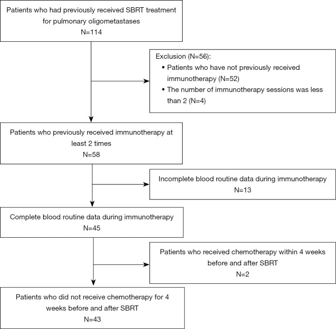
This retrospective cohort study focused on patients with pulmonary oligometastatic tumors receiving SBRT combined with immunotherapy. Data were collected from the Radiotherapy Department of The Affiliated Hospital of Soochow University between October 2018 and October 2022. The inclusion criteria were as follows: (I) patients aged 18 years or older with pathologically confirmed primary lesions, presence of 1–5 measurable lung metastases; (II) all metastases were examined by chest enhanced computed tomography (CT) or positron emission tomography-CT (PET-CT) multiple times. The diagnosis was made jointly by the clinician and the radiologist; (III) the primary tumor and extrapulmonary metastases were stable or showed no signs of activity; (IV) administration of immunotherapy at least twice after SBRT; (V) availability of complete blood routine data throughout treatment in the hospital, and follow-up for more than 1 month after immunotherapy. Meanwhile, the exclusion criteria were the following: (I) incomplete blood routine data; (II) patients with serious and uncontrollable medical disease; (III) administration of fewer than two rounds of immunotherapy after SBRT due to personal reasons or severe adverse immune reactions (Figure 1); (IV) to minimize the effect of myelosuppression after chemotherapy on the neutrophil-to-lymphocyte ratio (NLR) value, patients who received chemotherapy within 4 weeks before and after SBRT were also excluded. This study was conducted in accordance with the Declaration of Helsinki (as revised in 2013). It was approved by the medical ethics committee of The Affiliated Hospital of Soochow University [No. 440(2024)]. The ethics committee waived the necessity of informed consent due to the retrospective analysis of routine data. Patient records/information were anonymized and deidentified prior to analysis.
Figure 1.
Flowchart of patient inclusion and exclusion. SBRT, stereotactic body radiation therapy.
The determination of sample size and the collection of clinical data
In order to ensure the statistical efficacy of the sample size, we used G*Power (version 3.1) to calculate the effective sample size needed for our study, and finally set the total sample size to 34 (19). The patients’ baseline information was collected and recorded as follows: (I) general information, including age, sex, Eastern Cooperative Oncology Group (ECOG) score, and smoking history; (II) tumor-related information, including the primary tumor site, pathological type, duration of SBRT, number of target areas, maximum diameter of lung metastasis, planning target volume (PTV), SBRT dose segmentation scheme, and whether antitumor drugs such as chemotherapy, targeted therapy, and immunotherapy were received after SBRT; (III) hematology data, including blood routine indicators 4 weeks before and after SBRT. In instances where multiple blood routine data were available, the values closest to the start time of SBRT (used as blood indicators 4 weeks before radiotherapy) and the values closest to the end of SBRT were recorded. Absolute lymphocyte count (ALC) and neutrophil count (NE) were collected from blood routine data. The NLR is calculated by dividing the NE by the ALC, which are measured simultaneously in each patient’s routine blood examination.
Stereotactic radiotherapy
In this study, 43 patients received local radiotherapy for pulmonary oligometastases, with stereotactic radiotherapy being the chosen modality. During simulated positioning, all patients were fixed in a supine position, lying flat on a vacuum mat, with their hands behind their heads. CT scanning commenced after the respiratory rhythm was stabilized. The scan encompassed the area from the incisor to the lower margin of the second lumbar vertebra, with a scanning layer thickness of 3 mm. Tumor targets of all patients were mapped with the Eclipse 13.6 planning system (Varian Medical Systems, Palo Alto, CA, USA). In combination with the patient’s positron emission tomography-CT (PET-CT) and other imaging data, clinicians delineated the target area and the organs at risk (OARs) on the localization CT scan images. The gross tumor volume (GTV) was outlined within the lung window (window width 1,600, window position −600), while the PTV was formed by expanding 5 mm beyond the GTV. The outline of the OARs included the lungs, trachea, heart and large blood vessels, skin, and spinal cord. According to the number, size, and location of the lesions, as well as the patient’s physical condition and tolerance, clinicians employed corresponding dose segmentation schemes, including 5 Gy × 10 f, 9 Gy × 5 f, 8 Gy × 8 f, 8 Gy × 7 f, 8 Gy × 6 f, 8 Gy × 5 f, 7 Gy × 8 f, 7 Gy × 7 f, 7 Gy × 6 f, 6 Gy × 10 f, 6 Gy × 8 f, 6 Gy × 5 f, 5 Gy × 8 f, 5 Gy × 6 f, and 5 Gy × 5 f. The presence of multiple dose segmentation modes was not conducive to making comparisons between groups. With reference to relevant literature, we employed the Linear-Quadric (LQ) model equation to uniformly convert physical doses in various dose segmentation modes into biologically effective doses (BEDs) using the following formula: BED = n × d × [1 + d/(α/β)], where n is the number of irradiations, d is the single irradiation dose, α/β is the irradiation dose when the killing contribution caused by single-strand breaks and double-strand breaks in the tissue is equal, and α/β is generally set to 10 for solid tumors.
Patient follow-up
All patients were followed up until their death through telephone follow-up and hospital visits. The deadline for the follow-up period was June 30, 2022. The follow-up process included an examination of the patient’s local recurrence, distant metastasis, survival, and incidence of radiotherapy-related adverse reactions. After the completion of SBRT treatment, patients underwent the initial chest CT reexamination so that their condition at 1 month could be evaluated. Subsequent chest CT scans were scheduled every 3 months for the first two years and then every 6 months thereafter. OS was defined as the time between the end of pulmonary oligometastatic SBRT and the date of tumor-related death or the follow-up deadline. Progression-free survival (PFS) was defined as the time between the end of oligometastatic SBRT and the date of the first occurrence of disease progression, including local recurrence, pulmonary progression, distant progression, or the follow-up deadline. LC was defined as the time between the end of pulmonary oligometastatic SBRT and the date of emergence of new lesions within or at the edge of the PTV or the recurrence of the original lesion.
Statistical methods
For baseline data, quantitative variables are expressed as median and interquartile range (IQR), and categorical variables are expressed as counts and percentages. Wilcoxon paired rank sum test was used to compare the changes of ALC, NE and NLR before and after SBRT. Kaplan-Meier (KM) method was used for survival analysis and corresponding survival curve was generated. Cox proportional hazard regression model was used for univariate and multifactor analysis, and it was used to calculate the hazard ratio (HR) and 95% confidence interval (95% CI). Results with P value <0.05 were considered statistically significant. The receiver operating characteristic (ROC) curve of NLR after SBRT was plotted, and the corresponding optimal truncation value was calculated using the Youden index. The corresponding KM survival curve was plotted respectively for the group with NLR greater than or equal to the cut-off value and the group with NLR less than the cut-off value after SBRT, and the difference between the two groups was compared by log-rank method. P value <0.05 was considered as statistically significant. Spearman rank correlation analysis was used to determine the relationship between PTV and ALC before and after SBRT and NE after SBRT. Linear regression was used to analyze the relationship between ALC and clinical factors after SBRT. Statistical analysis was performed using GraphPad Prism version 8.4.0 (GraphPad Software) to plot the survival curve.
Results
Clinical characteristics of patients
We reviewed the clinical data of 43 patients with pulmonary oligometastatic tumors who received SBRT combined with immunotherapy (Table 1). ALC, NE, NLR and other relevant indicators were collected before and after SBRT. The study cohort comprised 31 (72.09%) males and 12 (27.91%) females, with 26 (60.47%) patients younger than 65 years old and 17 (39.53%) patients aged 65 years and above. ECOG scores were 0–1 in 30 (69.77%) cases and higher than 1 in 13 (30.23%) cases. There were 14 (32.56%) cases of primary lung cancer, 7 (16.28%) cases of primary colorectal cancer, 15 (34.88%) cases of primary esophageal cancer, 7 (16.28%) cases of primary head and neck malignant tumors, and 7 cases of other tumors, including 4 cases of cervical cancer, 1 case of endometrial cancer, 1 case of kidney cancer, and 1 case of thymus cancer. The primary tumor pathological type was squamous cell carcinoma in 24 (55.81%) cases and non-squamous cell carcinoma in 19 (44.19%) cases. Among the 39 (90.70%) patients who underwent chemotherapy, the interval between chemotherapy and SBRT was more than 4 weeks, and 4 (9.30%) patients had not undergone chemotherapy prior to SBRT. Of the 43 patients, a total of 55 metastatic lesions were treated with SBRT, 35 patients were single metastases in the lung, 5 patients were irradiated with 2 metastases, 2 patients were irradiated with 3 metastases, and 1 patient was irradiated with 4 metastases. Patients with ≥2 nodules were analyzed for survival by nodule with the largest diameter. There were 29 patients with maximum lesion diameter <2 cm and 14 patients with maximum lesion diameter ≥2 cm. In patients with multiple irradiated lesions, the PTV volume was defined as the sum of the PTV volume of multiple lesions without overlapping. The median PTV volume was 27.80 cm2, and the interquartile interval was 19.7–59.2 cm3. The median BED is 100 Gy, and the interquartile interval is 80–100 Gy.
Table 1. Clinical characteristics of 43 patients.
| Characteristics | N (%) |
|---|---|
| Age (years) | |
| <65 | 26 (60.47) |
| ≥65 | 17 (39.53) |
| Sex | |
| Female | 12 (27.91) |
| Male | 31 (72.09) |
| ECOG score | |
| >1 | 13 (30.23) |
| 0–1 | 30 (69.77) |
| Smoking history | |
| Yes | 19 (44.19) |
| No | 24 (55.81) |
| Primary tumor | |
| Lung cancer | 14 (32.56) |
| Esophagus cancer | 15 (34.88) |
| Colorectal cancer | 7 (16.28) |
| Head and neck cancer | 7 (16.28) |
| Pathological type | |
| Squamous carcinoma | 24 (55.81) |
| Nonsquamous carcinoma | 19 (44.19) |
| Chemotherapy | |
| Yes | 39 (90.70) |
| No | 4 (9.30) |
| Targeted therapy | |
| Yes | 28 (65.12) |
| No | 15 (34.88) |
| Time interval between radiotherapy and immunotherapy (days) | |
| ≤21 | 22 (51.16) |
| >21 | 21 (48.84) |
| SBRT target size (cm) | |
| ≥2 | 14 (32.56) |
| <2 | 29 (67.44) |
| Number of targets | |
| 1 | 35 (81.40) |
| >1 | 8 (18.60) |
| BED (Gy), median [IQR] | 100 [80–100] |
| PTV (cm2), median [IQR] | 27.80 [19.7–59.2] |
ECOG, Eastern Cooperative Oncology Group; SBRT, stereotactic body radiotherapy; BED, biologically effective dose; PTV, planning target volume; IQR, interquartile range.
Analysis of ALC, NE, and NLR changes before and after SBRT
The Wilcoxon paired rank sum test was used to compare the changes of ALC, NE, and NLR before and after SBRT, and the results showed that changes in both ALC and NLR were statistically significant (both P values <0.001), while the change of NE was not statistically significant, as illustrated in Table 2. To determine whether the timing of blood drawing after SBRT affected the NLR value, the Mann-Whitney test was employed. The analysis revealed that varying the blood drawing time after radiotherapy did not yield statistically significant differences in the NLR value after SBRT, as shown in Table 3.
Table 2. Changes in ALC, NE, and NLR before and after SBRT.
| Index | Before SBRT | After SBRT | Amplitude of change | P |
|---|---|---|---|---|
| ALC | 1.22 (0.96–1.42) | 0.98 (0.64–1.12) | −22.73 | <0.001 |
| NE | 3.09 (2.38–4.18) | 2.94 (2.19–4.05) | −9.46 | 0.58 |
| NLR | 2.71 (2.13–3.64) | 3.16 (2.25–5.34) | 38.04 | <0.001 |
Values are presented as median (range). ALC, absolute lymphocyte count; NE, neutrophil count; NLR, neutrophil-to-lymphocyte ratio; SBRT, stereotactic body radiotherapy.
Table 3. Relationship between blood drawing time after radiotherapy and NLR after SBRT.
| Index | 1–2 weeks (n=23) | 3–4 weeks (n=20) | Z | P |
|---|---|---|---|---|
| NLR | 3.06 (2.23–4.46) | 3.27 (2.46–6.25) | −0.546 | 0.58 |
Values are presented as median (range). NLR, neutrophil-to-lymphocyte ratio; SBRT, stereotactic body radiotherapy.
Survival analysis
At the cutoff of June 30, 2022, the median follow-up time was 22.00 (IQR, 14.00–31.00) months. The median survival time was 38.00 (IQR, 17.24–58.76) months, and the 1-year and 2-year OS rates were 83.46% and 60.99%, respectively. The median PFS was 27.00 (17.84–36.13) months, and the 1-year and 2-year PFS rates were 69.92% and 54.25%, respectively. Additionally, the 1-year and 2-year LC rates were 90.3% and 87.5%, respectively (Figures 2-4).
Figure 2.
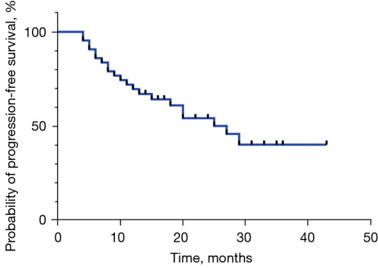
Progression-free survival curve of 43 patients.
Figure 3.
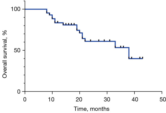
Overall survival curve of 43 patients.
Figure 4.
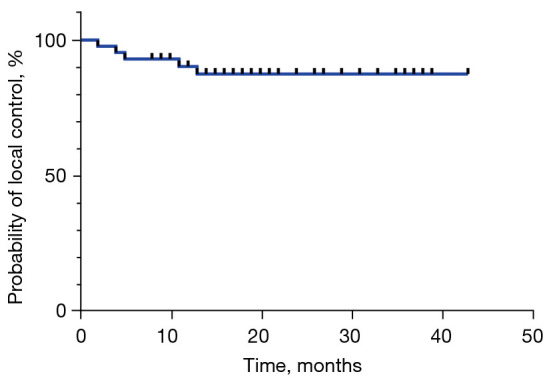
Local control rate curve of 43 patients.
Analysis of the prognostic factors of OS in SBRT combined with immunotherapy
Cox regression was used to analyze the association with OS of age, ECOG score, primary tumor site, primary tumor pathological type, smoking status, BED, number of SBRT target areas, administration of targeted therapy postradiotherapy, administration of chemotherapy postradiotherapy, PTV, SBRT interval immunotherapy time, ALC, NLR before SBRT, and ALC and NLR after SBRT. Multivariate analysis revealed NLR to be an independent prognostic factor for OS after radiotherapy [hazard ratio (HR) =1.10, 95% CI: 1.01–1.9; P=0.02]. Furthermore, an SBRT interval immunotherapy duration of ≤21 days, indicative of simultaneous SBRT combined with immunotherapy, was associated with longer OS (HR =0.24, 95% CI: 0.06–0.89; P=0.03), as shown in Table 4.
Table 4. Univariate and multivariate Cox analysis of overall survival.
| Variables | Univariate analysis | Multivariate analysis | |||||
|---|---|---|---|---|---|---|---|
| HR | 95% CI | P | HR | 95% CI | P | ||
| Age (<65 years vs. ≥65 years) | 0.50 | 0.16–1.55 | 0.22 | ||||
| ECOG score (≤1 vs. >1) | 1.61 | 0.51–5.02 | 0.41 | ||||
| Primary tumor | 0.99 | 0.66–1.46 | 0.94 | ||||
| Pathological type (squamous vs. nonsquamous) | 0.72 | 0.27–1.92 | 0.50 | ||||
| Smoking history (yes vs. no) | 0.96 | 0.35–2.62 | 0.94 | ||||
| BED | 1.00 | 0.97–1.04 | 0.91 | ||||
| Number of targets (1 vs. >1) | 1.43 | 0.32–6.35 | 0.63 | ||||
| Chemotherapy (yes vs. no) | 0.76 | 0.17–3.41 | 0.72 | ||||
| Targeted therapy (yes vs. no) | 3.07 | 0.87–10.81 | 0.08 | ||||
| SBRT target size (<2 vs. ≥2 cm) | 1.28 | 0.41–3.97 | 0.67 | ||||
| PTV | 1.03 | 1.00–1.06 | 0.07 | ||||
| Time interval between radiotherapy and immunotherapy ≤21 vs. >21 days | 0.18 | 0.05–0.63 | 0.007 | 0.24 | 0.06–0.89 | 0.03 | |
| ALC before SBRT | 0.38 | 0.11–1.28 | 0.11 | ||||
| NLR before SBRT | 1.23 | 0.89–1.71 | 0.20 | ||||
| ALC after SBRT | 0.07 | 0.01–0.37 | 0.002 | ||||
| NLR after SBRT | 1.14 | 1.06–1.23 | <0.001 | 1.10 | 1.01–1.9 | 0.02 | |
HR, hazard ratio; CI, confidence interval; ECOG, Eastern Cooperative Oncology Group; BED, biologically effective dose; SBRT, stereotactic body radiotherapy; PTV, planning target volume; ALC, absolute lymphocyte count; NLR, neutrophil-to-lymphocyte ratio.
According to the ROC curve analysis, an optimal NLR cutoff value of 4.12 was identified, yielding the largest Youden index of 0.826, with a sensitivity is 68.75 and a specificity of 88.89. The patients were divided into two groups based on the cutoff value. KM survival analysis was used to analyze the OS outcomes for both groups, and the difference was found to be statistically significant. The results showed that the group with NLR ≤4.12 after SBRT exhibited a longer median survival time (log-rank P=0.001), as shown in Figures 5,6.
Figure 5.
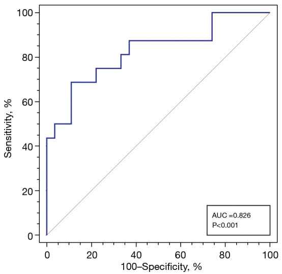
ROC curve of OS prediction by NLR after SBRT. ROC, receiver operating characteristic; AUC, area under the ROC curve; OS, overall survival; NLR, neutrophil-to-lymphocyte ratio; SBRT, stereotactic body radiotherapy.
Figure 6.
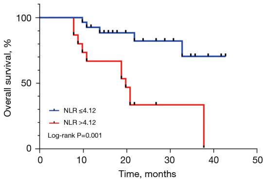
OS curves of different NLRs after SBRT. OS, overall survival; NLR, neutrophil-to-lymphocyte ratio; SBRT, stereotactic body radiotherapy.
Correlation analysis between PTV and ALC after SBRT
Spearman rank correlation analysis was employed to analyze the relationship between the PTV and various parameters, specifically ALC after SBRT, ALC before SBRT, and NE after SBRT. There was no statistically significant correlation between PTV and ALC before SBRT, and NE after SBRT. However, PTV was negatively correlated with ALC after SBRT (r=−0.41; P=0.006), and the difference was statistically significant (Figures 7-9).
Figure 7.
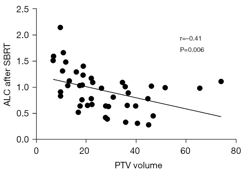
Correlation between ALC and PTV volume after SBRT. ALC, absolute lymphocyte count; PTV, planning target volume; SBRT, stereotactic body radiotherapy.
Figure 8.
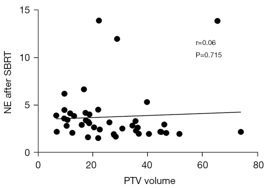
Correlation between NE and PTV volume after SBRT. NE, neutrophil count; PTV, planning target volume; SBRT, stereotactic body radiotherapy.
Figure 9.
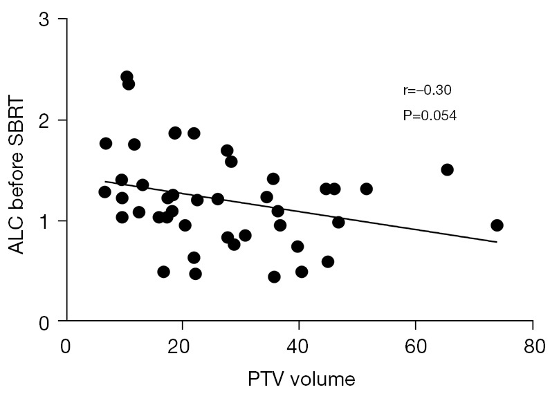
Correlation between ALC and PTV volume before SBRT. ALC, absolute lymphocyte count; PTV, planning target volume; SBRT, stereotactic body radiotherapy.
After the normality test, the ALC count after SBRT conformed to a normal distribution (Kolmogorov-Smirnov: Z=0.635, P=0.81). Consequently, linear regression was employed to analyze the association of various factors with ALC after SBRT, including age, primary tumor site, pathological type of primary tumor, administration of chemotherapy post-radiotherapy, administration of targeted therapy post-radiotherapy, PTV, number of SBRT target areas, immunological interval after radiotherapy, and ALC count before SBRT. The analysis revealed a positive correlation between ALC before SBRT and ALC after SBRT (P<0.001). Furthermore, PTV was negatively correlated with ALC after SBRT (P=0.04), and the difference was statistically significant, as shown in Table 5.
Table 5. Univariate and multivariate linear regression analysis of ALC after SBRT.
| Variables | Univariate analysis | Multivariate analysis | |||||
|---|---|---|---|---|---|---|---|
| B | 95% CI | P | B | 95% CI | P | ||
| Age | 0.08 | −0.17 to 0.34 | 0.50 | ||||
| Primary tumor | −0.04 | −0.14 to 0.06 | 0.42 | ||||
| Pathological type | 0.04 | −0.21 to 0.29 | 0.76 | ||||
| Chemotherapy | 0.15 | −0.28 to 0.58 | 0.48 | ||||
| Targeted therapy | −0.08 | −0.34 to 0.18 | 0.55 | ||||
| PTV | −0.01 | −0.02 to 0 | <0.001 | −0.01 | −0.01 to 0 | 0.04 | |
| Number of targets | −0.13 | −0.45 to 0.19 | 0.41 | ||||
| SBRT target size | 0.01 | −0.26 to 0.27 | 0.95 | ||||
| Time interval between radiotherapy and immunotherapy | 0.21 | −0.03 to 0.45 | 0.07 | ||||
| ALC before SBRT | 0.54 | 0.33 to 0.74 | <0.001 | 0.26 | 0.26 to 0.68 | <0.001 | |
ALC, absolute lymphocyte count; SBRT, stereotactic body radiotherapy; CI, confidence interval; PTV, planning target volume.
Discussion
An increasing number of studies support radiotherapy for improving the immunogenicity of tumor cells. Notably, SBRT has emerged as a prominent modality due to its potential effect on immune function, regulation of the TME, and promotion of the body’s antitumor immune response. For example, it can enhance the release of antigens for immune recognition, regulate the immune mechanism of tumors, and promote the infiltration of immune cells (20). The TME is immunosuppressive and involves multiple mechanisms for achieving the immune escape of tumor cells, which is the main drug resistance mechanism associated with ICIs. SBRT can change tumor cells from a “cold” to “hot” state, rendering them more sensitive to ICI treatment, by regulating the TME. Therefore, SBRT combined with immunotherapy has a synergistic effect, which has been confirmed by multiple preclinical studies (21). At present, many clinical trials investigating the combination of treatment SBRT and ICIs in the treatment of advanced tumors are underway (22,23). Spaas and his team conducted a phase II randomized controlled study, the CHEER study, which investigated SBRT combined with immunotherapy in multiple stage IV tumors. The primary endpoint was PFS, and the secondary endpoints included OS, objective response rate, LC, quality of life, and safety. The study is ongoing, with preliminary results suggesting that SBRT combined with immunotherapy across various advanced solid tumors could not only improve the LC rate but also effectively control the tumor lesions in the irradiation field (24).
In our study, we retrospectively analyzed 43 patients treated with SBRT combined with immunotherapy and analyzed the efficacy through LC, PFS, OS, and other survival indicators. The results showed that the 1- and 2-year LC rates were 90.3% and 87.5%, respectively, the 1-year and 2-year OS rates were 83.5% and 61.0%, respectively, and the 1-year and 2-year PFS rate was 69.9% and 54.3%, respectively; no adverse reactions of grade 3 or higher were observed. In terms of LC rate, compared with the existing SBRT-related studies on pulmonary oligometastatic tumors, the data in our study were similar or achieved a higher LC rate (25). For example, in the study of Sharma et al., the 2-year LC rate of SBRT in the treatment of lung oligometastases was 85%, while in our study it was 87.5% (26). This suggests that SBRT combined with immunotherapy has a high LC rate and survival rate, along with acceptable safety. The results of multivariate analysis for OS indicated that patients who received concurrent radiotherapy and immunotherapy (interval ≤21 days) had longer OS than those who received sequential radiotherapy (interval >21 days). This result was similar to that in previous research. The retrospective study of Woody et al. showed that patients who received SBRT before immunotherapy had notably worse OS outcomes than those who received concurrent or subsequent immunotherapy. This may be attributed to SBRT’s potential to enhance tumor immunogenicity and sensitize ICIs by regulating the TME (27). At present, the optimal dose segmentation mode and the optimal interval time of SBRT combined with ICIs remain unclear. The SAFRON II trial compared the efficacy of single segmentation (28 Gy/1 f) and multi-segmentation (12 Gy/4 f) SBRT in the treatment of pulmonary oligometastatic tumors, and there was no significant difference in OS or disease-free survival between the two groups (28). Preclinical data suggest that immunotherapy is most effective in preventing delayed presentation of antigens in an immune-tolerant setting when combined in parallel or in close sequence with SBRT; however, this has not been proven by clinical studies. Therefore, more large-scale retrospective and prospective studies are imperative to confirm these findings. Our study also showed that NLR is an independent prognostic factor for OS after SBRT. The higher the NLR after SBRT, the poorer the patient’s OS. NLR is defined as the ratio of peripheral blood NE to lymphocyte count. This ratio reflects an enhanced neutrophil-dependent inflammatory response and/or a reduced lymphocyte-mediated antitumor immune response. Platelet-to-lymphocyte ratio (PLR) and peripheral blood lymphocyte-to-monocyte ratio (LMR) are also indicators reflecting the body’s inflammatory response (29,30). Studies have shown that systemic immune-inflammation index (SII), NLR, PLR, LMR, and other inflammatory indicators have certain predictive value in the prognosis of various tumor types, among which peripheral blood SII, NLR and PLR are considered to be negatively correlated with OS and LMR positively correlated with tumor prognosis (31-34). This predictive value is particularly pronounced in patients receiving immunotherapy and radiotherapy, as lymphocyte count is closely associated with the level of radiotherapy and immunotherapy. For example, a retrospective study showed that SII could predict the resection rate and OS of esophageal squamous cell carcinoma patients after neoadjuvant radiotherapy. Low SII (less than or equal to 916.6×10⁹/L) was associated with longer OS. a higher SII was associated with a lower resect ability rate (34). Notably, a high NLR has been shown to be an independent risk factor for survival in patients receiving immunotherapy. Moreover, a meta-analysis suggested that NLR could be used as a prognostic indicator for SBRT patients (35). Analysis results from three phase I–II trials at MD Anderson Cancer Center showed that postradiotherapy ALC predicted the distant effects and survival outcomes of patients receiving combined immunotherapy with radiotherapy. Although these preliminary findings need to be confirmed with additional data, they point to the importance of monitoring ALC in patients receiving combined immunotherapy with radiotherapy and the potential of manipulating this parameter to induce a distant effect (36). In this study, the Wilcoxon paired rank sum test was used to compare the changes in ALC, NE, and NLR before and after SBRT, and the results showed that there were statistically significant changes in ALC (P<0.0001) and NLR (P=0.007) but not in NE. This suggests that the primary factor influencing the change in NLR after SBRT is the reduction of ALC after SBRT; that is, the depletion of lymphocytes, which has also been confirmed by a previous study (37). Lymphocytes play a key role in cancer surveillance. Cytotoxic T cells and natural killer cells are the key mediators of anti-tumor response, activated B cells activate tumor-infiltrating lymphocytes to obtain anti-tumor activity, and lymphocyte depletion reflects the impaired anti-tumor response (38). Programmed cell death protein 1 (PD-1) antibodies induce immunogenic cell death by directly activating cytotoxic T lymphocytes (39,40). Therefore, an increased NLR after SBRT may be interpreted as SBRT-induced lymphocytopenia, with SBRT reducing the body’s response to ICIs and resulting in shortened survival. Although there was no statistically significant difference in NE before and after SBRT in our study, related studies have reported that neutrophils can secrete signaling molecules that promote tumor angiogenesis and escape while inhibiting cytotoxic T lymphocytes, indicating that neutrophils also play a regulatory role in tumor immune response (41,42). Recently, the prognostic value of NLR has been demonstrated in clinical trials involving patients treated with ICIs (43). The study results of Li et al. showed that in patients with advanced solid tumors receiving immunotherapy, an NLR ≥5 during immunotherapy predicted worse OS (44). However, few studies have explored the ability of NLR to predict the prognosis of patients receiving SBRT. A retrospective study with a small sample showed that the higher the NLR and PLR were after SBRT, the worse the survival outcomes of patients with hepatocellular carcinoma. Both the NLR and PLR increased significantly after SBRT and slowly decreased to the pre-SBRT value 6 months later (45).
Our study involved certain limitations that should be mentioned. First, our sample size was small and might have introduced a degree of selection bias. Second, since most patients receiving SBRT combined with immunotherapy lacked previous peripheral blood lymphocyte subsets analyses, indicators related to lymphocyte subsets were excluded, limiting the ability to further determine the correlation between SBRT and lymphocytopenia.
Recently, it has been reported that the PTV may be related to the reduction of lymphocyte count after radiotherapy. Meanwhile, some radiotherapy parameters, such as lung V5, mean lung dose, and heart V5, can also predict radiation-related lymphocytopenia (46,47). However, there are few studies investigating the prediction of PTV for the efficacy of SBRT combined with immunotherapy. It has been reported that in patients with pancreatic cancer receiving SBRT combined with immunotherapy, PTV could predict the reduction of ALC after SBRT, and the reduction of ALC after SBRT was related to shorter OS, with PTV also being associated with OS (38). Chen et al.’s study showed that when combined with immunotherapy, SBRT may better preserve lymphocytes and improve survival outcomes as compared to traditional conventional fractionated radiotherapy (48). In our study, Spearman rank correlation analysis showed that PTV was negatively correlated with ALC after SBRT. Moreover, linear regression analysis also showed that PTV was correlated with ALC reduction after SBRT, aligning with previous findings (38). However, in the OS-related Cox regression analysis, there was no statistical significance between PTV and OS prognosis. This may be due to the small sample size, which failed to fully reflect the difference between groups. Notably, at the data level, the HR in the Cox univariate analysis was greater than 1, indicating a risk factor association, suggesting that a large PTV may be associated with poor OS. Narrowing the PTV range could potentially mitigate the shortening of OS indirectly caused by lymphocytopenia. Further prospective studies are needed to demonstrate the correlation between dynamic changes in PTV, NLR, and survival outcomes in patients treated with SBRT combined with immunotherapy.
Conclusions
According to our preliminary research findings, it can be preliminarily posited that SBRT combined with immunotherapy can achieve satisfactory LC, long PFS and OS, and improve the quality of life of patients with pulmonary oligometastatic tumors, with the side effects being controllable. SBRT and drug therapy, especially immunotherapy, can complement one another in the treatment of lung metastases and enhance clinical efficacy. However, additional prospective randomized controlled studies are needed to confirm their combined clinical value. Furthermore, investigations are needed to identify the optimal segmentation mode of SBRT, determine the interval time of combined immunotherapy, optimize the timing of treatment, and develop individualized and comprehensive treatment plans for patients with multi-tumor lung metastases and other types of tumor.
Supplementary
The article’s supplementary files as
Acknowledgments
Funding: This work was supported by the grants from the National Natural Science Foundation of China (Nos. 81970138 and 82270165), the National Key R&D Program of China (No. 2022YFC2502703), the Jiangsu Province Natural Science Foundation of China (No. BK20221235), the Translational Research Grant of NCRCH (No. 2020ZKMB05), the Jiangsu Province “333” Project, Social Development Project of the Science and Technology Department of Jiangsu (No. BE2021649), the Boxi Clinical Research Project (No. BXLC008), and the Gusu Key Medical Talent Program (No. GSWS2019007).
Ethical Statement: The authors are accountable for all aspects of the work in ensuring that questions related to the accuracy or integrity of any part of the work are appropriately investigated and resolved. This study was conducted in accordance with the Declaration of Helsinki (as revised in 2013). It was approved by the medical ethics committee of The Affiliated Hospital of Soochow University [No. 440(2024)]. The ethics committee waived the necessity of informed consent due to the retrospective analysis of routine data. Patient records/information were anonymized and deidentified prior to analysis.
Footnotes
Reporting Checklist: The authors have completed the STROBE reporting checklist. Available at https://tlcr.amegroups.com/article/view/10.21037/tlcr-24-588/rc
Conflicts of Interest: All authors have completed the ICMJE uniform disclosure form (available at https://tlcr.amegroups.com/article/view/10.21037/tlcr-24-588/coif). The authors have no conflicts of interest to declare.
Data Sharing Statement
Available at https://tlcr.amegroups.com/article/view/10.21037/tlcr-24-588/dss
References
- 1.Hellman S, Weichselbaum RR. Oligometastases. J Clin Oncol 1995;13:8-10. 10.1200/JCO.1995.13.1.8 [DOI] [PubMed] [Google Scholar]
- 2.Izmailov T, Ryzhkin S, Borshchev G, et al. Oligometastatic Disease (OMD): The Classification and Practical Review of Prospective Trials. Cancers (Basel) 2023;15:5234. 10.3390/cancers15215234 [DOI] [PMC free article] [PubMed] [Google Scholar]
- 3.Boda-Heggemann J, Frauenfeld A, Weiss C, et al. Clinical outcome of hypofractionated breath-hold image-guided SABR of primary lung tumors and lung metastases. Radiat Oncol 2014;9:10. 10.1186/1748-717X-9-10 [DOI] [PMC free article] [PubMed] [Google Scholar]
- 4.Baumann P, Nyman J, Hoyer M, et al. Outcome in a prospective phase II trial of medically inoperable stage I non-small-cell lung cancer patients treated with stereotactic body radiotherapy. J Clin Oncol 2009;27:3290-6. 10.1200/JCO.2008.21.5681 [DOI] [PubMed] [Google Scholar]
- 5.Timmerman RD, Paulus R, Pass HI, et al. Stereotactic Body Radiation Therapy for Operable Early-Stage Lung Cancer: Findings From the NRG Oncology RTOG 0618 Trial. JAMA Oncol 2018;4:1263-6. 10.1001/jamaoncol.2018.1251 [DOI] [PMC free article] [PubMed] [Google Scholar]
- 6.Klement RJ, Hoerner-Rieber J, Adebahr S, et al. Stereotactic body radiotherapy (SBRT) for multiple pulmonary oligometastases: Analysis of number and timing of repeat SBRT as impact factors on treatment safety and efficacy. Radiother Oncol 2018;127:246-52. 10.1016/j.radonc.2018.02.016 [DOI] [PubMed] [Google Scholar]
- 7.Cao Y, Chen H, Sahgal A, et al. The impact of local control on widespread progression and survival in oligometastasis-directed SBRT: Results from a large international database. Radiother Oncol 2023;186:109769. 10.1016/j.radonc.2023.109769 [DOI] [PubMed] [Google Scholar]
- 8.Tree AC, Khoo VS, Eeles RA, et al. Stereotactic body radiotherapy for oligometastases. Lancet Oncol 2013;14:e28-37. 10.1016/S1470-2045(12)70510-7 [DOI] [PubMed] [Google Scholar]
- 9.Mayinger M, Kotecha R, Sahgal A, et al. Stereotactic Body Radiotherapy for Lung Oligo-metastases: Systematic Review and International Stereotactic Radiosurgery Society Practice Guidelines. Lung Cancer 2023;182:107284. 10.1016/j.lungcan.2023.107284 [DOI] [PubMed] [Google Scholar]
- 10.Turchan WT, Chmura SJ. The role of immunotherapy in combination with oligometastasis-directed therapy: a narrative review. Ann Palliat Med 2021;10:6028-44. 10.21037/apm-20-1528 [DOI] [PubMed] [Google Scholar]
- 11.Lee Y, Auh SL, Wang Y, et al. Therapeutic effects of ablative radiation on local tumor require CD8+ T cells: changing strategies for cancer treatment. Blood 2009;114:589-95. 10.1182/blood-2009-02-206870 [DOI] [PMC free article] [PubMed] [Google Scholar]
- 12.Ashrafizadeh M, Farhood B, Eleojo Musa A, et al. Abscopal effect in radioimmunotherapy. Int Immunopharmacol 2020;85:106663. 10.1016/j.intimp.2020.106663 [DOI] [PubMed] [Google Scholar]
- 13.Yang R, Sun L, Li CF, et al. Galectin-9 interacts with PD-1 and TIM-3 to regulate T cell death and is a target for cancer immunotherapy. Nat Commun 2021;12:832. 10.1038/s41467-021-21099-2 [DOI] [PMC free article] [PubMed] [Google Scholar]
- 14.Zou W, Chen L. Inhibitory B7-family molecules in the tumour microenvironment. Nat Rev Immunol 2008;8:467-77. 10.1038/nri2326 [DOI] [PubMed] [Google Scholar]
- 15.Ying X, You G, Shao R. The analysis of the efficacy and safety of stereotactic body radiotherapy with sequential immune checkpoint inhibitors in the management of oligoprogressive advanced non-small cell lung cancer. Transl Cancer Res 2024;13:2408-18. 10.21037/tcr-23-2232 [DOI] [PMC free article] [PubMed] [Google Scholar]
- 16.Nguyen NP, Ali A, Vinh-Hung V, et al. Stereotactic Body Radiotherapy and Immunotherapy for Older Patients with Oligometastases: A Proposed Paradigm by the International Geriatric Radiotherapy Group. Cancers (Basel) 2022;15:244. 10.3390/cancers15010244 [DOI] [PMC free article] [PubMed] [Google Scholar]
- 17.Lin AJ, Roach M, Bradley J, et al. Combining stereotactic body radiation therapy with immunotherapy: current data and future directions. Transl Lung Cancer Res 2019;8:107-15. 10.21037/tlcr.2018.08.16 [DOI] [PMC free article] [PubMed] [Google Scholar]
- 18.Luke JJ, Lemons JM, Karrison TG, et al. Safety and Clinical Activity of Pembrolizumab and Multisite Stereotactic Body Radiotherapy in Patients With Advanced Solid Tumors. J Clin Oncol 2018;36:1611-8. 10.1200/JCO.2017.76.2229 [DOI] [PMC free article] [PubMed] [Google Scholar]
- 19.Faul F, Erdfelder E, Lang AG, et al. G*Power 3: a flexible statistical power analysis program for the social, behavioral, and biomedical sciences. Behav Res Methods 2007;39:175-91. 10.3758/bf03193146 [DOI] [PubMed] [Google Scholar]
- 20.Rodriguez-Ruiz ME, Vitale I, Harrington KJ, et al. Immunological impact of cell death signaling driven by radiation on the tumor microenvironment. Nat Immunol 2020;21:120-34. 10.1038/s41590-019-0561-4 [DOI] [PubMed] [Google Scholar]
- 21.Rückert M, Deloch L, Fietkau R, et al. Immune modulatory effects of radiotherapy as basis for well-reasoned radioimmunotherapies. Strahlenther Onkol 2018;194:509-19. 10.1007/s00066-018-1287-1 [DOI] [PubMed] [Google Scholar]
- 22.Bauml JM, Mick R, Ciunci C, et al. Pembrolizumab After Completion of Locally Ablative Therapy for Oligometastatic Non-Small Cell Lung Cancer: A Phase 2 Trial. JAMA Oncol 2019;5:1283-90. 10.1001/jamaoncol.2019.1449 [DOI] [PMC free article] [PubMed] [Google Scholar]
- 23.Theelen WSME, Peulen HMU, Lalezari F, et al. Effect of Pembrolizumab After Stereotactic Body Radiotherapy vs Pembrolizumab Alone on Tumor Response in Patients With Advanced Non-Small Cell Lung Cancer: Results of the PEMBRO-RT Phase 2 Randomized Clinical Trial. JAMA Oncol 2019;5:1276-82. 10.1001/jamaoncol.2019.1478 [DOI] [PMC free article] [PubMed] [Google Scholar]
- 24.Spaas M, Sundahl N, Hulstaert E, et al. Checkpoint inhibition in combination with an immunoboost of external beam radiotherapy in solid tumors (CHEERS): study protocol for a phase 2, open-label, randomized controlled trial. BMC Cancer 2021;21:514. 10.1186/s12885-021-08088-w [DOI] [PMC free article] [PubMed] [Google Scholar]
- 25.Yamamoto T, Niibe Y, Aoki M, et al. Analyses of the local control of pulmonary Oligometastases after stereotactic body radiotherapy and the impact of local control on survival. BMC Cancer 2020;20:997. 10.1186/s12885-020-07514-9 [DOI] [PMC free article] [PubMed] [Google Scholar]
- 26.Sharma A, Duijm M, Oomen-de Hoop E, et al. Factors affecting local control of pulmonary oligometastases treated with stereotactic body radiotherapy. Acta Oncol 2018;57:1031-7. 10.1080/0284186X.2018.1445285 [DOI] [PubMed] [Google Scholar]
- 27.Woody S, Hegde A, Arastu H, et al. Survival Is Worse in Patients Completing Immunotherapy Prior to SBRT/SRS Compared to Those Receiving It Concurrently or After. Front Oncol 2022;12:785350. 10.3389/fonc.2022.785350 [DOI] [PMC free article] [PubMed] [Google Scholar]
- 28.Siva S, Bressel M, Mai T, et al. Single-Fraction vs Multifraction Stereotactic Ablative Body Radiotherapy for Pulmonary Oligometastases (SAFRON II): The Trans Tasman Radiation Oncology Group 13.01 Phase 2 Randomized Clinical Trial. JAMA Oncol 2021;7:1476-85. 10.1001/jamaoncol.2021.2939 [DOI] [PMC free article] [PubMed] [Google Scholar]
- 29.Dong B, Zhu X, Chen R, et al. Derived Neutrophil-Lymphocyte Ratio and C-Reactive Protein as Prognostic Factors for Early-Stage Non-Small Cell Lung Cancer Treated with Stereotactic Body Radiation Therapy. Diagnostics (Basel) 2023;13:313. 10.3390/diagnostics13020313 [DOI] [PMC free article] [PubMed] [Google Scholar]
- 30.Zhou XL, Li YQ, Zhu WG, et al. Neutrophil-to-lymphocyte ratio as a prognostic biomarker for patients with locally advanced esophageal squamous cell carcinoma treated with definitive chemoradiotherapy. Sci Rep 2017;7:42581. 10.1038/srep42581 [DOI] [PMC free article] [PubMed] [Google Scholar]
- 31.Constantin GB, Firescu D, Voicu D, et al. The Importance of Systemic Inflammation Markers in the Survival of Patients with Complicated Colorectal Cancer, Operated in Emergency. Chirurgia (Bucur) 2020;115:39-49. 10.21614/chirurgia.115.1.39 [DOI] [PubMed] [Google Scholar]
- 32.Goto W, Kashiwagi S, Asano Y, et al. Predictive value of lymphocyte-to-monocyte ratio in the preoperative setting for progression of patients with breast cancer. BMC Cancer 2018;18:1137. 10.1186/s12885-018-5051-9 [DOI] [PMC free article] [PubMed] [Google Scholar]
- 33.Xiang Y, Zhang N, Lei H, et al. Neutrophil-to-lymphocyte ratio is a negative prognostic biomarker for luminal A breast cancer. Gland Surg 2023;12:415-25. 10.21037/gs-23-80 [DOI] [PMC free article] [PubMed] [Google Scholar]
- 34.Zhang H, Lin J, Huang Y, et al. The Systemic Immune-Inflammation Index as an Independent Predictor of Survival in Patients with Locally Advanced Esophageal Squamous Cell Carcinoma Undergoing Neoadjuvant Radiotherapy. J Inflamm Res 2024;17:4575-86. 10.2147/JIR.S463163 [DOI] [PMC free article] [PubMed] [Google Scholar]
- 35.Yu Y, Tan D, Liao C, et al. Neutrophil-to-lymphocyte ratio as a prognostic predictor for patients with cancer treated with stereotactic body radiation therapy: A meta-analysis. Mol Clin Oncol 2022;16:98. 10.3892/mco.2022.2531 [DOI] [PMC free article] [PubMed] [Google Scholar]
- 36.Chen D, Verma V, Patel RR, et al. Absolute Lymphocyte Count Predicts Abscopal Responses and Outcomes in Patients Receiving Combined Immunotherapy and Radiation Therapy: Analysis of 3 Phase 1/2 Trials. Int J Radiat Oncol Biol Phys 2020;108:196-203. 10.1016/j.ijrobp.2020.01.032 [DOI] [PubMed] [Google Scholar]
- 37.Reddy AV, Hill CS, Sehgal S, et al. Post-radiation neutrophil-to-lymphocyte ratio is a prognostic marker in patients with localized pancreatic adenocarcinoma treated with anti-PD-1 antibody and stereotactic body radiation therapy. Radiat Oncol J 2022;40:111-9. 10.3857/roj.2021.01060 [DOI] [PMC free article] [PubMed] [Google Scholar]
- 38.Chan KS, Shelat VG. The role of platelet-lymphocyte ratio in hepatocellular carcinoma: a valuable prognostic marker. Transl Cancer Res 2022;11:4231-4. 10.21037/tcr-22-2343 [DOI] [PMC free article] [PubMed] [Google Scholar]
- 39.Chen Y, Gao M, Huang Z, et al. SBRT combined with PD-1/PD-L1 inhibitors in NSCLC treatment: a focus on the mechanisms, advances, and future challenges. J Hematol Oncol 2020;13:105. 10.1186/s13045-020-00940-z [DOI] [PMC free article] [PubMed] [Google Scholar]
- 40.Welsh JW, Tang C, de Groot P, et al. Phase II Trial of Ipilimumab with Stereotactic Radiation Therapy for Metastatic Disease: Outcomes, Toxicities, and Low-Dose Radiation-Related Abscopal Responses. Cancer Immunol Res 2019;7:1903-9. 10.1158/2326-6066.CIR-18-0793 [DOI] [PMC free article] [PubMed] [Google Scholar]
- 41.Masucci MT, Minopoli M, Carriero MV. Tumor Associated Neutrophils. Their Role in Tumorigenesis, Metastasis, Prognosis and Therapy. Front Oncol 2019;9:1146. 10.3389/fonc.2019.01146 [DOI] [PMC free article] [PubMed] [Google Scholar]
- 42.Aarts CEM, Hiemstra IH, Béguin EP, et al. Activated neutrophils exert myeloid-derived suppressor cell activity damaging T cells beyond repair. Blood Adv 2019;3:3562-74. 10.1182/bloodadvances.2019031609 [DOI] [PMC free article] [PubMed] [Google Scholar]
- 43.Faget J, Peters S, Quantin X, et al. Neutrophils in the era of immune checkpoint blockade. J Immunother Cancer 2021;9:e002242. 10.1136/jitc-2020-002242 [DOI] [PMC free article] [PubMed] [Google Scholar]
- 44.Li M, Spakowicz D, Burkart J, et al. Change in neutrophil to lymphocyte ratio during immunotherapy treatment is a non-linear predictor of patient outcomes in advanced cancers. J Cancer Res Clin Oncol 2019;145:2541-6. 10.1007/s00432-019-02982-4 [DOI] [PMC free article] [PubMed] [Google Scholar]
- 45.Park Y, Chang AR. Neutrophil to Lymphocyte Ratio and Platelet to Lymphocyte Ratio in Hepatocellular Carcinoma Treated with Stereotactic Body Radiotherapy. Korean J Gastroenterol 2022;79:252-9. 10.4166/kjg.2022.021 [DOI] [PubMed] [Google Scholar]
- 46.Tang C, Liao Z, Gomez D, et al. Lymphopenia association with gross tumor volume and lung V5 and its effects on non-small cell lung cancer patient outcomes. Int J Radiat Oncol Biol Phys 2014;89:1084-91. 10.1016/j.ijrobp.2014.04.025 [DOI] [PubMed] [Google Scholar]
- 47.Upadhyay R, Venkatesulu BP, Giridhar P, et al. Risk and impact of radiation related lymphopenia in lung cancer: A systematic review and meta-analysis. Radiother Oncol 2021;157:225-33. 10.1016/j.radonc.2021.01.034 [DOI] [PubMed] [Google Scholar]
- 48.Chen D, Patel RR, Verma V, et al. Interaction between lymphopenia, radiotherapy technique, dosimetry, and survival outcomes in lung cancer patients receiving combined immunotherapy and radiotherapy. Radiother Oncol 2020;150:114-20. 10.1016/j.radonc.2020.05.051 [DOI] [PubMed] [Google Scholar]


