Abstract
Background
Seborrheic dermatitis (SD) is a common chronic inflammatory skin disease. In recent years, significant progress has been made in the field of SD, but there has been no bibliometric research yet. This study aims to use bibliometric methods to analyze the current research status and hot topics of SD, to understand further the research trends and future development prospects in this field.
Methods
Retrieve core literature on SD from the Web of Science database and conduct a detailed analysis using CiteSpace and VOSviewer software based on factors such as publication volume, countries (regions), research institutions, journals, authors, highly‐cited papers, and keywords.
Results
From 1996 to 2024, a total of 1436 publications were included in the bibliometric analysis. The number of publications has shown an increasing trend year by year. The USA is the leading country in this field of research. The University of California System is the primary research institution. The International Journal of Dermatology is the journal with the highest number of publications. The author Yang Won Lee has the highest number of publications, while the article “Seborrheic Dermatitis” (2004) by Gupta, A.K. has been cited the most. “Seborrheic dermatitis” is the most frequently occurring keyword. The main research hotspots and frontiers in SD are as follows: (1) The relationship between SD and other skin diseases is a popular research topic; (2) Malassezia and inflammation are current research hotspots in SD; and (3) Focusing on antifungal and anti‐inflammatory treatments for SD is the current frontier direction in this field.
Conclusion
This study is a summary of the current status and hot trends of SD research, which helps clinical doctors and researchers quickly understand the insights and valuable information of SD research and provides reference for clinical decision‐making and finding future research directions.
Keywords: bibliometric, CiteSpace, seborrheic dermatitis, visual analysis, VOSviewer
1. INTRODUCTION
Seborrheic dermatitis (SD) is a common chronic recurring inflammatory skin disease characterized by erythema, scaly patches, and a white or yellow “greasy” appearance of the scales. It usually affects areas of the body with abundant sebaceous glands, such as the scalp, chest, face, back, and groin. 1 SD can occur in people of all ages, including adults, adolescents, infants, etc. It is most common in young people, with infants mainly occurring on the scalp, while other populations can be affected in any area rich in sebaceous glands. 2 The scalp is the most common site of SD, characterized by the appearance of scales against a background of erythema, commonly referred to as scalp seborrheic dermatitis (SSD). SSD is the most common cause of scalp itching, which may lead to the progression of telogen alopecia and androgenetic alopecia. Its characteristic is the appearance of scales under the background of erythema. 3 The prevalence of this disease in the general population is 1%–3%, while it can reach 34%–83% in individuals with compromised immune systems. 4 Although the pathogenesis of SD is not fully understood, it mainly includes cutaneous microbial dysbiosis (driven by Malassezia yeast), inflammation, sebum production and skin barrier disruption. 5 Immunodeficiency (HIV), neurological (Parkinson's disease), alcoholic pancreatitis, cardiac disease are recognized risk factors for SD. 6 Due to the frequent occurrence of SD in the head, face, or other visible areas of the body, many patients experience feelings of inferiority and psychological distress, which seriously negatively affects their quality of life and incurs significant medical expenses. 7 The efficacy of existing treatment methods for SD is still controversial and requires long‐term treatment to maintain remission. 8 Therefore, it is necessary to identify new treatment targets, develop more effective treatment methods, and reduce related side effects in order to better manage these diseases.
Bibliometrics is the use of mathematics and statistics to quantitatively describe or display relationships between published works, to analyze published information and its related metadata. 9 , 10 CiteSpace and VOSviewer are commonly used software packages in visual analysis tools for analyzing bibliometric results in scientific drawing processes based on different algorithms. 11 This research method focuses on elements such as publication volume, country, author, institution, journal, highly‐cited literature, and keywords related to specific field research, providing readers with the changes and development trends of research hotspots and frontiers in the field, and identifying potential research hotspots that can provide references for future research. 12 Although the Web of Science Core Collection (WOSCC) has published much literature on SD, there is still a lack of bibliometric and visual analysis of SD‐related literature. Therefore, this study aims to review SD‐related articles published on WOSCC between 1996 and 2024, to help clinical doctors and researchers better understand the relevant research hotspots and frontiers in this field.
2. METHODS
2.1. Data sources and search strategy
The WOSCC is widely recognized by researchers as a premier bibliographic database, covering an extensive range of publications across multiple disciplines. It is considered the most suitable database for conducting bibliometric and visual analysis. 13 Consequently, WOS was chosen as the data source for this study. All data were collected on July 18, 2024, by searching the WOSCC for the literature published from 1996 to 2024. The search formula is TS = (Seborrheic Dermatitis OR Seborrheic Dermatitides OR Dermatitis Seborrheica OR Dermatitides, Seborrheic OR Seborrhea). The language restriction is “English”. The type of documents is set to “Articles” and “Review” (Figure 1). Since all the raw data utilized in this study were sourced from a public database, an ethical review was deemed unnecessary.
FIGURE 1.
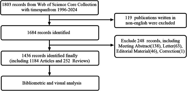
Publications screening flowchart.
2.2. Data analysis
CiteSpace (version 6.3.R1), VOSviewer (version 1.6.18), and Microsoft Excel 2019 were used for quantitative, bibliometric, and visual analysis. Download the information of English publications from WOSCC and save them separately in plain text format for analysis by various software. Excel 2019 was utilized to create a comprehensive graph illustrating the yearly growth trend of the literature. CiteSpace is a bibliometric visualization software developed by Chaomei Chen from Drexel University in Philadelphia, Pennsylvania, USA, which identifies relevant hotspots and trends within a given research field. VOSviewer is another quantitative analysis software developed by Nees Jan van Eck and Ludo Waltman from Leiden University in the Netherlands, designed to extract key information from numerous publications and construct bibliometric network maps. 14 , 15 In this study, we combined the use of two software tools, set appropriate parameters, and focused on bibliometric and visualization analysis of elements such as publication volume, countries (regions), research institutions, journals, authors, highly‐cited papers, and keywords in the field of SD research.
3. RESULTS
3.1. Quantitative analysis of publication
Figure 2 presents data exported from WoSCC and edited using Excel. According to the research retrieval strategy, a total of 1436 documents related to SD were included, comprising 1184 articles and 252 reviews. As shown in Figure 2, the publication volume on SD from 1996 to 2024 shows a phased upward trend. The first phase includes annual publication volumes of 22–39 papers, the second phase has 41–56 papers, and the third phase includes 62–78 papers. Over the past nearly 30 years, the publication level of SD‐related literature has remained relatively stable. The year 1997 had the lowest number of publications, with 22 papers, while 2022 had the highest, with 103 papers, ranking first in annual publication volume.
FIGURE 2.
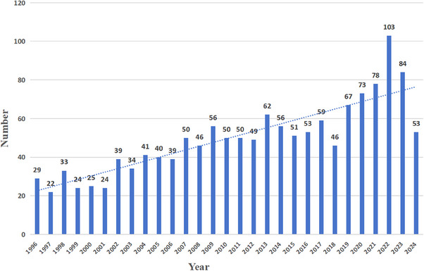
Trends in annual publications of seborrheic dermatitis from 1996 to 2024.
3.2. Country analysis
A total of 88 countries and regions have published articles related to SD. Table 1 shows the top 10 countries/regions ranked by the number of publications, citation frequency, and centrality. The top three countries by the number of articles published in this study are the USA (n = 367, 19.51%), Italy (n = 118, 6.27%), and China (n = 106, 5.05%). The betweenness centrality of a country/region measures its importance within the network. France has a centrality of 0.2, while the USA and Germany both have a centrality of 0.19. As shown in Figure 3, each circular node represents a country or region, with its size indicating the number of publications from that country/region. The thickness of the lines represents the strength of collaboration between countries/regions. The blue rings in the inner circle represent the earliest publications, while the red rings in the outer circle represent the most recent publications. The USA has the highest overall number of publications on SD, with a steady stream of new articles being published, indicating high centrality and a dominant position in the field. The USA collaborates with several countries worldwide, including Italy, China, Germany, England, Turkey, France, South Korea, and Japan.
TABLE 1.
Top 10 countries by the number of publications on seborrheic dermatitis.
| Rank | Country/region | Publications | Percentage (%) | Centrality |
|---|---|---|---|---|
| 1 | USA | 367 | 19.51 | 0.19 |
| 2 | ITALY | 118 | 6.27 | 0.07 |
| 3 | PEOPLES R CHINA | 106 | 5.05 | 0.03 |
| 4 | GERMANY | 95 | 4.89 | 0.19 |
| 5 | TURKEY | 91 | 4.84 | 0.01 |
| 6 | JAPAN | 81 | 4.31 | 0.04 |
| 7 | ENGLAND | 77 | 4.09 | 0.11 |
| 8 | SOUTH KOREA | 76 | 4.04 | 0 |
| 9 | FRANCE | 67 | 3.56 | 0.2 |
| 10 | INDIA | 60 | 3.19 | 0.05 |
FIGURE 3.
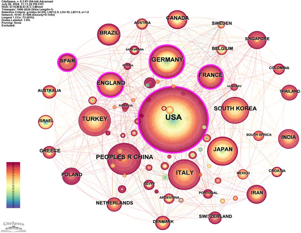
Visualization of countries researching seborrheic dermatitis.
3.3. Institutional analysis
A total of 562 academic institutions have contributed to the publication of SD‐related research. Table 2 lists the top 10 institutions ranked by the number of publications and centrality. The University of California System has the highest number of publications (n = 31, centrality = 0.16), followed by Konkuk University (n = 22, centrality = 0.01) and Chung Ang University (n = 21, centrality = 0.01). Among the top ten institutions, four are from Canada, two from the USA, two from South Korea, and one each from Greece and England. The University of California System and Procter & Gamble exhibit high centrality, indicating their significant roles in SD research. Figure 4 shows that the red outer rings represent recent publication volumes, highlighting these institutions as the most productive in the field. Additionally, Figure 4 illustrates close collaborations among institutions, forming a multi‐center cooperation model led by the University of California System, Procter & Gamble, Chung Ang University, and the National & Kapodistrian University of Athens.
TABLE 2.
Top 10 institutions researching seborrheic dermatitis.
| Rank | Affiliation | Country | Publications | Centrality |
|---|---|---|---|---|
| 1 | University of California System | USA | 31 | 0.16 |
| 2 | Konkuk University | SOUTH KOREA | 22 | 0.01 |
| 3 | Chung Ang University | SOUTH KOREA | 21 | 0.01 |
| 4 | University of Toronto | CANADA | 20 | 0.05 |
| 5 | Procter & Gamble | USA | 18 | 0.11 |
| 6 | National & Kapodistrian University of Athens | GREECE | 18 | 0.01 |
| 7 | Sunnybrook Research Institute | CANADA | 17 | 0 |
| 8 | Sunnybrook Health Science Center | CANADA | 17 | 0 |
| 9 | University of London | ENGLAND | 16 | 0.08 |
| 10 | Konkuk University Medical Center | CANADA | 16 | 0 |
FIGURE 4.
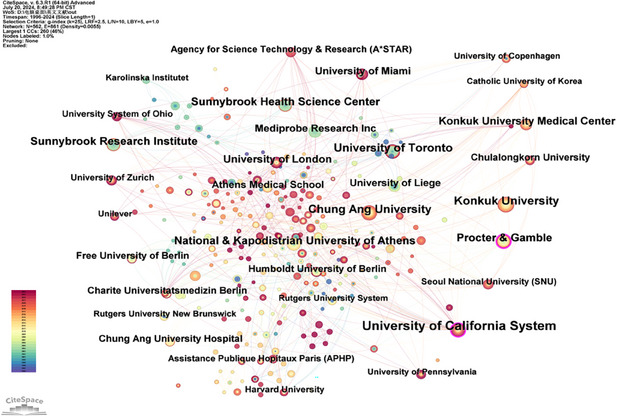
Co‐occurrence collaboration network of research institutions on seborrheic dermatitis.
3.4. Journal analysis
The impact factor (IF) and the Journal Citation Reports (JCR) quartiles are crucial indicators for assessing the influence of journals. Journals ranked within the top 25% by IF are classified into the first quartile (Q1), while those in the 25%−50% range fall into the second quartile (Q2). This study encompasses 456 journals that have published research related to SD. Table 3 lists the top 10 journals ranked by publication volume, citation count, average citations per item, JCR, and IF. Notably, the three journals with the highest publication volume are the International Journal of Dermatology (IF 3.5, Q1), the Journal of the American Academy of Dermatology (IF 12.8, Q1), and Clinics in Dermatology (IF 2.3, Q2), with citation counts of 1632, 2286, and 954, respectively. In terms of average citations per article, the leading journals are the Journal of the American Academy of Dermatology, Medical Mycology, and Dermatology. These journals not only publish a high volume of articles but also exhibit significant impact. Remarkably, six out of the top 10 journals have an average citation count exceeding 20 per article. Among these top 10 journals, 2 are in Q1, 6 in Q2, and 2 in Q3. The distribution of journals across JCR quartiles as shown in Table 3 highlights the ongoing scholarly interest in SD research and its publication in high‐quality journals, underscoring its substantial research value.
TABLE 3.
Top 10 journals by the number of publications on seborrheic dermatitis.
| Rank | Journal | Publications | Citations | Average citations per item | JCR | IF |
|---|---|---|---|---|---|---|
| 1 | International Journal of Dermatology | 59 | 1632 | 27.66 | Q1 | 3.5 |
| 2 | Journal of The American Academy of Dermatology | 44 | 2286 | 51.95 | Q1 | 12.8 |
| 3 | Clinics in Dermatology | 39 | 954 | 24.46 | Q2 | 2.3 |
| 4 | dermatology | 34 | 1148 | 33.76 | Q2 | 3 |
| 5 | Journal of Cosmetic Dermatology | 33 | 323 | 9.79 | Q2 | 2.3 |
| 6 | Medical Mycology | 29 | 1390 | 47.93 | Q2 | 2.7 |
| 7 | Journal of Dermatology | 28 | 869 | 31.04 | Q2 | 2.9 |
| 8 | Pediatric Dermatology | 28 | 452 | 16.14 | Q3 | 1.2 |
| 9 | Cutis | 25 | 506 | 20.24 | Q3 | 2.1 |
| 10 | Journal of Dermatological Treatment | 24 | 397 | 16.54 | Q2 | 2.9 |
Abbreviations: IF, impact factor; JCR, Journal Citation Reports.
Based on the visualization analysis of the journal collaboration network by VOSviewer, as illustrated in Figure 5, it is evident that the collaboration among journals has formed three distinct clusters. These clusters are prominently represented by the International Journal of Dermatology, Clinics in Dermatology, and Medical Mycology, respectively. There are close connections and cooperative relationships within the journals comprising each cluster. This reflects a positive and active academic exchange atmosphere in the field of SD research.
FIGURE 5.
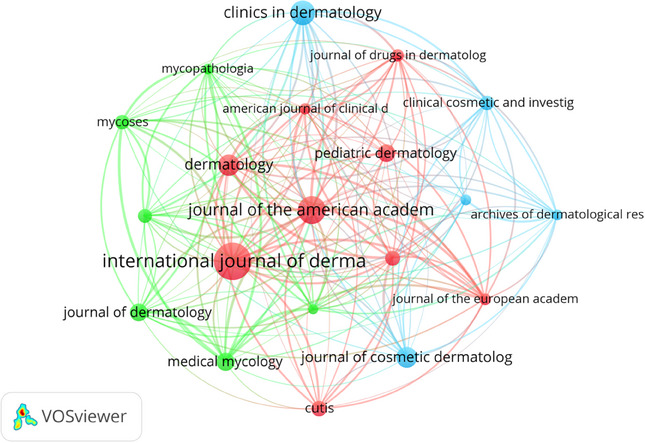
Journal co‐occurrence collaboration network.
3.5. Author analysis
Our search results identified a total of 785 authors who have published literature related to SD. Table 4 lists the top 10 authors by publication count, with each author having published at least seven papers. The top three authors are Lee, Yang Won (16 papers), Gupta, A.K. (15 papers), and Ahn, Kyu Joong (13 papers). Among the top ten authors, three are from Korea, two are from Belgium, and the others are from India, Singapore, Germany, and Scotland. Figure 6 illustrates the co‐authorship collaboration network map based on the publication count of each author. Lee, Yang Won, the most prolific author in the SD field, has close collaborations with Gupta, A.K., Jung, Won Hee, Kim, Beon Joon, Dawson, T.L., and Choe, Yong Beom. The network connections reveal that Schwartz, James R., and Bergfeld, Wilma F., as well as Angelis, Andrea, and Supuran, Claudiu, have formed their own academic teams. Belgian scholars Piérard, G.E., and Piérard‐Franchimont, C. also show a strong connection. Although Dawson, Thomas L. ranks fourth in publication count, his connections with other scholars in the field are relatively limited.
TABLE 4.
Top 10 authors by the number of publications on seborrheic dermatitis.
| Rank | Author | Affiliation | Country | Publications |
|---|---|---|---|---|
| 1 | Lee, Yang Won | Pukyong National University | SOUTH KOREA | 16 |
| 2 | Gupta, A.K. | All India Institute of Medical Sciences (AIIMS) New Delhi | INDIA | 15 |
| 3 | Ahn, Kyu Joong | Konkuk University School of Medicine | SOUTH KOREA | 13 |
| 4 | Dawson, Thomas L. | Medical University of South Carolina | SINGAPORE | 10 |
| 5 | Piérard, G.E. | University of Liege | BELGIUM | 8 |
| 6 | Piérard‐franchimont, C. | University of Liege | BELGIUM | 8 |
| 7 | Faergemann, J. | University of Gothenburg | SWEDEN | 7 |
| 8 | Jung, Won Hee | Chung Ang University | SOUTH KOREA | 7 |
| 9 | Batra, R. | Weill Cornell Medicine | GERMANY | 7 |
| 10 | Bond, R. | University of Edinburgh | SCOTLAND | 7 |
FIGURE 6.
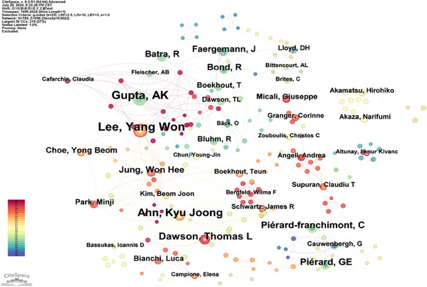
Author co‐occurrence collaboration network.
Our literature search identified 785 authors who have published on SD. Table 4 details the top ten authors by publication volume, with each author having published at least seven relevant papers. Among these, Lee, Yang Won leads with 16 publications, followed by Gupta, A.K. with 15, and Ahn, Kyu Joong with 13. Of these top ten prolific authors, three hail from South Korea, including Lee, Yang Won, Ahn, Kyu Joong, and Jung, Won Hee; two are from Belgium, namely Piérard, G.E. and Piérard‐Franchimont, C.; the remaining authors are from various countries across the globe. Figure 6 presents a detailed co‐authorship network map based on the publication volumes of these authors.
3.6. Analysis of highly‐cited articles
High citation counts of scholarly articles typically denote authoritative research and quality, playing a crucial role in advancing academic disciplines and enhancing researchers' reputations. According to Table 5, the top ten most‐cited articles feature prominent research such as “Seborrheic Dermatitis” (Q1, IF 8.4), which has garnered 157 citations. This is followed by “A new yeast, Malassezia yamatoensis, isolated from a patient with SD, and its distribution in patients and healthy subjects” (Q4, IF 1.9) with 132 citations, and “Molecular analysis of Malassezia microflora in SD patients: Comparison with other diseases and healthy subjects” (Q1, IF 5.7) with 125 citations. Of the top ten cited works, six articles are categorized under Q1, three under Q2, and one under Q3, indicating significant academic influence. The top five articles each boast over 100 citations, underscoring their impact. Particularly noteworthy is the article “Seborrheic Dermatitis” by Naldi, L., published in the prestigious New England Journal of Medicine with an impressive IF of 96.2. This publication highlights the extensive focus SD research receives in high‐impact journals, reflecting the scientific community's appreciation for groundbreaking research. These high‐impact journals play a central role in disseminating pivotal research findings, which are indispensable for driving scientific progress and fostering the development of innovative treatments in this field.
TABLE 5.
Top 10 highly‐cited articles on seborrheic dermatitis.
| Rank | First author | Title | Year | Citations | JCR | IF |
|---|---|---|---|---|---|---|
| 1 | Gupta, A.K. | Seborrheic dermatitis | 2004 | 157 | Q1 | 8.4 |
| 2 | Sugita, T. | A new yeast, Malassezia yamatoensis, isolated from a patient with seborrheic dermatitis, and its distribution in patients and healthy subjects | 2004 | 132 | Q4 | 1.9 |
| 3 | Tajima, M. | Molecular analysis of Malassezia microflora in Seborrheic dermatitis patients: Comparison with other diseases and healthy subjects | 2008 | 125 | Q1 | 5.7 |
| 4 | Dessinioti, C. | Seborrheic dermatitis: Etiology, risk factors, and treatments: Facts and controversies | 2013 | 112 | Q2 | 2.3 |
| 5 | Gaitanis, G. | AhR ligands, Malassezin, and indolo [3,2‐b] carbazole are selectively produced by Malassezia furfur strains isolated from seborrheic dermatitis | 2008 | 108 | Q1 | 5.7 |
| 6 | Naldi, L. | Seborrheic dermatitis | 2009 | 89 | Q1 | 96.2 |
| 7 | Gupta, A.K. | Etiology and management of seborrheic dermatitis | 2004 | 88 | Q2 | 3 |
| 8 | Clark, G.W. | Diagnosis and treatment of seborrheic dermatitis | 2015 | 74 | Q1 | 3.8 |
| 9 | Schwartz, J.R. | A comprehensive pathophysiology of dandruff and seborrheic dermatitis—towards a more precise definition of scalp health | 2013 | 74 | Q1 | 3.5 |
| 10 | Gupta, A.K. | Seborrheic dermatitis | 2003 | 68 | Q2 | 2.2 |
Abbreviations: IF, impact factor; JCR, Journal Citation Reports.
3.7. Analysis of keywords
3.7.1. Keyword co‐occurrence analysis
The prevalence of high‐frequency keywords and their centrality in the literature highlights the focal points of interest among researchers over a period, indicating the hotspots and frontiers of study. 16 Table 6 lists the top 20 most frequent keywords. The keyword “seborrheic dermatitis” (n = 616, centrality: 0.15) appears most frequently, followed by “atopic dermatitis” (n = 249, centrality: 0.19), “skin” (n = 223, centrality: 0.15), “pityriasis versicolor” (n = 115, centrality: 0.07), and “dandruff” (n = 107, centrality: 0.09). Other notable keywords include “double‐blind” (centrality: 0.15), “identification” (centrality: 0.04), “prevalence” (centrality: 0.12), and “efficacy” (n = 76, centrality: 0.12), indicating their popularity in SD‐related research. A total of 704 keywords were retrieved in this study. Keywords co‐occurrence is depicted in Figure 7A, where larger nodes represent higher frequencies of keyword occurrences. The lines between nodes indicate the degree of relevance between keywords. The diagram reveals that high‐frequency keywords related to SD include “atopic dermatitis”, “skin”, “pityriasis versicolor”, “dandruff”, “double‐blind”, “identification”, “prevalence”, “efficacy”, “expression”, “Malassezia furfur”, “yeast”, and “psoriasis”.
TABLE 6.
Top20 high‐frequency keywords.
| Rank | Keywords | Frequency | Centrality | Degree |
|---|---|---|---|---|
| 1 | Seborrheic dermatitis | 616 | 0.15 | 112 |
| 2 | Atopic dermatitis | 249 | 0.19 | 129 |
| 3 | Skin | 223 | 0.15 | 97 |
| 4 | Pityriasis versicolor | 115 | 0.07 | 92 |
| 5 | Dandruff | 107 | 0.09 | 89 |
| 6 | Double‐blind | 102 | 0.15 | 107 |
| 7 | Identification | 78 | 0.04 | 74 |
| 8 | Prevalence | 77 | 0.12 | 76 |
| 9 | Efficacy | 76 | 0.11 | 89 |
| 10 | Expression | 57 | 0.09 | 74 |
| 11 | Disease | 57 | 0.07 | 56 |
| 12 | Malassezia furfur | 53 | 0.09 | 90 |
| 13 | Yeast | 52 | 0.01 | 44 |
| 14 | Psoriasis | 49 | 0.08 | 67 |
| 15 | Children | 49 | 0.05 | 63 |
| 16 | In vitro | 47 | 0.08 | 72 |
| 17 | Stratum corneum | 46 | 0.03 | 48 |
| 18 | Human skin | 45 | 0.05 | 74 |
| 19 | Management | 45 | 0.05 | 57 |
| 20 | Acne vulgaris | 44 | 0.08 | 65 |
FIGURE 7.
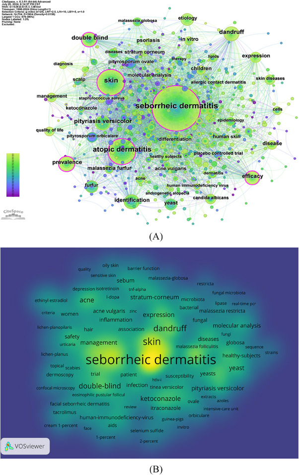
(A) Co‐occurrence network of keywords. (B) VOSviewer keywords density map.
To better understand the current research landscape in the field of SD, we also conducted a co‐occurrence analysis using VOSviewer, setting a threshold for keyword appearances at a minimum of five occurrences. The high‐frequency keyword density map shown in Figure 7B illustrates that the larger and brighter the area, the higher the density, indicating more frequent appearances of the keywords.
3.7.2. Keywords clustering analysis
Cluster labels were derived from significant noun phrases extracted using the log‐likelihood ratio algorithm from keywords. 17 As shown in Figure 8, we categorized the keywords into 11 groups, each represented by a distinct color. The categories are as follows: #0 acne vulgaris, #1 women, #2 androgenetic alopecia, #3 pityriasis versicolor, #4 identification, #5 allergic contact dermatitis, #6 double‐blind, #7 human HIV virus, #8 atopic dermatitis, #9 2% ketoconazole cream, and #10 skin cancer.
FIGURE 8.
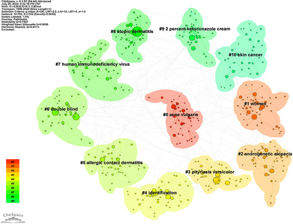
Keywords clustering map.
3.7.3. Keyword timeline analysis
In VOSviewer, the co‐occurrence network of keywords is visualized to reveal their relationships. 18 As shown in Figure 9, using VOSviewer's visualization overlay technique, the temporal evolution of keywords is displayed. In this visualization, purple clusters represent the earliest keywords, green indicates slightly later keywords, and yellow denotes the most recent keywords. Initially, “ketoconazole”, “yeasts”, “itraconazole”, “susceptibility”, and “infection” were central in the purple cluster. Subsequently, in the green cluster, keywords such as “seborrheic dermatitis”, “dandruff”, “skin”, “acne”, “molecular analysis”, “stratum‐corneum”, and “sebum” gained prominence. In recent years, “oily skin”, “microbiota”, “dermoscopy”, “sensitive skin”, and “lichen planopilaris” have emerged as new focal points in the field. Therefore, we utilized the CiteSpace tool for a clearer analysis of these keywords' timelines. Figure 10 displays the timeline visualization of the evolution of significant keywords in SD research.
FIGURE 9.
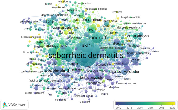
Keywords temporal weight map.
FIGURE 10.
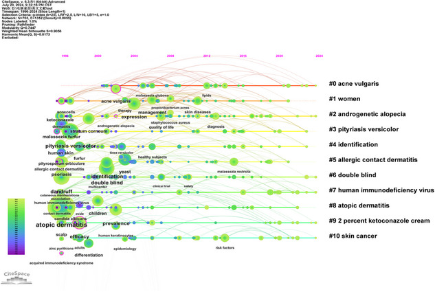
Timeline visualization analysis of keywords.
3.7.4. Keyword burst analysis
Keyword burst analysis is a powerful tool that enables researchers to intuitively understand the hot topics, core concepts, and research trends within a specific field over different time intervals. 19 Figure 11 presents a keyword burst map, where light blue lines indicate time intervals, blue lines mark the periods when keywords first appeared, and red lines highlight the burst periods of these keywords. The detection of burst keywords is based on a significant increase in their publication frequency in specific years, irrespective of their overall usage frequency. 20 From 1996 to 2006, the prominent keywords included “placebo‐controlled trials”, “double‐blind”, and “healthy subjects”, reflecting that research during this period focused primarily on clinical controlled studies. Over time, from 2009 to 2019, the keywords shifted to “microbiota”, “molecular analysis”, “risk factors”, and “etiology”, indicating that SD‐related research transitioned from clinical studies to an emphasis on exploring the disease's pathogenesis. This shift signifies a deeper understanding of the disease's occurrence and development among researchers. After 2020, keywords such as “etiology”, “fungal”, “dandruff”, “staphylococcus aureus”, and “psoriasis” emerged as prominent, reflecting a research focus on clarifying the causes and typical clinical symptoms of SD. This provides direction for further research and aids in optimizing the clinical diagnosis and treatment strategies for SD.
FIGURE 11.
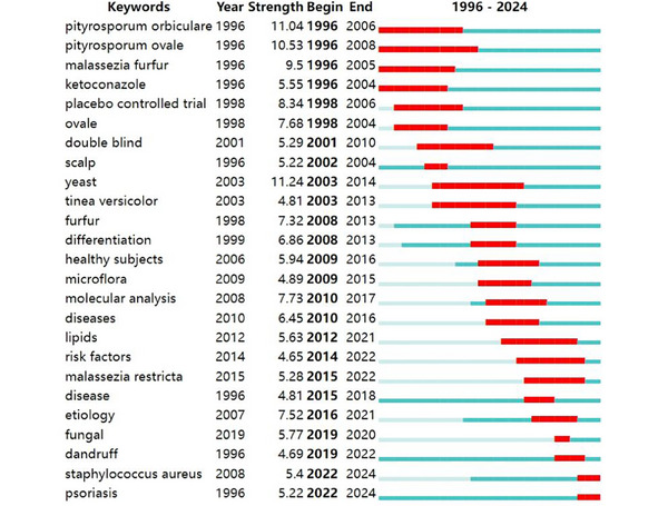
Top 25 keywords with the strongest citation bursts.
4. DISCUSSION
4.1. General information
This study conducted a bibliometric and visual analysis of 1436 publications related to SD research from 1996 to 2024. Overall, the number of publications on SD has shown a general upward trend year by year, indicating that dermatologists and researchers are increasingly focusing on the study of this condition.
The USA, Italy, and China are the three countries with the highest number of publications, each having published over 100 articles, accounting for approximately 30.83% of the total. These figures indicate that these countries are most concerned with the evolution and research in the field of SD. As the world's largest economy, the United States, wielding substantial financial resources, not only invests heavily in research related to SD but also collaborates with multiple countries and institutions, amassing a total of 367 publications, thereby cementing its leadership in the global SD research arena. France has the highest centrality, followed by the USA and Germany, indicating their significance within the network.
Among the top ten research institutions, Canada boasts four, while both the United States and South Korea are represented by two each. Among them, the four Canadian institutions have collectively published 70 papers, demonstrating Canada's high output in the field. From an individual institutional perspective, the University of California System and Procter & Gamble in the USA, both leading in publication volume and centrality, further affirm the pivotal role of the USA in the field of SD research. Simultaneously, the relatively close connections observed among the various institutions indicate that academic cooperation and communication between them are quite frequent.
Lee, Yang Won, the most prolific author in the SD field, has close collaborations with Gupta, A.K., Jung, Won Hee, Kim, Beon Joon, Dawson, T.L., and Choe, Yong Beom. The network connections reveal that Schwartz, James R., and Bergfeld, Wilma F., as well as Angelis, Andrea, and Supuran, Claudiu, have formed their own academic teams. Belgian scholars Piérard, G.E., and Piérard‐Franchimont, C. also show a strong connection. Although Dawson, Thomas L. ranks fourth in publication count, his connections with other scholars in the field are relatively limited. Highly‐cited literature is often regarded as a classic work in a specific disciplinary field, laying a solid foundation for the knowledge system and theoretical basis of that field. 21 Interestingly, we found that three out of the top ten articles with the highest citation frequency were titled “Seborrheic derivatis”, which is also the most frequently used core keyword. Among them, three of the authors are Gupta, A.K., who ranks first in highly‐cited articles in the SD field and second in total publication volume. It is evident that using the disease name alone as the title of an article makes it more easily searchable, studied, and cited by relevant scholars and researchers.
The majority of articles on SD are published in the International Journal of Dermatology (IF = 3.5, Q1), currently the most popular journal in this field of research. Among the top ten journals by publication volume, the Journal of the American Academy of Dermatology (IF = 12.8, Q1) holds the highest IF and also ranks as the journal with the most citations and the highest average citations per item. From the perspective of JCR categorization, two journals are classified as Q1, six as Q2, and two as Q3, clearly indicating that these are high‐quality international journals that provide substantial support and a platform for research on SD.
In summary, cooperation among countries, institutions, journals, or authors facilitates overcoming the scientific challenges and obstacles in the field of SD, achieving consensus on academic viewpoints, and fully leveraging their respective strengths and characteristics, which also indicates that some scientific questions and technical difficulties related to SD require collaborative resolution.
4.2. Hotspots and frontiers
4.2.1. Close relationship between SD and other skin diseases
Through careful analysis and mining of keyword information, we discovered that SD frequently co‐occurs with various skin diseases, including atopic dermatitis (AD), pityriasis versicolor, dandruff, psoriasis, acne vulgaris, androgenetic alopecia, and allergic contact dermatitis. Skin infections caused by Malassezia are characterized by a high prevalence, frequent recurrence, and a significant impact on the physical and mental health of patients. 22
Malassezia is the primary pathogen for SD, AD, and pityriasis versicolor. However, SD and AD are both multifactorial endogenous dermatoses that commonly affect the face. 23 , 24 , 25 Dandruff, a chronic inflammatory skin disease, shares the characteristic of recurrent oily scales with SD, often accompanied by erythema and itching. An imbalance in the scalp microbiome is a major contributing factor. 26 , 27 Psoriasis exhibits clinical features very similar to SD, especially when only the scalp is affected, making differential diagnosis challenging due to hair interference, subtle vascular features, and thick white scales. 28 , 29 Additionally, SD and acne vulgaris may be linked to abnormal sebum secretion and Demodex mite infection, which can trigger immune system reactivation, inflammation, and follicular changes. 30 , 31 The association between androgenetic alopecia and SD has also been confirmed through microscopic examination. 32 Furthermore, many SD patients are diagnosed with allergic contact dermatitis, showing unique allergen profiles related to personal care products, particularly hair care items. This could be due to topical treatments for SD or differing allergen immune responses. 33 These findings enhance our understanding of the pathophysiology of SD, providing a scientific basis for clinical diagnosis and treatment.
Additionally, the prevalence of SD among HIV‐positive patients ranges from 30% to 83%. SD often manifests early in the course of HIV infection and may serve as an initial clinical marker of HIV infection. 34 Parkinson's disease patients also exhibit a high prevalence of SD, ranging from 18.6% to 59%, with a positive correlation between the prevalence and both age and severity of motor symptoms. This warrants further investigation to determine if there are other yet undiscovered diseases associated with SD. 35
4.2.2. Close relationship between SD, Malassezia, and inflammation
For a long time, the yeast of the genus Malassezia has been considered the primary trigger in the pathogenesis of SD, dominating the dysbiosis of the skin microbiota and consistently being a focal point of research in the SD field. 27 , 36 This aligns with the findings of our bibliometric study, which revealed a high publication volume, highly‐cited literature, and high‐frequency keywords related to this topic. Malassezia is one of the resident bacteria on the human skin surface, a lipophilic yeast‐like fungus that can infect the skin through direct tissue invasion in its filamentous form, or indirectly induce SD by promoting immune and metabolic processes in its yeast form. 22 , 37 In 1996, the taxonomy of the genus Malassezia was revised based on molecular phylogenetic analysis, using molecular biology methods to accurately identify the Malassezia communities on the skin, marking the first step toward elucidating the role of Malassezia in the development of SD. 38 With the deepening of extensive research into the molecular mechanisms of SD, it has been discovered that the inflammatory process of SD is mediated by metabolites of fungi of the genus Malassezia, particularly the free fatty acids released from triglycerides. 38 , 39 Malassezia has been demonstrated to activate the NLRP3 inflammasome through the dectin‐1 and Syk signaling cascade, leading to the release of pro‐inflammatory cytokines IL‐1β and IL‐18, and altering the host cytokine expression profile to induce SD. Concurrently, lesional SD skin exhibits a shift towards a pro‐inflammatory signaling environment, with increased immunoreactivity of inflammatory cytokines such as IL‐1β, IL‐2, IL‐4, IL‐6, IL‐8, IL‐10, IL‐12, and IL‐17, which bind to their respective receptors and activate the NF‐κB, MAPK, and C/EBP pathways, all closely related to inflammation, thereby exacerbating the inflammatory response in SD. 35 , 40 , 41 , 42 In summary, the close relationship between SD and Malassezia, as well as inflammation, underscores and determines that antifungal and anti‐inflammatory strategies are pivotal and central to the treatment approach for SD.
4.2.3. SD treatment focused on antifungal and anti‐inflammatory strategies
Currently, the focus of SD treatment is to eliminate the visible symptoms of the skin condition, alleviate associated symptoms characterized primarily by itching, and restore the overall health of the scalp and skin. 7 , 8 This bibliometric study reveals that the high‐frequency keywords, highly‐cited articles, and prolific authors primarily emphasize treatments for SD involving antifungal agents such as Ketoconazole, 43 Bifonazole, 44 Ciclopirox Olamine, 45 and Zinc Pyrithione, 46 which inhibit fungal cell wall synthesis. Corticosteroids such as Desonide 47 is used to reduce inflammation and alleviate skin irritation. Using immunomodulators such as Pimecrolimus 48 and Tacrolimus 49 to inhibit cytokine production by T‐lymphocytes. Other treatment options include Lithium gluconate/succinate 50 and Metronidazole. 51 Currently, the most commonly used clinical treatments involve topical antifungals to inhibit fungal cell wall synthesis and anti‐inflammatory medications. 8 When selecting a treatment plan, it is essential to consider factors such as the patient's age, affected area, efficacy, side effects, and compliance. From an age perspective, infants are typically treated with moisturizers that help loosen scales (e.g., mineral oil, olive oil, or petroleum jelly), while adolescents and adults may use over‐the‐counter shampoos and topical antifungals, calcineurin inhibitors, and corticosteroids. Considering the affected area, the scalp can be treated with over‐the‐counter anti‐dandruff shampoos containing selenium sulfide, zinc pyrithione, or coal tar, while the face and body can be treated with topical antifungals, corticosteroids, and calcineurin inhibitors. 7
5. ADVANTAGES AND SHORTCOMINGS
To our knowledge, this is the first bibliometric analysis of SD using data from an English database, which helps clinical doctors and researchers engaged in skin disease research to quickly understand the research trends and hotspots of this disease. Firstly, although the WOSCC is optimally suited for bibliometric analyses, its predilection for journals published in English may result in a significant oversight of research contributions from fields prevalent in countries like China, Russia, and France, where there exists a tradition of publishing in the native languages, potentially leading to a severe underestimation of their influence and contributions. Secondly, despite the inevitability of recently published studies manifesting lower citation frequencies, their significance might largely go unrecognized; consequently, the representation of emerging hotspots and frontiers in this analysis could be substantially underrepresented. Finally, owing to the constraints imposed by bibliometric and visualization software analysis capabilities, a substantial amount of information may remain inadequately analyzed and summarized, meriting further exploration and excavation in the future.
6. CONCLUSIONS
This study utilized bibliometric and visualization techniques to analyze relevant research in the field of SD in the WOSCC database. Our research results reveal in detail the factors of publication volume, national (regional) authors, institutions, journals, authors, highly‐cited articles, and keywords in this field. In summary, the main research hotspots and frontiers of SD are as follows: (1) the relationship between SD and other skin diseases is a hot research topic; (2) Malassezia and inflammation are currently research hotspots in SD; and (3) the focus of research on antifungal and anti‐inflammatory treatments for SD is currently at the forefront of this field. This can help clinical doctors and researchers quickly understand the insights and valuable information of SD research, providing references for clinical decision‐making and predicting future research directions.
CONFLICT OF INTEREST STATEMENT
The authors declare no conflicts of interest.
ACKNOWLEDGMENTS
We extend our gratitude to all authors involved in the bibliometric and visualization studies on seborrheic dermatitis. This study was supported by the National Key Research and Development Program of China (No. 2019YFC1709900) and Natural Science Foundation of Jilin Province (No. YDZJ202301ZYTS140).
Yan H, Zhang S, Sun W, et al. A bibliometric and visual analysis of the research status and hotspots of seborrheic dermatitis based on web of science. Skin Res Technol. 2024;30:e70048. 10.1111/srt.70048
Huixin Yan and Shaobo Zhang contributed equally to this study.
Contributor Information
Xingquan Wu, Email: wuxingquan2005@163.com.
Bailin Song, Email: czdsongbailin@126.com.
DATA AVAILABILITY STATEMENT
The data that support the findings of this study are available on request from the corresponding author.
REFERENCES
- 1. Jackson JM, Alexis A, Zirwas M, Taylor S. Unmet needs for patients with seborrheic dermatitis. J Am Acad Dermatol. 2024;90(3):597‐604. doi: 10.1016/j.jaad.2022.12.017 [DOI] [PubMed] [Google Scholar]
- 2. Dall'Oglio F, Nasca MR, Gerbino C, Micali G. An overview of the diagnosis and management of seborrheic dermatitis. Clin Cosmet Investig Dermatol. 2022;15:1537‐1548. doi: 10.2147/CCID.S284671 [DOI] [PMC free article] [PubMed] [Google Scholar]
- 3. Leroy AK, Cortez de Almeida RF, Obadia DL, Frattini S, Melo DF. Scalp seborrheic dermatitis: what we know so far. Skin Appendage Disord. 2023;9(3):160‐164. doi: 10.1159/000529854 [DOI] [PMC free article] [PubMed] [Google Scholar]
- 4. Mustarichie R, Rostinawati T, Pitaloka DAE, Saptarini NM, Iskandar Y. Herbal therapy for the treatment of seborrhea dermatitis. Clin Cosmet Investig Dermatol. 2022;15:2391‐2405. doi: 10.2147/CCID.S376700 [DOI] [PMC free article] [PubMed] [Google Scholar]
- 5. Mangion SE, Mackenzie L, Roberts MS, Holmes AM. Seborrheic dermatitis: topical therapeutics and formulation design. Eur J Pharm Biopharm Off J Arbeitsgemeinschaft Pharm Verfahrenstechnik EV. 2023;185:148‐164. doi: 10.1016/j.ejpb.2023.01.023 [DOI] [PubMed] [Google Scholar]
- 6. Elgash M, Dlova N, Ogunleye T, Taylor SC. Seborrheic dermatitis in skin of color: clinical considerations. J Drugs Dermatol JDD. 2019;18(1):24‐27. [PubMed] [Google Scholar]
- 7. Seborrheic Dermatitis and Dandruff: A Comprehensive Review—PubMed. Accessed July 26, 2024. https://pubmed.ncbi.nlm.nih.gov/27148560/
- 8. Clark GW, Pope SM, Jaboori KA. Diagnosis and treatment of seborrheic dermatitis. Am Fam Physician. 2015;91(3):185‐190. [PubMed] [Google Scholar]
- 9. Ninkov A, Frank JR, Maggio LA. Bibliometrics: methods for studying academic publishing. Perspect Med Educ. 2022;11(3):173‐176. doi: 10.1007/s40037-021-00695-4 [DOI] [PMC free article] [PubMed] [Google Scholar]
- 10. Bai F, Huang Z, Luo J, et al. Bibliometric and visual analysis in the field of traditional Chinese medicine in cancer from 2002 to 2022. Front Pharmacol. 2023;14:1164425. doi: 10.3389/fphar.2023.1164425 [DOI] [PMC free article] [PubMed] [Google Scholar]
- 11. Azra MN, Wong LL, Aouissi HA, et al. Crayfish research: a global scientometric analysis using CiteSpace. Anim Open Access J MDPI. 2023;13(7):1240. doi: 10.3390/ani13071240 [DOI] [PMC free article] [PubMed] [Google Scholar] [Retracted]
- 12. Wan Y, Shen J, Ouyang J, et al. Bibliometric and visual analysis of neutrophil extracellular traps from 2004 to 2022. Front Immunol. 2022;13:1025861. doi: 10.3389/fimmu.2022.1025861 [DOI] [PMC free article] [PubMed] [Google Scholar]
- 13. Wang G, Chen Y, Liu X, Ma S, Jiang M. Global research trends in prediabetes over the past decade: bibliometric and visualized analysis. Medicine (Baltimore). 2024;103(3):e36857. doi: 10.1097/MD.0000000000036857 [DOI] [PMC free article] [PubMed] [Google Scholar]
- 14. Zhang W, Li M, Li X, Wang X, Liu Y, Yang J. Global trends and research status in ankylosing spondylitis clinical trials: a bibliometric analysis of the last 20 years. Front Immunol. 2023;14:1328439. doi: 10.3389/fimmu.2023.1328439 [DOI] [PMC free article] [PubMed] [Google Scholar]
- 15. Yang Y, Zheng X, Lv H, et al. A bibliometrics study on the status quo and hot topics of pathogenesis of psoriasis based on Web of Science. Skin Res Technol Off J Int Soc Bioeng Skin ISBS Int Soc Digit Imaging Skin ISDIS Int Soc Skin Imaging ISSI. 2024;30(1):e13538. doi: 10.1111/srt.13538 [DOI] [PMC free article] [PubMed] [Google Scholar]
- 16. Ma K, Luo C, Du M, et al. Advances in stem cells treatment of diabetic wounds: a bibliometric analysis via CiteSpace. Skin Res Technol Off J Int Soc Bioeng Skin ISBS Int Soc Digit Imaging Skin ISDIS Int Soc Skin Imaging ISSI. 2024;30(4):e13665. doi: 10.1111/srt.13665 [DOI] [PMC free article] [PubMed] [Google Scholar]
- 17. Wei T, Jin Q. Research trends and hotspots in exercise interventions for liver cirrhosis: a bibliometric analysis via CiteSpace. Medicine (Baltimore). 2024;103(28):e38831. doi: 10.1097/MD.0000000000038831 [DOI] [PMC free article] [PubMed] [Google Scholar]
- 18. Yao L, Zhang L, Tai Y, et al. Visual analysis of obesity and hypothyroidism: a bibliometric analysis. Medicine (Baltimore). 2024;103(1):e36841. doi: 10.1097/MD.0000000000036841 [DOI] [PMC free article] [PubMed] [Google Scholar]
- 19. Zhou X, Kang C, Hu Y, Wang X. Study on insulin resistance and ischemic cerebrovascular disease: a bibliometric analysis via CiteSpace. Front Public Health. 2023;11:1021378. doi: 10.3389/fpubh.2023.1021378 [DOI] [PMC free article] [PubMed] [Google Scholar]
- 20. Chen J, Luo C, Ju P, et al. A bibliometric analysis and visualization of acupuncture and moxibustion therapy for herpes zoster and postherpetic neuralgia. Skin Res Technol Off J Int Soc Bioeng Skin ISBS Int Soc Digit Imaging Skin ISDIS Int Soc Skin Imaging ISSI. 2024;30(6):e13815. doi: 10.1111/srt.13815 [DOI] [PMC free article] [PubMed] [Google Scholar]
- 21. Tang F, Jiang C, Chen J, Wang L, Zhao F. Global hotspots and trends in Myofascial Pain Syndrome research from 1956 to 2022: a bibliometric analysis. Medicine (Baltimore). 2023;102(12):e33347. doi: 10.1097/MD.0000000000033347 [DOI] [PMC free article] [PubMed] [Google Scholar]
- 22. Wang K, Cheng L, Li W, et al. Susceptibilities of Malassezia strains from pityriasis versicolor, Malassezia folliculitis and seborrheic dermatitis to antifungal drugs. Heliyon. 2020;6(6):e04203. doi: 10.1016/j.heliyon.2020.e04203 [DOI] [PMC free article] [PubMed] [Google Scholar]
- 23. Ramos‐E‐Silva M, Sampaio AL, Carneiro S. Red face revisited: endogenous dermatitis in the form of atopic dermatitis and seborrheic dermatitis. Clin Dermatol. 2014;32(1):109‐115. doi: 10.1016/j.clindermatol.2013.05.032 [DOI] [PubMed] [Google Scholar]
- 24. Jung WH. Alteration in skin mycobiome due to atopic dermatitis and seborrheic dermatitis. Biophys Rev. 2023;4(1):011309. doi: 10.1063/5.0136543 [DOI] [PMC free article] [PubMed] [Google Scholar]
- 25. Gholami M, Mokhtari F, Mohammadi R. Identification of Malassezia species using direct PCR‐ sequencing on clinical samples from patients with pityriasis versicolor and seborrheic dermatitis. Curr Med Mycol. 2020;6(3):21‐26. doi: 10.18502/cmm.6.3.3984 [DOI] [PMC free article] [PubMed] [Google Scholar]
- 26. Tao R, Li R, Wang R. Skin microbiome alterations in seborrheic dermatitis and dandruff: a systematic review. Exp Dermatol. 2021;30(10):1546‐1553. doi: 10.1111/exd.14450 [DOI] [PubMed] [Google Scholar]
- 27. Lin Q, Panchamukhi A, Li P, et al. Malassezia and Staphylococcus dominate scalp microbiome for seborrheic dermatitis. Bioprocess Biosyst Eng. 2021;44(5):965‐975. doi: 10.1007/s00449-020-02333-5 [DOI] [PubMed] [Google Scholar]
- 28. Yu Z, Kaizhi S, Jianwen H, Guanyu Y, Yonggang W. A deep learning‐based approach toward differentiating scalp psoriasis and seborrheic dermatitis from dermoscopic images. Front Med. 2022;9:965423. doi: 10.3389/fmed.2022.965423 [DOI] [PMC free article] [PubMed] [Google Scholar]
- 29. Tao R, Li R, Wan Z, Wu Y, Wang R. Skin microbiome signatures associated with psoriasis and seborrheic dermatitis. Exp Dermatol. 2022;31(7):1116‐1118. doi: 10.1111/exd.14618 [DOI] [PubMed] [Google Scholar]
- 30. Boughton B, Mackenna RM, Wheatley VR, Wormall A. The fatty acid composition of the surface skin fats ('sebum’) in acne vulgaris and seborrheic dermatitis. J Invest Dermatol. 1959;33:57‐64. doi: 10.1038/jid.1959.122 [DOI] [PubMed] [Google Scholar]
- 31. Aktaş Karabay E, Aksu Çerman A. Demodex folliculorum infestations in common facial dermatoses: acne vulgaris, rosacea, seborrheic dermatitis. An Bras Dermatol. 2020;95(2):187‐193. doi: 10.1016/j.abd.2019.08.023 [DOI] [PMC free article] [PubMed] [Google Scholar]
- 32. Bhoyrul B. A simple technique to distinguish fibrosing alopecia in a pattern distribution from androgenetic alopecia and concomitant seborrheic dermatitis. J Am Acad Dermatol. 2022;86(1):163‐165. doi: 10.1016/j.jaad.2020.12.077 [DOI] [PubMed] [Google Scholar]
- 33. Silverberg JI, Hou A, Warshaw EM, et al. Allergens in patients with a diagnosis of seborrheic dermatitis, North American Contact Dermatitis Group data, 2001–2016. J Am Acad Dermatol. 2022;86(2):460‐463. doi: 10.1016/j.jaad.2021.09.039 [DOI] [PubMed] [Google Scholar]
- 34. Chatzikokkinou P, Sotiropoulos K, Katoulis A, Luzzati R, Trevisan G. Seborrheic dermatitis—an early and common skin manifestation in HIV patients. Acta Dermatovenerol Croat ADC. 2008;16(4):226‐230. [PubMed] [Google Scholar]
- 35. Tomic S, Kuric I, Kuric TG, et al. Seborrheic Dermatitis Is Related to Motor Symptoms in Parkinson's Disease. J Clin Neurol Seoul Korea. 2022;18(6):628‐634. doi: 10.3988/jcn.2022.18.6.628 [DOI] [PMC free article] [PubMed] [Google Scholar]
- 36. Wikramanayake TC, Borda LJ, Miteva M, Paus R. Seborrheic dermatitis‐Looking beyond Malassezia. Exp Dermatol. 2019;28(9):991‐1001. doi: 10.1111/exd.14006 [DOI] [PubMed] [Google Scholar]
- 37. Li J, Feng Y, Liu C, et al. Presence of malassezia hyphae is correlated with pathogenesis of seborrheic dermatitis. Microbiol Spectr. 2022;10(1):e0116921. doi: 10.1128/spectrum.01169-21 [DOI] [PMC free article] [PubMed] [Google Scholar]
- 38. Tajima M, Sugita T, Nishikawa A, Tsuboi R. Molecular analysis of Malassezia microflora in seborrheic dermatitis patients: comparison with other diseases and healthy subjects. J Invest Dermatol. 2008;128(2):345‐351. doi: 10.1038/sj.jid.5701017 [DOI] [PubMed] [Google Scholar]
- 39. Vincenzi C, Tosti A. Efficacy and tolerability of a shampoo containing broad‐spectrum cannabidiol in the treatment of scalp inflammation in patients with mild to moderate scalp psoriasis or seborrheic dermatitis. Skin Appendage Disord. 2020;6(6):355‐361. doi: 10.1159/000510896 [DOI] [PMC free article] [PubMed] [Google Scholar]
- 40. Ghalili S, David E, Ungar B, et al. IL‐12/23‐targeting in seborrheic dermatitis patients leads to long‐lasting response. Arch Dermatol Res. 2023;315(10):2937‐2940. doi: 10.1007/s00403-023-02680-9 [DOI] [PubMed] [Google Scholar]
- 41. Kerr K, Darcy T, Henry J, et al. Epidermal changes associated with symptomatic resolution of dandruff: biomarkers of scalp health. Int J Dermatol. 2011;50(1):102‐113. doi: 10.1111/j.1365-4632.2010.04629.x [DOI] [PubMed] [Google Scholar]
- 42. Perkins MA, Cardin CW, Osterhues MA, Robinson MK. A non‐invasive tape absorption method for recovery of inflammatory mediators to differentiate normal from compromised scalp conditions. Skin Res Technol Off J Int Soc Bioeng Skin ISBS Int Soc Digit Imaging Skin ISDIS Int Soc Skin Imaging ISSI. 2002;8(3):187‐193. doi: 10.1034/j.1600-0846.2002.20337.x [DOI] [PubMed] [Google Scholar]
- 43. Massiot P, Clavaud C, Thomas M, et al. Continuous clinical improvement of mild‐to‐moderate seborrheic dermatitis and rebalancing of the scalp microbiome using a selenium disulfide‐based shampoo after an initial treatment with ketoconazole. J Cosmet Dermatol. 2022;21(5):2215‐2225. doi: 10.1111/jocd.14362 [DOI] [PMC free article] [PubMed] [Google Scholar]
- 44. Shemer A, Nathansohn N, Kaplan B, Weiss G, Newman N, Trau H. Treatment of scalp seborrheic dermatitis and psoriasis with an ointment of 40% urea and 1% bifonazole. Int J Dermatol. 2000;39(7):532‐534. doi: 10.1046/j.1365-4362.2000.00986-3.x [DOI] [PubMed] [Google Scholar]
- 45. Starova A, Aly R. The safety and efficacy of ciclopirox olamine for the treatment of seborrheic dermatitis. Expert Opin Drug Saf. 2005;4(2):235‐239. doi: 10.1517/14740338.4.2.235 [DOI] [PubMed] [Google Scholar]
- 46. Lorette G, Ermosilla V. Clinical efficacy of a new ciclopiroxolamine/zinc pyrithione shampoo in scalp seborrheic dermatitis treatment. Eur J Dermatol EJD. 2006;16(5):558‐564. [PubMed] [Google Scholar]
- 47. Treatment of scalp and facial seborrheic dermatitis with desonide hydrogel 0.05%—PubMed. Accessed July 26, 2024. https://pubmed.ncbi.nlm.nih.gov/20967179/ [PMC free article] [PubMed]
- 48. Ozden MG, Tekin NS, Ilter N, Ankarali H. Topical pimecrolimus 1% cream for resistant seborrheic dermatitis of the face: an open‐label study. Am J Clin Dermatol. 2010;11(1):51‐54. doi: 10.2165/11311160-000000000-00000 [DOI] [PubMed] [Google Scholar]
- 49. Meshkinpour A, Sun J, Weinstein G. An open pilot study using tacrolimus ointment in the treatment of seborrheic dermatitis. J Am Acad Dermatol. 2003;49(1):145‐147. doi: 10.1067/mjd.2003.450 [DOI] [PubMed] [Google Scholar]
- 50. Dréno B, Blouin E, Moyse D. [Lithium gluconate 8% in the treatment of seborrheic dermatitis]. Ann Dermatol Venereol. 2007;134(4 Pt 1):347‐351. doi: 10.1016/s0151-9638(07)89189-7 [DOI] [PubMed] [Google Scholar]
- 51. Seckin D, Gurbuz O, Akin O. Metronidazole 0.75% gel vs. ketoconazole 2% cream in the treatment of facial seborrheic dermatitis: a randomized, double‐blind study. J Eur Acad Dermatol Venereol JEADV. 2007;21(3):345‐350. doi: 10.1111/j.1468-3083.2006.01927.x [DOI] [PubMed] [Google Scholar]
Associated Data
This section collects any data citations, data availability statements, or supplementary materials included in this article.
Data Availability Statement
The data that support the findings of this study are available on request from the corresponding author.


