1. PATIENT PRESENTATION
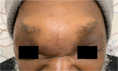
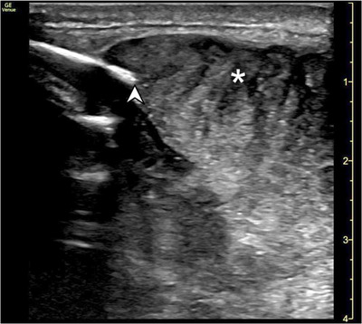
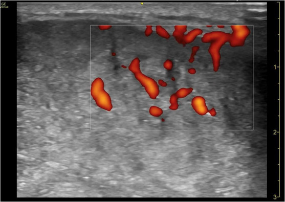
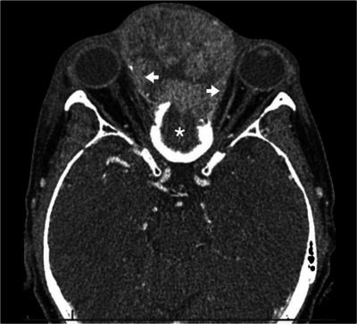
A 60‐year‐old male presented to the emergency department with the complaint of “sinus infection and facial swelling.” The patient endorsed worsening symptoms for 1 month including nasal congestion, bloody nasal drainage, facial swelling and pain, and new onset diplopia. Physical examination was notable for forehead swelling extending to the periorbital area bilaterally (Figure 1). Point‐of‐care ultrasound was performed (Figures 2 and 3) and identified a heterogeneous highly vascular soft tissue mass with associated defect of the frontal bone. Computed tomography of the head and maxillofacial structures was obtained (Figure 4), confirming the diagnosis of a large anterior soft tissue mass with destruction of the frontal bone and mass effect on the orbits.
FIGURE 1.

Photograph demonstrating forehead swelling with bilateral periorbital mass effect noted.
FIGURE 2.

Point‐of‐care ultrasound view of patient's anterior forehead demonstrating discontinuity and erosion of frontal bone (arrowhead) with protrusion of heterogeneous soft tissue mass (asterisk).
FIGURE 3.

Point‐of‐care power doppler ultrasound view of patient's forehead demonstrating extensive vascularity within the mass.
FIGURE 4.

Computed tomography in axial plane with view of orbits and cribriform demonstrating large mass centered at the cribriform plate (asterisk) involving the frontal and ethmoid sinuses as well as left nasal cavity with extensive mass effect with lateral displacement of the orbits (arrows).
2. DIAGNOSIS: SINONASAL SQUAMOUS CELL CARCINOMA
The patient underwent endoscopic biopsy, demonstrating squamous cell carcinoma originating from the skull base. In patients presenting with forehead swelling, point‐of‐care ultrasound (POCUS) provides a rapid imaging modality for superficial soft tissue masses. 1 , 2 Given the broad differential for this presentation, POCUS can facilitate the evaluation of skin and soft tissue infections, soft tissue, and bony or vascular pathology. Importantly, the use of Doppler ultrasound can prevent inadvertent incision of occult vascular structures. 3 POCUS has aided the diagnosis of Pott's puffy tumor, a rare disorder which may present with forehead swelling due to an underlying abscess associated with frontal bone osteomyelitis. 4 In evaluation of forehead masses, ultrasound can expedite further investigation by providing characterization of substance, vascularity, and compressibility. 5 POCUS examination in this patient rapidly facilitated appropriate additional imaging, consultation and diagnosis, and the avoidance of harmful bedside procedures.
CONFLICT OF INTEREST STATEMENT
The authors declare conflicts of interest.
Williams Chen MC, Passons SM, Gullett JP, Thompson MA, Pigott DC, Burleson SL. Man with forehead swelling. JACEP Open. 2024;5:e13256. 10.1002/emp2.13256
Meetings: 2024 Poster Session, Emerald Coast Conference, Sandestin, FL, USA.
REFERENCES
- 1. Aparisi Gómez MP, Errani C, Lalam R, et al. The role of ultrasound in the diagnosis of soft tissue tumors. Semin Musculoskelet Radiol. 2020;24(2):135‐155. [DOI] [PubMed] [Google Scholar]
- 2. Catalano O, Varelli C, Sbordone C, et al. A bump: what to do next? Ultrasound imaging of superficial soft‐tissue palpable lesions. J Ultrasound. 2020;23(3):287‐300. [DOI] [PMC free article] [PubMed] [Google Scholar]
- 3. Burleson SL, Cirillo FN, Gibson CB, Gullett JP, Pigott DC. Superficial temporal artery pseudoaneurysm diagnosed by point‐of‐care ultrasound. Clin Pract Cases Emerg Med. 2019;3(1):77‐78. doi: 10.5811/cpcem.2018.11.40958 [DOI] [PMC free article] [PubMed] [Google Scholar]
- 4. Acuña J, Shockey D, Adhikari S. The use of point‐of‐care ultrasound in the diagnosis of Pott's puffy tumor: a case report. Clin Pract Cases Emerg Med. 2021;5(4):422‐424. [DOI] [PMC free article] [PubMed] [Google Scholar]
- 5. Kim HW, Yoo SY, Oh S, Jeon TY, Kim JH. Ultrasonography of pediatric superficial soft tissue tumors and tumor‐like lesions. Korean J Radiol. 2020;21(3):341‐355. [DOI] [PMC free article] [PubMed] [Google Scholar]


