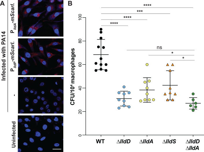
Fig 6.
Expression of lldA and contributions of lld genes during macrophage infection. (A) Fluorescence images of RAW264.7 macrophages infected with the PlldA-mScarlet strain (top), the PlldP-mScarlet strain, or WT PA14, and of an uninfected control (bottom). 4′,6-Diamidino-2-phenylindole (DAPI) fluorescence is shown in blue, and mScarlet fluorescence is shown in red. Scale bar is 10 µm. (B) Intracellular burden of P. aeruginosa WT and indicated mutant strains in RAW264.7 macrophages 3 hours post-infection and subjected to the gentamicin protection assay. Each dot represents one replicate, and error bars represent standard deviation. *P < 0.05, ***P < 0.001, and ****P < 0.0001; ns = not significant.

