Abstract
Acquired benign tracheoesophageal fistulas and bronchoesophageal fistulas (TEF) are typically associated with granulomatous mediastinal infections, 75% of which are iatrogenic. Candida albicans and Actinomyces are commonly occurring organisms, but are uncommon etiologies of TEF. Normal colonization and the slow growth characteristics of some species of these agents rarely result in infection, mycetoma, and broncholithiasis, and thus, delays in diagnosis and treatment are likely. Few reports describe C. albicans or Actinomyces spp. as the etiology of TEF or broncholithiasis. Herein, we report a case of benign acquired TEF secondary to coinfection of Candida and Actinomyces complicated by the formation of an actinomycetoma and broncholithiasis and a comprehensive literature review to highlight the unique nature of this presentation and offer a diagnostic algorithm for diagnosis and treatment of TEFs. Following a presentation of three months of productive cough, choking sensation, night sweats, and weight loss, a bronchoscopy revealed a fistulous connection between the esophagus and the posterior right middle lobe. Pathology identified a calcified fungus ball and a broncholith secondary to the co-infection of Candida and Actinomyces. This unique presentation of Candida and Actinomyces co-infection and the associated diagnostic algorithm are presented as education and a useful tool for clinicians.
Keywords: bronchoesophageal fistula, Candida albicans, Actinomyces, co-infection, benign acquired TEF
Introduction
Tracheoesophageal fistulas and broncoesophageal fistulas (TEF) may be congenital or acquired [1–3]; acquired TEF has been noted in advanced esophageal or respiratory malignancies [1, 2, 4], as a surgical complication, or as a result of traumatic or prolonged intubation [1–4]. Non-surgical causes include granulomatous etiologies, most notably tuberculosis, which has been the most common cause of benign acquired TEFs; however, iatrogenic causes make up 75% of all cases [2, 5–7]. Candida albicans and Actinomyces spp., when co-infecting, can form an actinomycetoma, which has not been previously reported as an etiology of benign acquired TEF [8, 9], or broncholithiasis, from erosion and extrusion of chronically inflamed or granulomatous tissue within the tracheobronchial tree [4, 5, 7]. Tuberculosis and histoplasmosis infections account for most cases of broncholith formation; few reports describe actinomycetoma as the etiology of broncholith and subsequent TEF formation. Herein, following institutional review board (IRB) approval, we present a case of a patient with a tracheoesophageal fistula secondary to an actinomycetoma that was further complicated by the formation of a broncholith, demonstrating an unusual complication of bronchopulmonary Candida and Actinomyces co-infection.
We also present a comprehensive literature review to better educate clinicians regarding this unique pathology's presentation, diagnosis, and treatment, other infectious causes of TEF, and known presentations of Candida and Actinomyces co-infection.
Ethics
This report was approved by the Prisma Health IRB as non-human research; consent for publication was received from the patient.
Case report
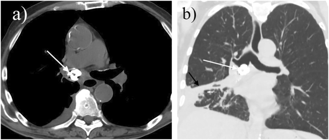
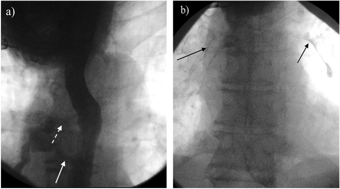
A 77-year-old male with a medical history significant only for recurrent pneumonia and no other known chronic medical conditions presented to his primary care provider multiple times over a year for complications related to recurrent aspiration, bronchitis, and dysphagia. He eventually presented to the emergency department (ED) with complaints of three months of productive cough, a sensation of choking, night sweats, and 5 kg of unintended weight loss. On exam, the patient was tachycardic and hypoxic, and a chest radiograph taken in the ED showed a consolidation in the patient's right middle and right lower lobe. The patient was treated with ceftriaxone and azithromycin and discharged home with ten-days of oral antibiotics; however, the patient continued to worsen and experienced increased dyspnea, which prompted a cat scan (CT) of the abdomen and pelvis that showed a focal calcified endobronchial lesion in the right mainstem bronchus (Fig. 1a and b). This finding prompted investigation and debulking of the lesion via bronchoscopy, revealing a fistulous connection between the esophagus and the right mainstem bronchus. Imaging via esophagogram confirmed a fistulous tract between the esophagus and the posterior right middle lobe (Fig. 2a and b). Pathology from the biopsy during bronchoscopy revealed a calcified fungus ball, filamentous bacteria with cultures positive for Actinomyces, and a broncholith secondary to Candida infection (Fig. 3a and b). Due to the complexity of this presentation and the culture results, surgical treatment commenced; a right thoracotomy with right lower lobe wedge resection and repair of the TEF using an intercostal muscle flap was completed.
Fig. 1.
Non-contrast computed tomography examination of the chest in axial (a) and coronal planes (b). A broncholith is present within the right mainstem bronchus (white arrow). Note is made of post obstructive right lower lobe atelectasis (Fig. 1b; black arrow)
Fig. 2.
Upper gastrointestinal examination with water soluble contrast. (a) Contrast opacifies a communication from the esophagus to the right mainstem bronchus (broken white arrow) consistent with bronchial esophageal fistula. (b). Delayed image demonstrating contrast within the central bronchial tree (black arrows) bilaterally
Fig. 3.
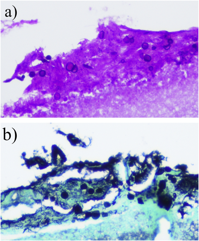
Histology consistent with Candida. (a) Periodic acid Schiff (PAS) stain (400×) illustrating necrotic tissue and yeast forms consistent with Candida species. PAS allows for identification of basement membranes, glycogen, and mucopolysaccarides groups so that the morphology can be determined. (b) Grocott-Gomori methenamine silver (GMS; 400X), a specific stain to detect fungi by binding to the polysaccharides in the cell walls, identified yeast forms consistent with Candida species
The postoperative course was uncomplicated, and the patient was extubated on postoperative day one (POD1) and transferred out of intensive care on POD2. By POD8, the patient resumed a modified oral diet, and based on the microbial cultures, was treated with a six-month course of amoxicillin-clavulanate and fluconazole. At a follow-up visit, the patient was eating well and had a resolution of sputum production and daytime dyspnea. A repeat bronchoscopy performed two months later showed normal mucosa without the presence of a fistula tract.
Discussion
TEF are abnormal connections between the esophagus and the upper respiratory tract defined as either congenital or acquired, with the latter divided into malignant or benign entities (Fig. 4) [2, 3]. Most acquired TEFs in adults arise from advanced esophageal or bronchogenic malignancy [2, 3]. While benign TEFs are most commonly caused by granulomatous mediastinal infections, such as tuberculosis, 75% are secondary to complications of mechanical ventilation, traumatic intubation, or airway suctioning [1–3, 6, 10, 11]. An increased incidence of infection, such as immunosuppression, diabetes, and corticosteroid use, or prior airway infections are considered risk factors for TEF development [2, 3]. While our patient was not immunocompromised, he did have a significant tobacco smoking history (>50 pack-years), as well as a history of recurrent pneumonia, which may have contributed to the TEF [2].
Fig. 4.
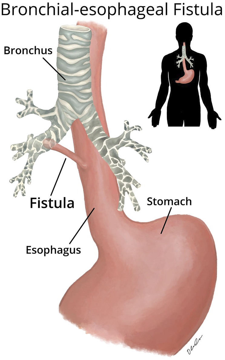
Illustration of tracheoesophageal fistulas (TEF). Note the abnormal connection between the esophagus and the upper respiratory tract
TEFs are often asymptomatic and diagnosed as incidental findings on CT or bronchoscopy, but patients with TEF who are symptomatic often follow an insidious and progressive course [1–3]. Symptoms commonly include cough (56%), aspiration (37%), fever (25%), and dysphagia (19%), and may result in recurrent pneumonia, ARDS, and sepsis in those not diagnosed and treated correctly [2, 3]. Benign TEFs can often be definitively corrected with surgical intervention; however if a concurrent or underlying infection is not treated appropriately, there are higher rates of failure, recurrence, and complications [1–3]. Thus, the prompt and correct identification of the fistula and its etiology is paramount in treating those with TEFs.
Candida and actinomyces
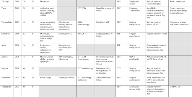
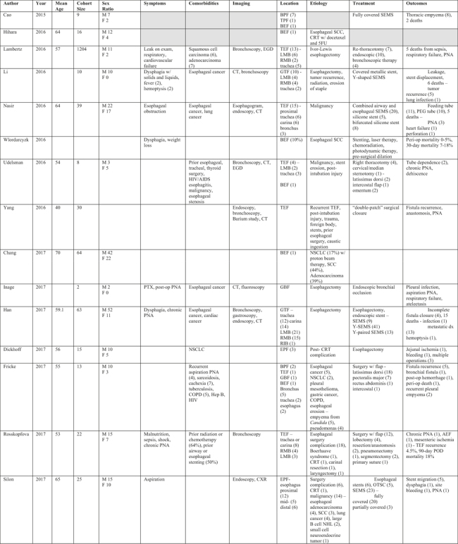
The most common infectious cause of benign acquired TEF is granulomatous infection, most often tuberculosis (Tables 1 and 2) [2, 3, 5–7]. Individual case reports noted 35.5% (n = 11) of fistula cases were attributed to infectious causes, with tuberculosis being the most common (72.7%, n = 8; Table 1). Other less common causes include agents of mucormycosis, herpes simplex infections, and Candida spp. infection, often in the setting of a compromised immune system or diabetes [12–14]. There have only been 12 published cases of TEF caused by Candida (Tables 3 and 4); seven individual reports and two reviews note Candida co-infection with Actinomyces, six of which confirmed the infections by culture (Tables 5 and 6). Candida and Actinomyces species can be found in the oral mucosa, often found in dental plaques and among normal mouth flora, typically causing infection in individuals with impaired mucosal barriers or compromised immune systems [15, 16]. While isolated Candida infections can occur, Actinomyces rarely is the sole culprit of infection, instead, it is often part of a microbial infection in conjunction with other species like Streptococcus, Fusobacterium, or Candida [15, 16]. While there are several reports of Candida and Actinomyces co-infection resulting in esophageal or pulmonary infection, only one noted the formation of a fistula (Table 5) [17]. Like tuberculosis, actinomycosis causes chronic granulomatous inflammation, which may have contributed to the fistula formation in our patient [15, 16]. Actinomyces is a known etiology of mycetoma, chronic granulomatous infections that are classically difficult to treat and require prolonged therapy [8, 9]. These are characterized as either fungal (eumycetoma) or bacterial (actinomycetoma), as in the case of our patient, and classically present with small, painless subcutaneous nodules that develop purulent discharge [8, 9]. Our patient was unusual, not only in the presentation of their actinomycetoma but in its co-infection with Candida and its complication by the formation of a broncholith and subsequent fistula, which, to our knowledge, has not been previously described.
Table 1.
Case reports of documented Bronchial Esophageal Fistula

|
|
Grey boxes indicate information not available in the report; ARDS – Acute Respiratory Distress Syndrome; AML – Acute Myeloid Leukemia; AmpB – Amphotericin B; CRT – Chemoradiation Therapy; GSS – Gastrograffin Swallow Study; OTSC – Over-The-Scope Clipping; PNA – Pneumonia; SCC – Squamous Cell Carcinoma; SEMS – Self-Expanding Metallic Stent; VATS – Video-Assisted Thoracic Surgery; LLL – Left Lower Lobe, LMB – Left Main Bronchus, RML – Right Middle Lobe, RLL – Right Lower Lobe; TEF – Tracheoesophageal Fistula; TBF – Tracheobronchial Fistula; BEF – Bronchioesophageal Fistula; EMF – Esophageal-Mediastinal Fistula; EPF – Esophageal-Pulmonary Fistula; GTF – Gastro-Tracheal Fistula; GBF – Gastro-Bronchial Fistula.
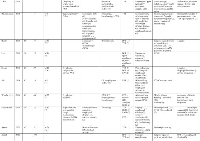
Table 2.
Review articles reporting Bronchial Esophageal Fistula
|
|
Grey boxes indicate information not available in the report; CRT – Chemoradiation Therapy; EVAC – Endoluminal Vacuum; NHL – Non-Hodgkin Lymphoma; NSCLC – Non-Small Cell Lung Cancer; OTSC – Over-The-Scope Clipping; PNA – Pneumonia; PTX – Pneumothorax; SCC – Squamous Cell Carcinoma; SEMS – Self-Expanding Metallic Stent; RIB – Right Intermediate Bronchus; RMB – Right Main Bronchus; LMB – Left Main Bronchus; TEF – Tracheoesophageal Fistula; TBF – Tracheobronchial Fistula; TPF – Tracheopulmonary Fistula; BEF – Bronchioesophageal Fistula; BPF – Bronchopulmonary Fistula; EPF – Esophageal-Pulmonary Fistula; GTF – Gastro-Tracheal Fistula; GBF – Gastro-Bronchial Fistula; AEF – Aorto-Esophageal Fistula.
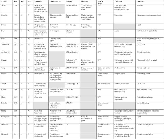
Table 3.
Case Reports of documented Candida Fistulas
|
Grey boxes indicate information not available in the report; AmpB – Amphotericin B; PNA – Pneumonia; PUD – Peptic Ulcer Disease; TEF – Tracheoesophageal Fistula; PEF – Pericardial-Esophageal Fistula; EPF – Esophageal-Pulmonary Fistula; ABF – Aorto-Bronchial Fistula.
Table 4.
Review articles of Candida Fistulas

|
Grey boxes indicate information not available in the report; CRT – Chemoradiation Therapy; PPI – Proton-Pump Inhibitor; TEF – Tracheoesophageal Fistula; BEF – Bronchioesophageal Fistula.
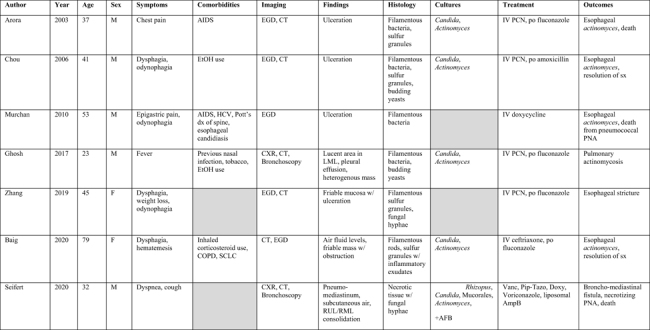
Table 5.
Case Reports of documented Actinomyces/Candida Co-infection
|
Grey boxes indicate information not available in the report; AFB – Acid-Fast Bacillus; PCN – Penicillin; PNA – Pneumonia; SCLC – Small Cell Lung Cancer; RML – Right Middle Lobe; RUL – Right Upper Lobe; LML – Left Middle Lobe.
Table 6.
Review articles of Actinomyces/Candida Co-infection

|
Grey boxes indicate information not available in the report; AmpB – Amphotericin B.
Actinomycetoma and broncholithiasis
Broncholithiasis is caused by calcification of material that enters the tracheobronchial tree, often due to granulomatous infection material that migrates from surrounding lymphatic structures [4, 5]. This can result in airway obstruction and inflammation, and while most often asymptomatic, they can be complicated by recurrent pneumonia and TEF formation [4, 5]. In fact, it was found that the most common infectious causes were fungal and mycobacterial infections, with Histoplasma being the most common etiology in the United States and other less common pathogens being tuberculosis, Actinomyces, and Aspergillus [4]. Despite the association with actinomycosis, there has not been a reported case of the formation of an actinomycetoma with Candida co-infection further complicated by broncholithiasis and TEF, representing a novel presentation for this pathology.
Diagnosis of TEF
Currently, there are no formal guidelines to establish or confirm the diagnosis of TEF. However, most experts agree that either esophagography or endoscopy is necessary to diagnosis and assist with pre-operative planning [2, 18]. The diagnosis of TEF is most often made using a combination of upper endoscopy and bronchoscopy. The literature review noted that 67.7% (n = 21) of patients were initially evaluated with CT, followed by bronchoscopy (41.9%, n = 13) and an esophagogastroduodenoscopy (EGD; 35.5%, n = 11; Table 1) [1, 2, 18]. CT initially only revealed a broncholith in our patient, but follow-up upper endoscopy revealed a fistulous communication between the right mainstem bronchus and the esophagus (Fig. 2a and b). While not necessary, radiographic imaging with chest radiography is often obtained in the setting of aspiration pneumonia or other symptoms suggestive of consolidation, and this may show non-specific signs of aspiration-related changes, lung masses, or widening of the mediastinum in the setting of granulomatous disease [1–3, 18]. CT imaging of the chest may be used to evaluate for signs of fistula or abnormalities in the mediastinum or aerodigestive tract; currently, there is a lack of data regarding the sensitivity, specificity, or predictive values of CT imaging in patients with known TEF [2]. CT imaging is useful, however, and plays a crucial role in diagnosing broncholithiasis, as it can give information on the location and degree of obstruction. While CT has good sensitivity for the early detection of bone involvement in those with more classic mycetoma of the lower extremity, in those cases, it is less frequently used as part of the routine diagnostic work-up in favor of culture and histology [4, 8]. In our patient, non-contrast CT of the chest revealed a broncholith within the right mainstream bronchus with evidence of post-obstructive atelectasis in the right lower lobe (Fig. 2).
Treatment of infections in TEF
While treatment of TEFs is often aimed at treating the fistulous communication, concomitant efforts need to be made to treat the underlying cause. Although infectious causes of benign acquired TEF are not uncommon, the treatment and diagnosis are not well described. The most commonly described infectious cause of TEF was tuberculosis infection, which, once confirmed as secondary to infection with Mycobacterium tuberculosis, is typically treated with surgical repair and prolonged courses of combination antibiotics [2, 5–7, 19]. Other infectious causes that have been recently described include mucormycosis infection treated with liposomal amphotericin B and a case of silicotuberculosis infection [5, 17]. Broncholithiasis is managed surgically or conservatively based on patient symptoms and extent of disease, with definitive management consisting of bronchoscopic extraction or pulmonary resection of the affected lobe [4, 5]. Unlike broncholithiasis, surgical treatment alone of mycetoma is rarely successful, but does play a role in fungal disease [8]. The pharmacologic treatments vary based on the infectious agent; fungal mycetoma (eumycetoma) often requires combined surgical excision followed by prolonged therapy with itraconazole for 1–2 years as first-line therapy, whereas actinomycetomas are often amenable to antibiotic therapy alone [8]. Antimicrobial treatment options for actinomycetoma often include use of penicillins, aminoglycosides, trimethoprim-sulfamethoxazole, or dapsone alone or in combination with one another, partially depending on the organisms determined to be driving the process [8]. A commonly used regimen in our review was use of IV penicillin for Actinomyces involvement, with oral fluconazole when there is concern for fungal coinfection. A period of more intensive treatment can then be followed by oral amoxicillin (Table 5).
Although reported as an infectious cause of benign acquired TEF, there are currently no concise guidelines on treatment for patients with TEF secondary to Candida infection. Of the cases that noted fistulas attributed to Candida infection, 15 reported treatment, and of those, eight were treated with combined surgery and medical therapy (44.4%) with an associated 25% mortality. Another four cases were treated with medical therapy alone (22.2%), with an associated 50% mortality and three patients were treated with surgery alone (16.7%), associated with a 100% mortality (Table 3). Of those that received medical therapy, the most commonly given antimicrobial agent was fluconazole (n = 8) followed by amphotericin B (n = 4), consistent with other reports [20]. To date, complications following treatment for infectious causes of fistulae have not been reported (Table 4). Only one reported case of an Actinomyces and Candida co-infection that resulted in a fistula has been reported; treatment was a combination of vancomycin, voriconazole, piperacillin-tazobactam, and liposomal amphotericin B and was complicated by Mucorales and Rhizopus infection, resulting in necrotizing pneumonia [17]. Our patient's definitive therapy was a combination of amoxicillin-clavulanate and fluconazole for six months based on the Actinomyces and Candida co-infection; a wedge resection and flap reconstruction was also completed without any perioperative morbidity. Follow-up esophagography suggested no leak from the fistula site, and all symptoms have resolved.
Diagnostic Algorithm
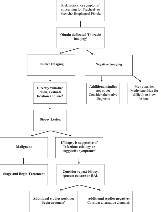
Based on the comprehensive literature review and the current management practices of benign acquired TEF, a diagnostic algorithm is proposed to assist in better diagnose those with relevant symptoms and history (Fig. 5). Once diagnostic imaging confirms the presence of a TEF, the anatomy should be further evaluated with esophageal and bronchial endoscopic visualization. Endoscopic evaluation allows for visualization and characterization of the fistula, can be used to dislodge debris obstructing the fistula site and assist in obtaining crucial biopsies of the lesion to help determine the underlying etiology of the TEF, guiding its treatment [1, 2, 4, 9, 10, 17, 18].
Fig. 5.
Diagnostic algorithm
1Risk factors include history of esophageal or bronchial malignancy, prior surgery or stenting, HIV/AIDS, TB, prior radiation
2Symptoms suggestive of TEF or EBF include cough (56%), aspiration (37%), fever (25%), dysphagia (19%), PNA (5%), hemoptysis (5%), or worsening of cough with swallowing liquids and solids 87% (Ono's Sign)
3Imaging includes Esophagography (1st line), barium preferred over gastrograffin (70% accurate), CT is preferred in those that cannot swallow, those that are ventilated or intubated that present with continued air-leaks despite well-inflated cuff, abdominal bloating, loss of tidal volume or worsening oxygenation
4Positive imaging should be followed up with Endoscopy or Bronchoscopy (flexible or rigid), as both will directly visualize lesion, evaluate location and size of lesion and can be used to get biopsy
5Symptoms suggestive of infectious etiology include additional symptoms of weight loss, night sweats or infection refractory to standard therapy
6Common infectious causes include tuberculosis, mucormycosis, histoplasmosis, syphilis, aspergillosis, each with specific biopsy findings, risk factors and treatments
Traditionally, TEF is repaired with an endoscopic approach, preferable to open surgical approaches, as endoscopy has a much lower risk of fistula complications [1, 3, 21]. Benign TEFs are often amenable to definitive surgical intervention compared to those with TEF secondary to malignancy, which often have multiple comorbidities that make them poor surgical candidates [1–3]. The primary endoscopic technique is esophageal or airway stenting, using self-expanding plastic stents that primarily serve as a bridge to definitive surgical repair, especially in those with concomitant tracheal stenosis in order to maintain airway patency [1, 12, 18, 22, 23]. Stenting can also be used in those with benign TEF that are not surgical candidates, opting for a single stent in those with distal fistula sites, or dual stenting those with more proximal or mid-esophagus fistulae [1, 12, 18, 22, 24–26]. Although self-expanding metallic stents (SEMS) are usually preferred for those with malignant fistulae, there is a growing body of evidence that prefers fully covered SEMS in those with complicated benign TEFs or those who are not surgical candidates [1, 12, 18, 22, 24, 26]. Definitive surgical correction involves division and closure of the TEF in conjunction with airway restoration using a muscle flap interposition based on the defect's size and absence of airway stenosis [1, 12, 27, 28]. Differences in single vs. double-layer closure techniques and in the tissue used to close the defects have been reported; intercostal muscles and latissimus dorsi muscle flaps are the most commonly used [1, 12, 27, 28]. Due to the complexity and associated comorbidities, patients with acquired TEFs often receive stenting and surgical repair. However, surgical management for tracheal and esophageal defects remains controversial [2, 3, 12, 27, 28]. Stent placement has been shown to prevent aspiration pneumonia, and often, oral intake can be resumed after complete surgical closure of the fistula. However, most patients require nutritional support via gastrostomy, jejunostomy, or parenteral nutrition [3, 18, 22]. Furthermore, there are reported complications of endoscopic stenting, the most common being stent migration, bleeding, perforation, and, paradoxically, the formation of new TEFs [3, 18, 22]. Of the 31 individual cases of fistula reviewed, 32.3% were secondary to surgery or stenting (Table 1). Although considered a definitive treatment, fistula recurrence often complicates surgical repair, and can have an operative mortality rate reported as high as 11% from post-op emphysema, pneumonia, and hemorrhage [3, 26–28].
Overall, TEF should be suspected in patients with known risk factors or symptoms, such as recurrent bronchitis or pneumonia, or persistent coughing following nutritional intake. Management by a multidisciplinary team is essential and includes imaging and endoscopic findings for diagnosis, histologic analysis for differentiation between malignant and benign entities, evaluation of infectious etiologies and appropriate treatments for optimal outcomes. Fungal or bacterial infections causing benign TEF, while uncommon but not rare, need to be considered in diagnosis and treated quickly and appropriately for optimal outcomes.
Footnotes
Disclosures: The authors report no conflicts of interest.
The authors have no financial disclosures or financial support.
Dr. Devane is a consultant with Boston Scientific and TriSalus Life Sciences.
IRB approval was received from Prisma Health IRB committee (Protocol 2176412-1) and was deemed ‘non-human research’.
Contributions: The authors confirm contribution to the paper as follows:
Study conception and design: AMD.
Data collection: AT and RR.
Analysis and interpretation of results: CMGS, DPS, PK, AMD.
Draft manuscript preparation: AT, RR, OC.
Editing manuscript: CMGS, DPS, PK, AMD.
Illustration: OC.
All authors reviewed the results and approved the final version of the manuscript.
References
- 1.Bibas BJ, Guerreiro Cardoso PF, Minamoto H, Eloy-Pereira LP, Tamagno MF, Terra RM, et al. Surgical management of benign acquired tracheoesophageal fistulas: a ten-year experience. Ann Thorac Surg. 2016;102:1081–7. 10.1016/j.athoracsur.2016.04.029. [DOI] [PubMed] [Google Scholar]
- 2.Kim HS, Khemasuwan D, Diaz-Mendoza J, Mehta AC. Management of tracheo-oesophageal fistula in adults. Eur Respir Rev. 2020;29. 10.1183/16000617.0094-2020. [DOI] [PMC free article] [PubMed] [Google Scholar]
- 3.Zhou C, Hu Y, Xiao Y, Yin W. Current treatment of tracheoesophageal fistula. Ther Adv Respir Dis. 2017;11:173–80. 10.1177/1753465816687518. [DOI] [PMC free article] [PubMed] [Google Scholar]
- 4.Krishnan S, Kniese CM, Mankins M, Heitkamp DE, Sheski FD, Kesler KA. Management of broncholithiasis. J Thorac Dis. 2018;10(Suppl 28):S3419–27. 10.21037/jtd.2018.07.15. [DOI] [PMC free article] [PubMed] [Google Scholar]
- 5.Zhang H, Li L, Sun XW, Zhang CL. Silicotuberculosis with esophagobronchial fistula and broncholithiasis. Int J Occup Environ Med. 2017;8:50–5. 10.15171/ijoem.2017.822. [DOI] [PMC free article] [PubMed] [Google Scholar]
- 6.Indiran V. Tuberculous bronchoesophageal fistula presenting as intractable cough. Tuberk Ve Toraks. 2017;65:60–2. [PubMed] [Google Scholar]
- 7.Sayeed A, Alqurashi EH, Alzanbagi AB, Ghaleb NAB. Tuberculosis presenting as broncho-oesophageal fistula in a young healthy man. BMJ Case Rep. 2017;2017:bcr-2017-220821. 10.1136/bcr-2017-220821. [DOI] [PMC free article] [PubMed] [Google Scholar]
- 8.Relhan V, Mahajan K, Agarwal P, Garg VK. Mycetoma: an update. Indian J Dermatol. 2017;62:332–40. 10.4103/ijd.IJD_476_16. [DOI] [PMC free article] [PubMed] [Google Scholar]
- 9.Sande WWJ van de, Fahal AH, Goodfellow M, Mahgoub ES, Welsh O, Zijlstra EE. Merits and pitfalls of currently used diagnostic tools in mycetoma. Plos Negl Trop Dis. 2014;8:e2918. 10.1371/journal.pntd.0002918. [DOI] [PMC free article] [PubMed] [Google Scholar]
- 10.Balakrishnan A, Tapias L, Wright CD, Lanuti M, Gaissert HA, Mathisen DJ, et al. Surgical management of post-esophagectomy tracheo-bronchial-esophageal fistula. Ann Thorac Surg. 2018;106:1640–6. 10.1016/j.athoracsur.2018.06.076. [DOI] [PubMed] [Google Scholar]
- 11.Lambertz R, Hölscher AH, Bludau M, Leers JM, Gutschow C, Schröder W. Management of tracheo- or bronchoesophageal fistula after Ivor-Lewis esophagectomy. World J Surg. 2016;40:1680–7. 10.1007/s00268-016-3470-9. [DOI] [PubMed] [Google Scholar]
- 12.Debourdeau A, Gonzalez JM, Dutau H, Benezech A, Barthet M. Endoscopic treatment of nonmalignant tracheoesophageal and bronchoesophageal fistula: results and prognostic factors for its success. Surg Endosc. 2019;33:549–56. 10.1007/s00464-018-6330-x. [DOI] [PubMed] [Google Scholar]
- 13.Li Y, Ren K, Ye L, Ren J, Han X. Intermediate bronchial fistula caused by mediastinal drainage tube compression and fungal infection: a case report. J Cardiothorac Surg. 2019;14:190. 10.1186/s13019-019-1020-x. [DOI] [PMC free article] [PubMed] [Google Scholar]
- 14.Kanzaki R, Yano M, Takachi K, Ishiguro S, Motoori M. Candida esophagitis complicated by an esophago-airway fistula: report of a case. Surg Today. 2009;39:972–8. 10.1007/s00595-009-3958-0. [DOI] [PubMed] [Google Scholar]
- 15.Zhang AN, Guss D, Mohanty SR. Esophageal stricture caused by actinomyces in a patient with No apparent predisposing factors. Case Rep Gastrointest Med. 2019;2019:7182976. 10.1155/2019/7182976. [DOI] [PMC free article] [PubMed] [Google Scholar]
- 16.Ghosh P, Gupta I, Kar M, Nandi P, Naskar P. Co-Infection of Candida parapsilosis in a patient of pulmonary actinomycosis-A rare case report. J Clin Diagn Res JCDR. 2017;11:DD01–2. 10.7860/JCDR/2017/24268.9300. [DOI] [PMC free article] [PubMed] [Google Scholar]
- 17.Seifert S, Wiley J, Kirkham J, Lena S, Schiers K. Pulmonary mucormycosis with extensive bronchial necrosis and bronchomediastinal fistula: a case report and review. Respir Med Case Rep. 2020;30:101082. 10.1016/j.rmcr.2020.101082. [DOI] [PMC free article] [PubMed] [Google Scholar]
- 18.Silon B, Siddiqui AA, Taylor LJ, Arastu S, Soomro A, Adler DG. Endoscopic management of esophagorespiratory fistulas: a multicenter retrospective study of techniques and outcomes. Dig Dis Sci. 2017;62:424–31. 10.1007/s10620-016-4390-0. [DOI] [PubMed] [Google Scholar]
- 19.Alharbi SR. Tuberculous esophagomediastinal fistula with concomitant mediastinal bronchial artery aneurysm-acute upper gastrointestinal bleeding: a case report. World J Gastroenterol. 2019;25:2144–8. 10.3748/wjg.v25.i17.2144. [DOI] [PMC free article] [PubMed] [Google Scholar]
- 20.Mohamed AA, Lu X liang, Mounmin FA. Diagnosis and treatment of esophageal candidiasis: current updates. Can J Gastroenterol Hepatol. 2019;2019:e3585136. 10.1155/2019/3585136. [DOI] [PMC free article] [PubMed] [Google Scholar]
- 21.Akram MJ, Khalid U, Abu Bakar M, Ashraf MB, Butt FM, Khan F. Indications and clinical outcomes of fully covered self-expandable metallic tracheobronchial stents in patients with malignant airway diseases. Expert Rev Respir Med. 2020;14:1173–81. 10.1080/17476348.2020.1796642. [DOI] [PubMed] [Google Scholar]
- 22.Vachtenheim J, Lischke R. Esophageal bypass surgery as a definitive repair of recurrent acquired benign bronchoesophageal fistula. J Cardiothorac Surg. 2019;14:73. 10.1186/s13019-019-0902-2. [DOI] [PMC free article] [PubMed] [Google Scholar]
- 23.Włodarczyk JR, Kużdżał J. Safety and efficacy of airway stenting in patients with malignant oesophago-airway fistula. J Thorac Dis. 2018;10:2731–9. 10.21037/jtd.2018.05.19. [DOI] [PMC free article] [PubMed] [Google Scholar]
- 24.Cao M, Zhu Q, Wang W, Zhang TX, Jiang MZ, Zang Q. Clinical application of fully covered self-expandable metal stents in the treatment of bronchial fistula. Thorac Cardiovasc Surg. 2016;64:533–9. 10.1055/s-0034-1396681. [DOI] [PubMed] [Google Scholar]
- 25.Li TF, Duan XH, Han XW, Wu G, Ren JZ, Ren KW, et al. Application of combined-type Y-shaped covered metallic stents for the treatment of gastrotracheal fistulas and gastrobronchial fistulas. J Thorac Cardiovasc Surg. 2016;152:557–63. 10.1016/j.jtcvs.2016.03.090. [DOI] [PubMed] [Google Scholar]
- 26.Yang G, Li WM, Zhao JB, Wang J, Ni YF, Zhou YA, et al. A novel surgical method for acquired non-malignant complicated tracheoesophageal and bronchial-gastric stump fistula: the “double patch” technique. J Thorac Dis. 2016;8:3225–31. 10.21037/jtd.2016.11.80. [DOI] [PMC free article] [PubMed] [Google Scholar]
- 27.Fricke A, Bannasch H, Klein HF, Wiesemann S, Samson-Himmels P, Passlick B, et al. Pedicled and free flaps for intrathoracic fistula management. Eur J Cardio-Thorac Surg Off J Eur Assoc Cardio-Thorac Surg. 2017;52:1211–7. 10.1093/ejcts/ezx216. [DOI] [PubMed] [Google Scholar]
- 28.Rosskopfova P, Perentes JY, Schäfer M, Krueger T, Lovis A, Dorta G, et al. Repair of challenging non-malignant tracheo- or broncho-oesophageal fistulas by extrathoracic muscle flaps. Eur J Cardio-Thorac Surg Off J Eur Assoc Cardio-Thorac Surg 2017;51:844–51. 10.1093/ejcts/ezw435. [DOI] [PubMed] [Google Scholar]


