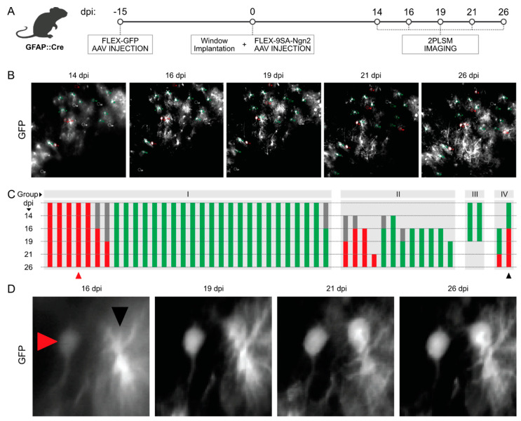
Figure 3.
Chronic in vivo imaging of FLEX-AAVs via 2-photon microscopy. (A) Scheme illustrating the timeline of the experimental procedures used for the in vivo imaging of GFAP::Cre mice injected with FLEX-AAVs. (B) Pictures of the imaged cortical area at different timepoints (14, 16, 19, 21, 26 dpi). (C) Classification of the cells observed during the chronic in vivo imaging according to their morphology and dynamic during the experiments. Astrocytes (green) and neurons (red) have been divided into four different groups: cells observed since the earliest timepoint (I), cells observed only from later timepoints (II), cells that disappeared before the completion of the experiment (III), and cells that underwent a morphological change (IV). Examples of non-converting and one of the few possibly converting cells are indicated with a red and a black triangle, respectively, in (D).

