Abstract
The integrin αvβ3 has been shown to function as one of the integrin receptors on cultured cells for foot-and-mouth disease virus (FMDV), and high-efficiency utilization of the bovine homolog of this integrin is dependent on the cysteine-rich repeat region of the bovine β3 subunit. In this study we have examined the role of the cytoplasmic domains of the αv and β3 subunits in FMDV infection. We have found that truncations or extensions of these domains of either subunit, including deletions removing almost all of the cytoplasmic domains, had little or no effect on the ability of the integrin to function as a receptor for FMDV. The lysosomotropic agent monensin inhibited viral replication in cells transfected with either intact or cytoplasmic domain-truncated αvβ3. In addition, viral replication in transfected cells was inhibited by an αvβ3 function-blocking antibody but not by function-blocking antibodies to three other RGD-directed integrins, suggesting that these integrins are not involved in the infectious process. These results indicate that alterations to the cytoplasmic domains of either subunit, which lead to the inability of the integrin receptor to function normally, do not abolish the ability of the integrin to bind and internalize this viral ligand.
Integrins are heterodimeric cell surface receptors consisting of α and β subunits that are involved in binding extracellular matrix proteins, cell-cell interactions, and signal transduction (20, 23). The two subunits interact noncovalently at the cell surface to bind their natural ligands via a ligand-binding region which is made up of elements of both subunits (16). Integrin subunits are type I membrane proteins consisting of a large N-terminal extracellular domain and smaller transmembrane and cytoplasmic domains. The cytoplasmic domains of the α and β integrin subunits, specifically certain sequence motifs within those domains, have been shown to be involved in inside-out and outside-in signal transduction, integrin activation, and conformational changes leading to alterations in ligand-binding affinities and connections to the cytoskeleton (6, 7, 18, 22, 27, 37, 38, 41, 44).
Foot-and-mouth disease virus (FMDV), an aphthovirus in the family Picornaviridae, utilizes the integrin αvβ3 as a receptor in cultured cells (4, 35, 36). We have recently molecularly cloned the bovine homolog of this integrin and shown that the high-efficiency utilization of the bovine integrin as a receptor for FMDV is dependent on sequences found within the cysteine-rich repeat region of the bovine β3 subunit extracellular domain (35). As part of an ongoing study of the roles that the various functional domains within the integrin subunits play in FMDV infection, we have examined subunits with altered cytoplasmic domains for their ability to retain viral receptor function.
Generation of bovine αv and β3 subunits with altered cytoplasmic domains.
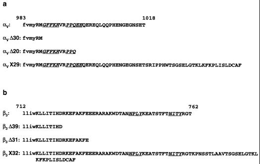
We generated two truncation mutants and one extension mutant for each of the subunits, as shown in Fig. 1, utilizing the plasmids pBovαvZEO and pBovβ3ZEO, which encode the bovine αv and β3 integrin subunits, respectively (35). pBovαvZEO was used as the template for a 20-cycle PCR with the N-terminal PCR primer 5′GGAAGGTGCCTACGAAGCTGAG3′ and the following C-terminal primers: for αvΔ30, 5′GGAATTCCTTACATCCTGTACATTACAA3′; for αvΔ20, 5′GGAATTCCTTATTGAGGTGGCCGTACACG3′; and for αvX29, 5′GGAATTCCTTAGTTTCAGAGTTTCCTTCGCC3′. Plasmids encoding the altered αv subunits were created utilizing BstEII and EcoRI sites shared by pBovαvZEO and the PCR products. pBovβ3ZEO was also used as the template for a 20-cycle PCR using the N-terminal PCR primer 5′CCACGCGTGGTGTGAGCTCCTG3′ and the following C-terminal primers: for β3Δ39, 5′CGGGATCCTTAGTCATGGATGGTGATGAG3′; for β3Δ31, 5′CGGGATCCTTAGGCTCTGGCTCTCTCTTC3′; and for β3X32, 5′CGGGATCC T TAAG TGCCCCGG TACG TGATAT TG3 ′. Plasmids encoding the altered β3 subunits were created utilizing MluI and BamHI sites shared by pBovβ3ZEO and the PCR products. All of the plasmid constructs were sequenced through the region that was subcloned.
FIG. 1.
Predicted amino acid sequences of wild-type and altered integrin subunit cytoplasmic domains. The sequences of the cytoplasmic domains are in uppercase letters. The last four residues of the transmembrane domains are in lowercase letters. Functionally important sequence motifs, as noted in the text, are italicized and underlined.
The amino acid sequences of the cytoplasmic domains of the wild-type and altered subunits are shown in Fig. 1. There is some question as to where the exact borders between the transmembrane and cytoplasmic domains occur in the integrin subunits. We have marked the border as lying at the YR junction for the αv subunit (Fig. 1a) and the WK junction for the β3 subunit (Fig. 1b). A recent study suggests that the first five residues of the αv subunit (RMGFF) and the first six residues of the β3 subunit (KLLITI) cytoplasmic domains, as we have represented them, are located within the cell membrane in the absence of interactions with intracellular proteins and become exposed to the cytoplasm upon binding to intracellular proteins, resulting in conformational changes leading to integrin activation and signaling (1). The mutant αv subunits are shown in Fig. 1a. The first, αvΔ30, retains only two cytoplasmic domain amino acids and is truncated before the GFFKR motif, which is conserved among all α subunits. This motif maintains integrins in a low-affinity state (8, 37, 49) and has been reported to be necessary for stabilization of the integrin αβ complex (48). This mutant is also lacking the PPQ(L)EE(DD) motif, which defines a β-turn in the α subunit cytoplasmic domain and is found in seven other α subunits. A human αvβ3 heterodimer with a truncated α subunit cytoplasmic domain lacking this motif cannot bind to either vitronectin or fibronectin (18). The second mutant, αvΔ20, contains the GFFKR motif and the five amino acids downstream of it but is truncated at the last residue of the PPQEE motif. The final mutant, αvX29, contains the complete cytoplasmic domain of the αv subunit with an additional 29 amino acids added to the C terminus.
The mutant β3 subunits are shown in Fig. 1b. The first of these, β3Δ39, retains only 8 amino acids while β3Δ31 retains 16 amino acids of the β3 cytoplasmic domain. Neither of these two constructs contain the NPL(X)Y or the NI(X)T(X)Y motifs that have been shown to be important for signal transduction, integrin affinity states, and interaction with cytoplasmic integrin-associated proteins (7, 9, 10, 13, 15, 17, 25, 28, 31, 32, 44, 46, 54). Both of these truncations, however, still retain the membrane-proximal region of the subunit cytoplasmic domain, which has also been reported to control ligand-binding affinity and to regulate signal transduction (21, 22). The third β3 subunit mutant, β3X32, retains both the NPLY and NITY motifs along with an additional 32 amino acids added to the C terminus of the cytoplasmic domain. Additions to the cytoplasmic domains of both the α and β subunits were made because the specific conformations of these domains appear to play a role in the way they control ligand affinity and signal transduction (21).
Analysis of integrins with altered cytoplasmic domains.
Coupled in vitro transcription-translations were performed to check that the mutant-encoding plasmids that were generated were encoding proteins of the expected sizes. In all cases, the relative sizes of the resulting altered integrin subunits compared to the corresponding wild-type subunits were as expected (not shown).
To analyze whether integrin subunits with truncated or extended cytoplasmic domains could still function as receptors for FMDV, we utilized a previously described transient-expression assay system in COS-1 cells (35). Cells were plated at a density of 105 cells/well on six-well plates the day prior to transfection. Transfections were performed with 2.0 μg of each integrin-encoding plasmid utilizing the transfection reagent FuGENE 6 (Roche Molecular Biochemicals) according to the manufacturer's instructions. Twenty-four hours after transfection, the cells in each well were trypsinized and replated onto two wells of a 24-well plate. After a further 18 h of incubation, one well for each transfected condition was infected with FMDV type A12, strain 119ab, at a multiplicity of infection (MOI) of 10 PFU/cell and labeled between 4 and 18 h after infection with [35S]methionine. The other well was fixed with acetone-methanol (50:50) and analyzed by immunohistochemistry for integrin expression using the anti-αvβ3 monoclonal antibody (MAb) LM609 (MAB1976; Chemicon International) as previously described (35). Only transfections in which equal amounts of immunostaining were detected in all experimental conditions were used to analyze the results of viral infection (35). To evaluate FMDV replication in the infected-radiolabeled cultures, cell lysates were prepared in 1% Triton X-100. Equal amounts of trichloroacetic acid-precipitable counts per minute were immunoprecipitated (IP), using a virus-specific MAb, and proteins were analyzed by sodium dodecyl sulfate-polyacrylamide gel electrophoresis (SDS-PAGE) using a 10% polyacrylamide gel as described (35).
The results in Fig. 2 are representative of one such transfection. Viral replication is evident in cells transfected with both wild-type bovine αv- and β3-expressing plasmids, as evidenced by the synthesis of viral proteins which are not present in nontransfected-infected cells. When cells are transfected with a wild-type αv subunit and any of the altered β3 subunits, viral replication appears to be unaffected. Similarly, when any of the three altered αv subunits are transfected with the wild-type β3 subunit, levels of infection reach those seen when both wild-type subunits are present. Viral replication was also unaffected when the two subunits with the shortest cytoplasmic domains (αvΔ30 and β3Δ39) were expressed together.
FIG. 2.
Analysis of viral protein synthesis in COS-1 cells transfected with integrin subunit cDNAs. Cells were transfected with plasmids encoding integrin subunits as shown. Transfected cells were infected with FMDV type A12 at an MOI of 10 PFU/cell and labeled between 4 and 18 h with [35S]methionine. Cell extracts were analyzed by IP and SDS–10% PAGE as described in the text. The locations of viral structural proteins IP from infected and labeled BHK-21 cells (lane M) are shown on the left. con, nontransfected-infected cells (control).
Viral replication mediated by integrins with truncated cytoplasmic domains in the presence of monensin.
We have previously shown that the lysosomotropic ionophore monensin inhibited the replication of representative strains of all seven serotypes of FMDV (2). Monensin interferes with proton and pH gradients and raises the pH of endocytic vesicles (33, 40). In the presence of monensin, the virus adsorbed to cells normally; however, it was unable to undergo the initial alteration of the 140S virion to 12S pentameric subunits, which probably occurs within acidified endocytic vesicles and results in the release of the genome RNA (2, 3). To examine whether virus utilizing expressed integrins with truncated cytoplasmic domains as receptors infected cells through an eclipse mechanism similar to that of virus utilizing intact receptors, we transfected cells with intact and cytoplasmic domain-truncated integrin subunits, followed by infection in the presence or absence of monensin.
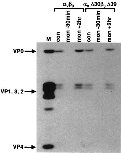
Cells were cotransfected with either αv and β3 subunits or αvΔ30 and β3Δ39 subunits. Forty-eight hours after transfection, cultures were incubated in the presence or absence of 50 μM monensin for 30 min prior to infection with FMDV type A12. Viral replication was determined by pulse-labeling cells with [35S]methionine between 5 and 6 h postinfection and analyzing cell extracts by IP and SDS-PAGE. The results are shown in Fig. 3. Cells cotransfected with intact integrin subunits and infected in the presence of monensin failed to synthesize viral proteins, indicating that the drug interfered with viral replication. Interestingly, cells cotransfected with cytoplasmic domain-truncated integrin subunits and infected in the presence of monensin also failed to synthesize viral proteins. In contrast, cells cotransfected with either intact or truncated integrins synthesized normal amounts of viral proteins when monensin was added at 2 h after infection, indicating that monensin inhibited an early event in viral replication, probably at the eclipse phase, as we have shown previously (2). Therefore, complete integrin subunit cytoplasmic domains are not necessary for internalization of virions into endocytic vesicles.
FIG. 3.
Analysis of transfected COS-1 cells infected in the presence of monensin. Cells were cotransfected with integrin subunits containing intact cytoplasmic domains or αvΔ30 and β3Δ39 as shown. Thirty minutes prior to infection, the medium was removed from some wells and replaced with medium containing 50 μM monensin (lanes mon −30min). Cells were infected in either the presence or absence (con) of monensin as noted. At 2 h after infection, the medium was removed from other wells and replaced with medium containing 50 μM monensin (lanes mon +2hr). At 4.5 h after infection, all cultures were incubated in minimal essential medium without l-methionine, with or without monensin, for 30 min. [35S]methionine (75 μCi) was added, and cells were labeled for 1 h. Cell extracts were prepared, and analysis of viral proteins by IP and SDS–10% PAGE was done as described in the text. The locations of marker FMDV structural proteins (lane M) are shown on the left.
Thus, bovine αvβ3 is able to function as a receptor for FMDV in the absence of motifs that are known to be required for the normal function of integrins in the context of their natural ligands. The truncations of the cytoplasmic domains of either the αv or β3 subunits and the addition of random amino acids to the subunits' cytoplasmic domains did not affect the ability of integrins to serve as receptors for FMDV, and the results show that when utilized as receptors, integrins altered in this manner were utilized as well as intact integrins.
Effect of integrin function-blocking antibodies on viral infection in transfected cells.
The internalization of natural integrin ligands has not been extensively studied. The NPXY motif, found in the cytoplasmic domains of all β subunits and other transmembrane receptors, has been reported to be required for internalization of bacteria mediated by β1 integrins (47), internalization mediated by the nonintegrin low-density lipoprotein receptor (12), and a signal for clathrin complex assembly (11). More recent results have shown that sequences surrounding the NPXY motif are important in association of the αvβ5 receptor with clathrin-coated pits via the β5 cytoplasmic domain (13). The internalization of vitronectin by αvβ3, however, appears to require a signal as a result of the ligation of the α5β1 integrin (39). This cross talk between the αvβ3 and α5β1 integrins requires the β3 cytoplasmic domain (5). Our results would rule out a cross talk mechanism of viral internalization, since important cytoplasmic domain motifs have been deleted from some of the subunit constructs. The results, however, might indicate that other cell surface molecules are acting as coreceptors for viral infection or that the expression of αvβ3 in COS cells is activating another receptor and the integrin itself is not involved in viral binding or internalization. Although, at this time, we have no evidence that a coreceptor, either integrin or nonintegrin, is required for infection, we examined the possibility that other RGD-directed integrins might be involved in the infectious process in αvβ3-transfected COS cells.
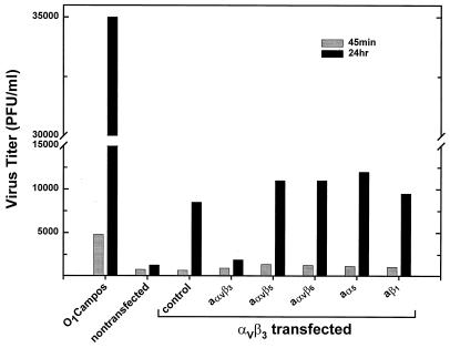
Cells were transfected with αvβ3 cDNAs, as described, and 30 min prior to infection with FMDV type A12, they were incubated with function-blocking antibodies to αvβ3, αvβ5, αvβ6, α5, or β1. All of these integrins interact with their natural ligands through an RGD sequence (42), and the αvβ6 integrin has recently been shown to be a receptor for FMDV (24), a result we have confirmed in our laboratory (not shown). In this experiment, we measured productive viral replication by determining the plaque titer of infectious virus immediately following a 45-min adsorption period and after 24 h of incubation. As a control, to determine the level of FMDV replication in COS cell cultures, a nontransfected culture was infected with an FMDV type O1 Campos variant containing a heparin-binding site in VP3 (43), which we have previously shown to require only the presence of cell surface heparan sulfate (HS), and not αvβ3, to infect cells (36). The results of this experiment are shown in Fig. 4.
FIG. 4.
Effect of anti-integrin function-blocking MAbs on viral replication in transfected COS-1 cells. Cells were cotransfected with αvβ3-encoding plasmids as described in the text. Thirty minutes prior to infection, paired transfected cell cultures were incubated at room temperature with the following function-blocking anti-integrin MAbs at a concentration of 25 μg/ml (all antibodies were from Chemicon International Inc): anti-αvβ3 (clone LM609; MAB1976), anti-αvβ5 (clone P1F6; MAB1961), anti-αvβ6 (clone 10D5; MAB2077Z), anti-α5 (clone CLB-705; MAB1986), and anti-β1 (clone 6S6; MAB2253). Transfected and nontransfected cultures were infected in pairs with type A12 at an MOI of 1 PFU/cell in the presence of the antibodies. One nontransfected culture pair was infected with the HS-binding O1 Campos variant vCRM4 (43) at an MOI of 1 PFU/cell. After an adsorption period of 45 min at 37°C, all cultures were washed with a low-pH buffer (25 mM MES [morpholineethanesulfonic acid, pH 5.5], 140 mM NaCl) to inactivate any nonadsorbed or noninternalized virus. After the addition of medium, one of the infected pairs was immediately frozen at −70°C to determine viral infectivity remaining at the end of the adsorption period (shaded bars). The other pair was incubated for 24 h at 37°C (solid bars) and then placed at −70°C. After thawing, cell debris was removed by centrifugation, and plaque titer was determined on BHK-21 cells.
In nontransfected COS cells, there was no increase in titer of type A12 at 24 h, indicating that viral replication did not take place in these cells, as we have previously shown in experiments utilizing detection of radioactively labeled viral proteins (35) (Fig. 2). In contrast, the heparin-binding type O1 variant showed an increase in titer of about 10-fold after 24 h in nontransfected cells, also confirming previously reported results (36). In αvβ3-transfected COS cells, the type A12 virus titer increased about 10-fold, again confirming results obtained by detection of viral proteins in infected cells (35) (Fig. 2). When transfected cells were treated with integrin function-blocking antibodies, only the antibody to αvβ3 and not those against any of the other integrins was capable of inhibiting viral replication (Fig. 4). The same experiment performed in cells transfected with the αvΔ30 and β3Δ39 cDNAs yielded nearly identical results (not shown). These results indicate that, in this transfection system, the αvβ3 integrin is absolutely required for productive viral infection and at least three other RGD-directed integrins (αvβ5, αvβ6, and α5β1) do not appear to be involved in this process. In addition, since the anti-β1 antibody has been shown to inhibit the function of at least two other β1 integrins (α2β1 and α4β1) (19), other integrins of this subclass are also probably not involved in viral infection. At this time, however, we cannot rule out a role for other cell surface molecules as coreceptors for either FMDV adsorption or internalization. We can, however, rule out HS as a coreceptor for type A12, as we have previously shown that this virus can replicate in αvβ3 cDNA-transfected HS-deficient CHO cells (36).
These results are similar to those reported for three other picornaviruses (poliovirus, rhinovirus 14, and coxsackievirus B3), all of whose single-subunit receptors appear to function normally in the absence of cytoplasmic domains (26, 45, 52). Human adenovirus (Ad) requires interaction with the integrin αvβ5 or αvβ3 for internalization into cells through a clathrin-coated pit pathway requiring dynamin (51, 53). We have not yet examined the role of dynamin in FMDV internalization, but in studies with two other picornaviruses, human rhinovirus 14 required dynamin for productive infection while poliovirus did not (14). Recently it has been shown that Ad internalization requires signaling through the focal adhesion kinase pathway involving phosphoinositide-3-OH kinase and GTP-binding proteins, all of which are activated following binding of integrins to their natural ligands (29, 30). In addition, the cytoplasmic domain of the β5 subunit of the αvβ5 integrin is essential for Ad-mediated gene delivery via host cell membrane penetration from endosomes (50). However, truncations of the β5 cytoplasmic domain, which still retains the NPXY motif, and do not allow Ad-mediated gene delivery do not abolish αvβ5-mediated Ad internalization (50). At least two picornavirus receptors, the coxsackievirus and adenovirus receptors, and ICAM-1 also do not require their transmembrane domains for receptor function (45, 52). Soluble human αvβ3 lacking both the transmembrane and cytoplasmic domains of both subunits can still bind to its natural ligands with high affinity (34); however, we have not examined either soluble or glycosylphosphatidylinositol-anchored αvβ3 for virus binding or the ability to act as a functional receptor.
The results we have presented, however, indicate that deletions of any of the important cytoplasmic domain motifs had little or no effect on receptor utilization by FMDV. In fact, the results showing that monensin still inhibited infection mediated by cytoplasmic domain-truncated αvβ3 suggest that these receptors are internalizing FMDV through the same mechanism as complete integrins. In addition, we have also shown that three other RGD-directed integrins do not appear to play any role in productive viral infection. Further studies will be necessary to delineate the exact mechanism by which intact and altered integrin subunits internalize virus.
Acknowledgments
We thank Michael LaRocco for excellent technical assistance.
REFERENCES
- 1.Armulik A, Nilsson I, von Heijne G, Johansson S. Determination of the border between the transmembrane and cytoplasmic domains of human integrin subunits. J Biol Chem. 1999;274:37030–37034. doi: 10.1074/jbc.274.52.37030. [DOI] [PubMed] [Google Scholar]
- 2.Baxt B. Effect of lysosomotropic compounds on early events in foot-and-mouth disease virus replication. Virus Res. 1987;7:257–271. doi: 10.1016/0168-1702(87)90032-3. [DOI] [PubMed] [Google Scholar]
- 3.Baxt B, Bachrach H L. Early interactions of foot-and-mouth disease virus with cultured cells. Virology. 1980;104:42–55. doi: 10.1016/0042-6822(80)90364-5. [DOI] [PubMed] [Google Scholar]
- 4.Berinstein A, Roivainen M, Hovi T, Mason P W, Baxt B. Antibodies to the vitronectin receptor (integrin αvβ3) inhibit binding and infection of foot-and-mouth disease virus to cultured cells. J Virol. 1995;69:2664–2666. doi: 10.1128/jvi.69.4.2664-2666.1995. [DOI] [PMC free article] [PubMed] [Google Scholar]
- 5.Blystone S D, Lindberg F P, LaFlamme S E, Brown E J. Integrin β3 cytoplasmic tail is necessary and sufficient for regulation of α5β1 phagocytosis by αvβ3 and integrin-associated protein. J Cell Biol. 1995;130:745–754. doi: 10.1083/jcb.130.3.745. [DOI] [PMC free article] [PubMed] [Google Scholar]
- 6.Blystone S D, Lindberg F P, Williams M P, McHugh K, Brown E J. Inducible tyrosine phosphorylation of the β3 integrin requires the αv integrin cytoplasmic tail. J Biol Chem. 1996;271:31458–31462. doi: 10.1074/jbc.271.49.31458. [DOI] [PubMed] [Google Scholar]
- 7.Blystone S D, Williams M P, Slater S E, Brown E J. Requirement of integrin β3 tyrosine 747 for β3 tyrosine phosphorylation and regulation of αvβ3 avidity. J Biol Chem. 1997;272:28757–28761. doi: 10.1074/jbc.272.45.28757. [DOI] [PubMed] [Google Scholar]
- 8.Briesewitz R, Kern A, Smilenov L B, David F S, Marcantonio E E. The membrane-cytoplasm interface of integrin α subunits is critical for receptor latency. Mol Biol Cell. 1996;7:1499–1509. doi: 10.1091/mbc.7.10.1499. [DOI] [PMC free article] [PubMed] [Google Scholar]
- 9.Calderwood D A, Zent R, Grant R, Jasper D, Rees G, Hynes R O, Ginsberg M H. The talin head domain binds to integrin β subunit cytoplasmic tails and regulates integrin activation. J Biol Chem. 1999;274:28071–28074. doi: 10.1074/jbc.274.40.28071. [DOI] [PubMed] [Google Scholar]
- 10.Chang D D, Wong C, Smith H, Liu J. ICAP-1, a novel β1 integrin cytoplasmic domain-associated protein, binds to a conserved and functionally important NPXY sequence motif of β1 integrin. J Cell Biol. 1997;138:1149–1157. doi: 10.1083/jcb.138.5.1149. [DOI] [PMC free article] [PubMed] [Google Scholar]
- 11.Chen W J, Goldstein J L, Brown M S. NPXY, a sequence often found in cytoplasmic tails, is required for coated pit-mediated internalization of the low density lipoprotein receptor. J Biol Chem. 1990;265:3116–3123. [PubMed] [Google Scholar]
- 12.Davis C G, Goldstein J L, Sudhof T C, Amderson R G W, Russell D W, Brown M S. Acid-dependent ligand dissociation and recycling of LDL receptor mediated by growth factor homology region. Nature. 1987;320:760–765. doi: 10.1038/326760a0. [DOI] [PubMed] [Google Scholar]
- 13.De Deyne P G, O'Neill A, Resneck W G, Dmytrenko G M, Pumplin D W, Bloch R J. The vitronectin receptor associates with clathrin-coated membrane domains via the cytoplasmic domain of its β5 subunit. J Cell Sci. 1998;111:2729–2740. doi: 10.1242/jcs.111.18.2729. [DOI] [PubMed] [Google Scholar]
- 14.De Tulleo L, Kirchhausen T. The clathrin endocytic pathway in viral infection. EMBO J. 1998;17:4585–4593. doi: 10.1093/emboj/17.16.4585. [DOI] [PMC free article] [PubMed] [Google Scholar]
- 15.Eigenthaler M, Höfferer L, Shattil S J, Ginsberg M H. A conserved sequence motif in the integrin β3 cytoplasmic domain is required for its specific interaction with β3-endonexin. J Biol Chem. 1997;272:7693–7698. doi: 10.1074/jbc.272.12.7693. [DOI] [PubMed] [Google Scholar]
- 16.Fernández C, Clark K, Burrows L, Schofield N R, Humphries M J. Regulation of the extracellular ligand binding activity of integrins. Front Biosci. 1998;3:684–700. doi: 10.2741/a313. [DOI] [PubMed] [Google Scholar]
- 17.Filardo E J, Brooks P C, Deming S L, Damsky C, Cheresh D A. Requirement of the NPXY motif in the integrin β3 subunit cytoplasmic tail for melanoma cell migration in vitro and in vivo. J Cell Biol. 1995;130:441–450. doi: 10.1083/jcb.130.2.441. [DOI] [PMC free article] [PubMed] [Google Scholar]
- 18.Filardo E J, Cheresh D A. A β turn in the cytoplasmic tail of the integrin αv subunit influences conformation and ligand binding of αvβ3. J Biol Chem. 1994;269:4641–4647. [PubMed] [Google Scholar]
- 19.Gao J X, Wilkins J, Issekutz A C. Migration of human polymorphonuclear leukocytes through a synovial fibroblast barrier is mediated by both β2 (CD11/CD18) integrins and the β1 (CD29) integrins VLA-5 and VLA-6. Cell Immunol. 1995;163:178–186. doi: 10.1006/cimm.1995.1114. [DOI] [PubMed] [Google Scholar]
- 20.González-Amaro R, Sánchez-Madrid F. Cell adhesion molecules: selectins and integrins. Crit Rev Immunol. 1999;19:389–429. [PubMed] [Google Scholar]
- 21.Haas T A, Plow E F. Development of a structural model for the cytoplasmic domain of an integrin. Protein Eng. 1997;10:1395–1405. doi: 10.1093/protein/10.12.1395. [DOI] [PubMed] [Google Scholar]
- 22.Hughes P E, O'Toole T E, Ylänne J, Shattil S J, Ginsberg M H. The conserved membrane-proximal region of an integrin cytoplasmic domain specifies ligand binding affinity. J Biol Chem. 1995;270:12411–12417. doi: 10.1074/jbc.270.21.12411. [DOI] [PubMed] [Google Scholar]
- 23.Hynes R O. Integrins: versatility, modulation, and signaling in cell adhesion. Cell. 1992;69:11–25. doi: 10.1016/0092-8674(92)90115-s. [DOI] [PubMed] [Google Scholar]
- 24.Jackson T, Sheppard D, Denyer M, Blakemore W, King A M Q. The epithelial integrin αvβ6 is a receptor for foot-and-mouth disease virus. J Virol. 2000;74:4949–4956. doi: 10.1128/jvi.74.11.4949-4956.2000. [DOI] [PMC free article] [PubMed] [Google Scholar]
- 25.Jenkins A L, Nannizzi-Alaimo L, Silver D, Sellers J R, Ginsberg M H, Law D A, Phillips D R. Tyrosine phosphorylation of the β3 cytoplasmic domain mediates integrin-cytoskeletal interactions. J Biol Chem. 1998;273:13878–13885. doi: 10.1074/jbc.273.22.13878. [DOI] [PubMed] [Google Scholar]
- 26.Koike S, Ise I, Nomoto A. Functional domains of the poliovirus receptor. Proc Natl Acad Sci USA. 1991;88:4104–4108. doi: 10.1073/pnas.88.10.4104. [DOI] [PMC free article] [PubMed] [Google Scholar]
- 27.Law D A, DeGuzman F R, Heiser P, Ministri-Madrid K, Kileen N, Phillips D R. Integrin cytoplasmic tyrosine motif is required for outside-in αIIbβ3 signaling and platelet function. Nature. 1999;401:808–811. doi: 10.1038/44599. [DOI] [PubMed] [Google Scholar]
- 28.Lerea K M, Cordero K P, Sakariassen K S, Kirk R I, Fried V A. Phosphorylation sites in the integrin β3 cytoplasmic domain in intact platelets. J Biol Chem. 1999;274:1914–1919. doi: 10.1074/jbc.274.4.1914. [DOI] [PubMed] [Google Scholar]
- 29.Li E, Stupack D, Bokoch G M, Nemerow G R. Adenovirus endocytosis requires actin cytoskeleton reorganization mediated by rho family GTPases. J Virol. 1998;72:8806–8812. doi: 10.1128/jvi.72.11.8806-8812.1998. [DOI] [PMC free article] [PubMed] [Google Scholar]
- 30.Li E, Stupack D, Klemke R, Cheresh D A, Nemerow G R. Adenovirus endocytosis via αv integrins requires phosphoinositide-3-OH kinase. J Virol. 1998;72:2055–2061. doi: 10.1128/jvi.72.3.2055-2061.1998. [DOI] [PMC free article] [PubMed] [Google Scholar]
- 31.Liu H-Y, Timmons S A, Lin Y-Z, Hawieger J. Identification of a functionally important sequence in the cytoplasmic tail of integrin β3 by using cell-permeable peptide analogs. Proc Natl Acad Sci USA. 1996;93:11819–11824. doi: 10.1073/pnas.93.21.11819. [DOI] [PMC free article] [PubMed] [Google Scholar]
- 32.Mastrangelo A M, Homan S H, Humphries M J, LaFlamme S E. Amino acid motifs required for isolated β cytoplasmic domains to regulate “in trans” β1 integrin conformation and function in cell attachment. J Cell Sci. 1999;112:217–229. doi: 10.1242/jcs.112.2.217. [DOI] [PubMed] [Google Scholar]
- 33.Maxfield F R. Weak bases and ionophores rapidly and reversibly raise the pH of endocytic vesicles in cultured mouse fibroblasts. J Cell Biol. 1982;95:676–681. doi: 10.1083/jcb.95.2.676. [DOI] [PMC free article] [PubMed] [Google Scholar]
- 34.Mehta R J, Diefenbach B, Brown A, Cullen E, Jonczyk A, Gussow D, Luckenbach G A, Goodman S L. Transmembrane-truncated αvβ3 integrin retains high affinity for ligand binding: evidence for an “inside-out” suppressor? Biochem J. 1998;330:861–869. doi: 10.1042/bj3300861. [DOI] [PMC free article] [PubMed] [Google Scholar]
- 35.Neff S, Mason P W, Baxt B. High-efficiency utilization of the bovine integrin αvβ3 as a receptor for foot-and-mouth disease virus is dependent on the bovine β3 subunit. J Virol. 2000;74:7298–7306. doi: 10.1128/jvi.74.16.7298-7306.2000. [DOI] [PMC free article] [PubMed] [Google Scholar]
- 36.Neff S, Carvalho D S, Rieder E, Mason P W, Blystone S D, Brown E J, Baxt B. Foot-and-mouth disease virus virulent for cattle utilizes the integrin αvβ3 as its receptor. J Virol. 1998;72:3587–3594. doi: 10.1128/jvi.72.5.3587-3594.1998. [DOI] [PMC free article] [PubMed] [Google Scholar]
- 37.O'Toole T E, Katagiri Y, Faull R J, Peter K, Tamura R, Quaranta V, Loftus J C, Shattil S J, Ginsberg M H. Integrin cytoplasmic domains mediate inside-out signal transduction. J Cell Biol. 1994;124:1047–1059. doi: 10.1083/jcb.124.6.1047. [DOI] [PMC free article] [PubMed] [Google Scholar]
- 38.Pfaff M, Liu S, Erie D J, Ginsberg M H. Integrin β cytoplasmic domains differentially bind to cytoskeletal proteins. J Biol Chem. 1998;273:6104–6109. doi: 10.1074/jbc.273.11.6104. [DOI] [PubMed] [Google Scholar]
- 39.Pijuan-Thompson V, Gladson C L. Ligation of integrin α5β1 is required for internalization of vitronectin by integrin αvβ3. J Biol Chem. 1997;272:2736–2743. doi: 10.1074/jbc.272.5.2736. [DOI] [PubMed] [Google Scholar]
- 40.Pressman B C. Biological applications of ionophores. Annu Rev Biochem. 1976;45:501–530. doi: 10.1146/annurev.bi.45.070176.002441. [DOI] [PubMed] [Google Scholar]
- 41.Puzon-McLaughlin W, Yednock T A, Takada Y. Regulation of conformation and ligand binding function of integrin α5β1 by the β1 cytoplasmic domain. J Biol Chem. 1996;271:16580–16585. doi: 10.1074/jbc.271.28.16580. [DOI] [PubMed] [Google Scholar]
- 42.Ruoslahti E. RGD and other recognition sequences for integrins. Annu Rev Cell Dev Biol. 1996;12:697–715. doi: 10.1146/annurev.cellbio.12.1.697. [DOI] [PubMed] [Google Scholar]
- 43.Sá-Carvalho D, Rieder E, Baxt B, Rodarte R, Tanuri A, Mason P W. Tissue culture adaptation of foot-and-mouth disease virus selects viruses that bind to heparin and are attenuated in cattle. J Virol. 1997;71:5115–5123. doi: 10.1128/jvi.71.7.5115-5123.1997. [DOI] [PMC free article] [PubMed] [Google Scholar]
- 44.Schaffner-Reckinger E, Gouon V, Melchior C, Plançon S, Kieffer N. Distinct involvement of β3 integrin cytoplasmic domain tyrosine residues 747 and 759 in integrin-mediated cytoskeletal assembly and phosphotyrosine signaling. J Biol Chem. 1998;273:12623–12632. doi: 10.1074/jbc.273.20.12623. [DOI] [PubMed] [Google Scholar]
- 45.Staunton D E, Gaur A, Chan P Y, Springer T A. Internalization of a major group human rhinovirus does not require cytoplasmic or transmembrane domains of ICAM-1. J Immunol. 1992;148:3271–3274. [PubMed] [Google Scholar]
- 46.Tahiliani P D, Singh L, Auer K L, LaFlamme S E. The role of conserved amino acid motifs within the integrin β3 cytoplasmic domain in triggering focal adhesion kinase phosphorylation. J Biol Chem. 1997;272:7892–7898. doi: 10.1074/jbc.272.12.7892. [DOI] [PubMed] [Google Scholar]
- 47.Tran Van Nhieu G, Krukonis E S, Reszka A A, Horwitz A F, Isberg R R. Mutations in the cytoplasmic domain of the integrin β chain indicate a role for endocytosis factors in bacterial internalization. J Biol Chem. 1996;271:7665–7672. doi: 10.1074/jbc.271.13.7665. [DOI] [PubMed] [Google Scholar]
- 48.Vallar L, Melchior C, Plançon S, Drobecq H, Lippens G, Regnault V, Kieffer N. Divalent cations differentially regulate integrin αIIb cytoplasmic tail binding to β3 and to calcium- and integrin-binding protein. J Biol Chem. 1999;274:17257–17266. doi: 10.1074/jbc.274.24.17257. [DOI] [PubMed] [Google Scholar]
- 49.Vinogradova O, Haas T, Plow E F, Qin J. A structural basis for integrin activation by the cytoplasmic tail of the αIIb-subunit. Proc Natl Acad Sci USA. 2000;97:1450–1455. doi: 10.1073/pnas.040548197. [DOI] [PMC free article] [PubMed] [Google Scholar]
- 50.Wang K, Guan T, Cheresh D A, Nemerow G R. Regulation of adenovirus membrane penetration by the cytoplasmic tail of integrin β5. J Virol. 2000;74:2731–2739. doi: 10.1128/jvi.74.6.2731-2739.2000. [DOI] [PMC free article] [PubMed] [Google Scholar]
- 51.Wang K, Huang S, Kapoor-Munshi A, Nemerow G. Adenovirus internalization and infection require dynamin. J Virol. 1998;72:3455–3458. doi: 10.1128/jvi.72.4.3455-3458.1998. [DOI] [PMC free article] [PubMed] [Google Scholar]
- 52.Wang X, Bergelson J M. Coxsackievirus and adenovirus receptor cytoplasmic and transmembrane domains are not essential for coxsackievirus and adenovirus infection. J Virol. 1999;73:2559–2562. doi: 10.1128/jvi.73.3.2559-2562.1999. [DOI] [PMC free article] [PubMed] [Google Scholar]
- 53.Wickham T J, Mathias P, Cheresh D A, Nemerow G R. Integrins αvβ3 and αvβ5 promote adenovirus internalization but not virus attachment. Cell. 1993;73:309–319. doi: 10.1016/0092-8674(93)90231-e. [DOI] [PubMed] [Google Scholar]
- 54.Ylänne J. Conserved functions of the cytoplasmic domains of integrin beta subunits. Front Biosci. 1998;3:877–886. doi: 10.2741/a329. [DOI] [PubMed] [Google Scholar]


