Abstract
We have previously demonstrated that an appropriately substituted four-membered-ring 1,2-diazacyclobutene is a useful compound in organic synthesis for the introduction of strained 1,2-diazetidine rings. In order to further explore the reactivity of this interesting heterocycle, we sought a method to improve upon the poor synthetic yield reported earlier. A novel route involving the synthesis of a similarly substituted 1,2-diazetidine compound followed by free-radical bromination and base-catalyzed debromination appeared promising. While there are some studies on the synthesis of the desired 1,2-diazetidine precursor, when we attempted its synthesis, we instead observed the exclusive formation of an eight-membered “dimer”-like compound. The structure of this compound was confirmed via single-crystal X-ray analysis. Fortunately, an alternative synthetic approach for the formation of the desired 1,2-diazetidine precursor proved successful, and the structure of the precursor has been confirmed via X-ray analysis. However, unfortunately, the required bromination step proved to be more challenging than expected, and ultimately, this route had to be abandoned since the anticipated improvement upon the original yield did not seem promising. Single-crystal X-ray analysis proved pivotal in properly identifying the structures of the synthesized compounds.
Keywords: 1,2-diazete; 1,2-dihydrodiazete; 1,2-diazacyclobutene; 1,2-diazetidine; 1,2-diazacyclobutane
1. Introduction
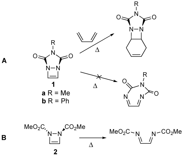
Several years ago, we introduced 1,2-diazacyclobutene 1a (Scheme 1) as a convenient dienophile for the integration of the strained 1,2-diazetidine motif into molecular structures via Diels–Alder reactions (see Scheme 1A) [1,2,3].
Scheme 1.
(A) Reactivity of diazetine 1 via Diels–Alder reactions to form 1,2-diazetidines, and its resistance to electrocyclic ring opening; (B) Thermally allowed electrocyclic ring opening of diazetine 2.
The fused urazole ring that is part of the structure of 1a proved critical to the thermal stability of the 1,2-diazacyclobutene ring system. In the absence of this fused ring, a thermally allowed conrotatory electrocyclic ring opening occurs, even at ambient temperatures, as has been reported for compound 2 (Scheme 1) [4]. The fused urazole ring prevents the ring opening process due to the strain that would be imposed on the resulting ring-opened product (see Scheme 1A) [1]. While 1a successfully engaged in Diels–Alder reactions with a variety of dienes, the utility of the synthetic method was limited by the low overall yield (15%) for the three-step synthesis of 1a [1].
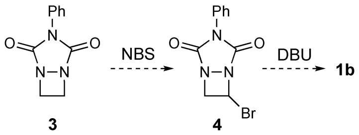
To further investigate the reactions of 1,2-diazacyclobutenes such as 1 in a more expansive manner, we realized that the development of a more robust, higher-yielding synthetic method was necessary. Therefore, we considered the possibility of synthesizing the N-Ph derivative 1b, as shown in Scheme 2. This novel synthetic route required the synthesis of the saturated 1,2-diazetidine compound 3. Earlier work found in the literature suggested that the synthesis of 3 was plausible [5,6,7]. Upon the successful synthesis of 3, we envisioned that routine free-radical bromination of 3 by NBS would afford 3-bromo-1,2-diazetidine 4. Finally, the base-catalyzed elimination of HBr from 4 using standard E2 conditions (e.g., DBU in DMF) would afford the desired product 1b. Assuming the synthesis of 3 (see below) and that of the two subsequent steps proceeded in reasonable yields, we hoped this new route might be of higher overall yield than the previous route followed for 1a. In this paper, we describe our attempts at the synthesis of compounds 3 and 4, which proved to be more challenging than anticipated, and illustrate the importance of X-ray crystallography for the confirmation of structures.
Scheme 2.
Proposed synthesis of 1b.
2. Results and Discussion
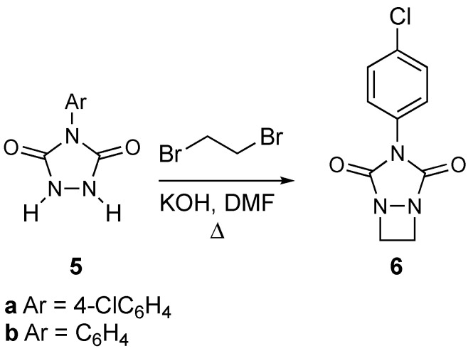
We began by attempting to synthesize 1,2-diazetidine 3. The synthesis of a derivative of 3 had already been reported (see 6 in Scheme 3) via the double nucleophilic substitution of the potassium salt formed from urazole 5a with 1,2-dibromoethane [5]. Although the reported yield of 6 was low (21% yield), the availability and low cost of the starting materials (we opted to use commercially available urazole 5b rather than 5a) and the operational simplicity for the synthesis of 3 made it an attractive approach. Following the previous literature, a solution of 4-phenyl urazole (5b), KOH, and 1,2-dibromoethane was heated in DMF to afford a small amount (9% yield) of a white solid product. We were heartened that the 1H and 13C NMR data appeared to be consistent with the structure of the anticipated product, 3, including a singlet for all four methylene protons in the 1H NMR spectrum (δ 4.14), and a total of six carbons in the 13C NMR spectrum, reflecting the symmetry of the ring system. However, we thought that the compound’s melting point of 252–253 °C was unexpectedly high for such a small molecule. On the other hand, compound 6 was also reported to have a high melting point (>280 °C) [5].
Scheme 3.
Literature procedure [5] for the formation of 6 (Ar = 4-ClC6H6) as extended towards the synthesis of 3.
Therefore, for conclusive verification of the structure, a crystal suitable for X-ray analysis was grown. Surprisingly, the X-ray analysis revealed the structure to be not that of the desired compound 3 but that of the “dimerized” eight-membered ring compound 7 instead (see Figure 1).
Figure 1.
(A) The structure of 7 and (B) an ORTEP representation of the X-ray crystal structure of 7 with thermal ellipsoids drawn at 50% probability. Selected bond lengths (Å): N(1)-N(2), 1.420; N(1)-C(1), 1.480; N(1)-C(2), 1.383, C(2)-N(3), 1.388; N(3)-C(3), 1.434; and C(2)-O(1), 1.208. Selected bond angles (°): C(1)-N(1)-N(2), 118.1; C(1)-N(1)-C(2), 118.2; and N(2)-N(1)-C(2), 108.0. Selected dihedral angles (°): N(2)-N(1)-C(2)-N(3), 10.8 and C(2)-N(3)-C(3)-C(4), 138.7.
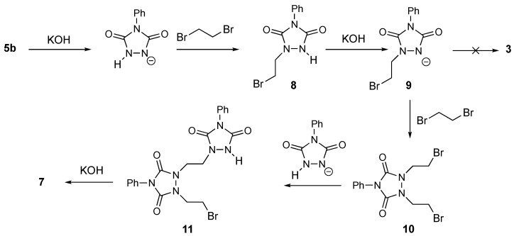
The formation of compound 7 can be rationalized as shown in Scheme 4. In the presence of the base KOH, N-phenylurazole 5b is deprotonated due to its low pKa value of approximately 5 [8]. Nucleophilic attack onto 1,2-dibromoethane affords the monoalkylated compound 8. Deprotonation of 8 affords anion 9. Anion 9 can either undergo intramolecular substitution to afford diazetidine 3, or it could react with a second molecule of dibromide to form 10. Apparently, a bimolecular reaction to form the relatively unstrained compound 10 is favored over the intramolecular cyclization that would form the strained structure of 3. This is also in line with the known general reluctance of compounds to undergo intramolecular reactions to afford four-membered rings [9]. Once formed, 10 can undergo further reaction to afford 11 followed by an intramolecular cyclization that yields the observed compound 7.
Scheme 4.
Proposed mechanism for the formation of dimer compound 7.
Attempts to circumvent the dimerization process via changes to the reaction conditions, such as running the reaction under more dilute conditions (in an attempt to discourage the formation of 10) or changing the base to Cs2(CO3)2 instead of KOH, failed to produce any product other than 7. Note that the structure for 7 is much more in line with the previously mentioned high melting point and further suggests that the structure for compound 6 may be misassigned in the literature.
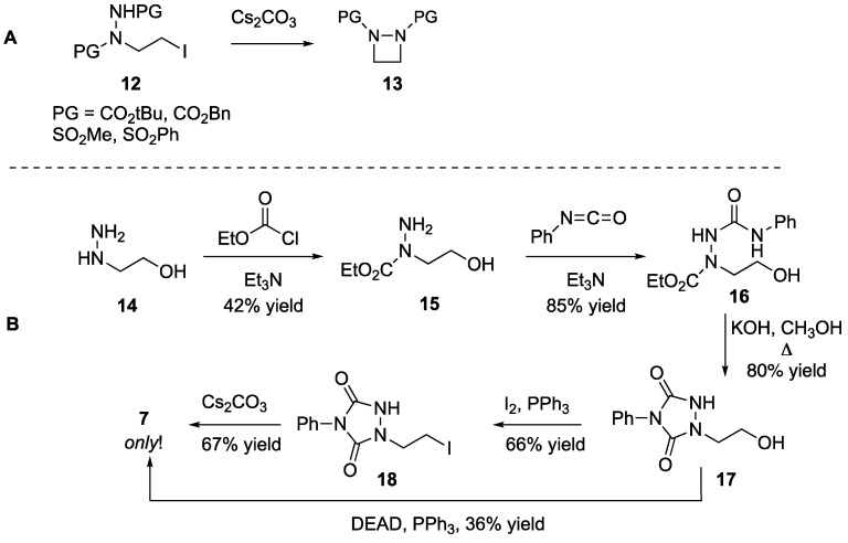
Given the failure of the above method to secure the desired compound 3, we took an alternative approach. Shipman had reported the syntheses of N,N-diprotected 1,2-diazetidines such as 13 via a cyclization reaction starting from halogenated precursor 12 (Scheme 5A) [6]. We considered the possibility of extending this methodology toward the synthesis of compound 3, as shown in Scheme 5B. Thus, the treatment of commercially available 2-hydroxyethylhydrazine 14 with ethyl chloroformate provided 15. A reaction between 15 with phenyl isocyanate gave 16, which could be easily cyclized to form 2-hydroxyethyl-N-phenylurazole 17. We attempted the final ring closure of 17 by two methods. First, iodination of alcohol 17 provided 18. Unfortunately, once more, the treatment of 18 according to the procedure worked out earlier by Shipman [6] for the synthesis of 13 did not yield the desired compound 3 and only yielded the “dimerized” 7. Similarly, an attempted direct Mitsunobu-type [10] intramolecular reaction of alcohol precursor 17 also only afforded 7.
Scheme 5.
(A) Shipman’s [6] cyclization of protected hydrazines (12) to form diazetidines (13). (B) Attempted extension of Shipman’s synthetic method to form 3.
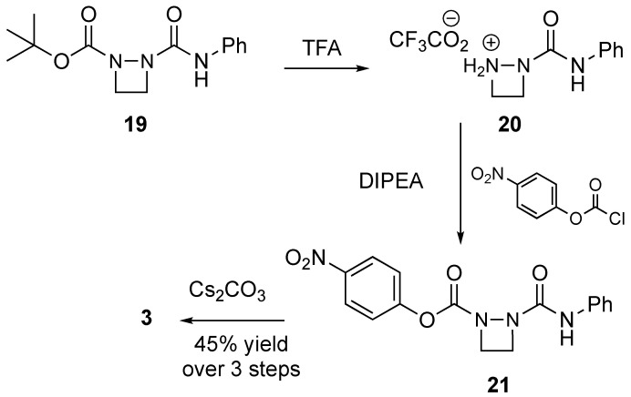
It was interesting that these cyclizations failed even though compound 13 could be formed from precursor compound 12. We surmised that the reason for the failure of the ring closure of 17 and 18 may be due to the rigidity of the urazole ring structure. The rigidity of the ring structure might inhibit the intramolecular nucleophilic attack that is necessary to form 3. Therefore, we worked out a new alternative synthetic scheme that would install the urazole ring structure after the cyclization process (see Scheme 6). Hence, starting with an already intact diazetidine compound 19 that had been previously reported by Shipman [7], we removed the BOC group with TFA. The treatment of the resulting crude trifluoroacetate salt 20 with 4-nitrophenyl chloroformate in the presence of the base diisopropylethylamine afforded 21. Crude 21 was then successfully cyclized with Cs2(CO3)2 to afford the desired diazetidine 3 as a white solid in 45% yield over three steps. The melting point of this compound, 173–174 °C, was much more in line with what would be expected.
Scheme 6.
Successful synthesis of 3 starting from previously reported compound 19 [7].
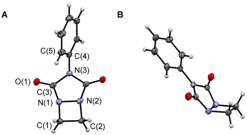
The 1H and 13C NMR spectra of both 3 and 7 are provided in the Supplementary Materials. Despite the significant difference in the structures of these two compounds, the spectra are very similar. Not surprisingly, the aromatic regions are nearly identical in both cases, and there is only a subtle shift of the signals for the methylene protons for 3 and 7 in the 1H NMR spectra (singlets at 4.47 and 4.14 ppm, respectively) and in the 13C NMR spectra (51.6 and 47.8 ppm, respectively). Therefore, wary of being deceived again, as in the case of compound 7, we verified the structure of 3 via X-ray analysis. This time, however, the data were in complete agreement with the structure of the desired compound 3 (Figure 2).
Figure 2.
ORTEP representation of the X-ray crystal structure of compound 3 with thermal ellipsoids drawn at 50% probability as visualized (A) from the front and (B) from the side of the structure. Selected bond lengths (Å): C(1)-C(2), 1.538; C(1)-N(1), 1.510; N(1)-N(2), 1.481; N(1)-C(3), 1.395; C(3)-O(1), 1.210; C(3)-N(3), 1.396; and N(3)-C(4), 1.435. Selected bond angles (°): C(1)-N(2)-C(3), 121.0; C(1)-N(1)-N(2), 91.5; and N(1)-C(3)-N(3), 105.9. Select dihedral angles (°): C(1)-C(2)-N(2)-N(1), 1.9 and C(3)-N(3)-C(4)-C(5), 53.2.
The N-N bond in the urazole ring of 3 (1.481 Å) was observed to be longer than the corresponding bond in dimer 7 (1.420 Å). It was also longer than the corresponding bond for 13 (PG = CO2tBu) of 1.450 Å [6], as well as previously reported 1a (1.470 Å) [11]. However, it was nearly identical to the bond length (1.480 Å) for the N-N bond of the similarly substituted diazetidine 22 (Figure 3) [12]. Interestingly, the bend of the urazole ring in relation to the four-membered ring, as measured by the C-N-(CO) bond angle, was 121.0° for 3, which was remarkably similar to that of both diazacyclobutene 1a (119.8°) and diazetidine 22 (122.6°), suggesting a preferred degree of nitrogen atom pyramidalization bound within a four-membered ring regardless of the type of carbon to which it is attached (i.e., SP2 in 2 versus SP3 in 3 and 22, respectively).
Figure 3.
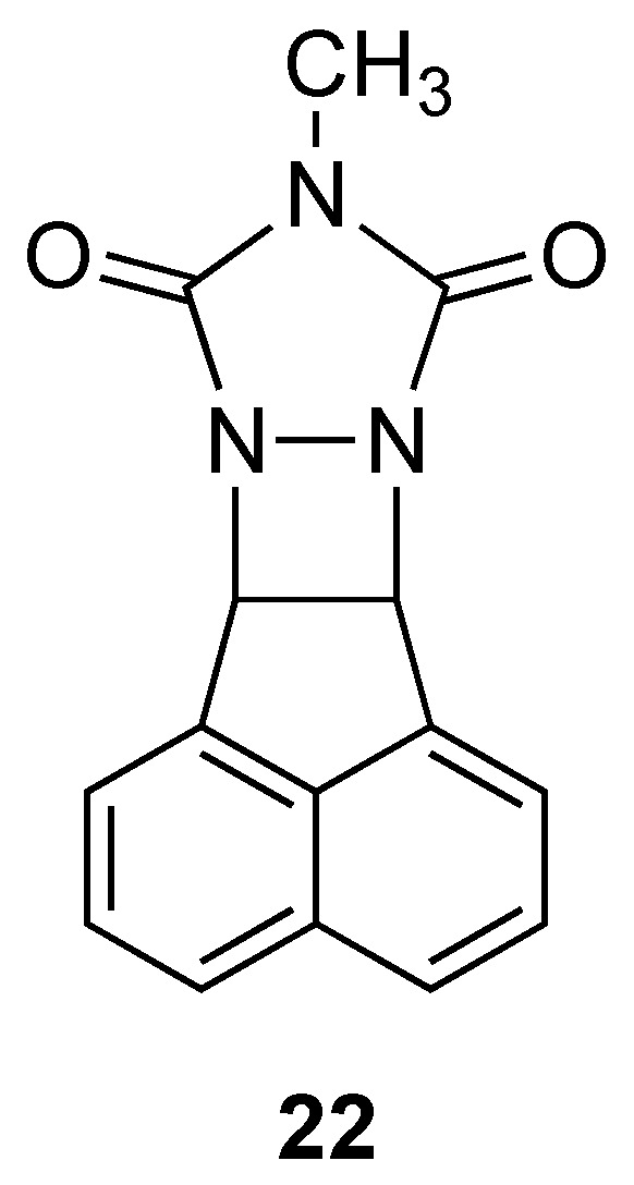
Structure of previously reported diazetidine 22.
Having finally obtained 3, we were free to pursue its free-radical bromination in pursuit of compound 4 (see Scheme 2). However, unfortunately, the bromination of this substrate proved difficult. Using standard bromination conditions (NBS, benzoyl peroxide, and heat [13]) upwards of 30% (estimated based on the 1H NMR spectrum) of the desired compound 4 could be formed based on the observation of chemical shifts consistent with the anticipated structure. In particular, the singlet representing the four methylene protons for compound 3 was transformed into three individual multiplets at 6.29 (CH-Br), 5.12, and 4.61 ppm, which was consistent with monobromination. In addition, conducting a 13C NMR experiment revealed two different saturated carbons (64.0 and 61.4 ppm) instead of the single carbon observed for compound 3 (51.6 ppm). The signal at 64.0 ppm was determined to be a CH carbon and the signal at 61.4 ppm was determined as a CH2 carbon by a DEPT experiment. However, unfortunately, attempts at further bromination did not appear to increase the yield and instead led to the formation of complex product mixtures. This was surprising since we expected that the nitrogen atom within the urazole ring would stabilize the radical intermediate formed during the bromination process and, thereby, promote the reaction [14]. However, in this case, it appears to have the opposite effect. Therefore, given the number of steps required to form 3, in addition to the reluctance of 3 to cleanly brominate to form 4, we felt it prudent to abandon this route towards the synthesis of diazetine 1b. However, other synthetic pathways are currently being pursued in our labs.
3. Materials and Methods
3.1. General Methods
Column chromatography was performed on a silica gel absorbent (234–400 mesh). Thin-layer chromatography was performed on silica gel plates that were pre-coated with a fluorescent indicator. Developed TLC plates were visualized by ultraviolet light. 1H and 13C NMR spectra were obtained on a JEOL NMR spectrometer at 400 and 200 MHz, respectively. All chemical shifts are reported in units of parts per million downfield from TMS. Reported high-resolution mass spectra (HRMS) data were acquired via the sampling technique of electron spray ionization utilizing an LTQ-FTMS hybrid mass spectrometer. Unless otherwise stated in the Experimental Procedures section, all compounds were purchased from commercial sources and used as received.
3.2. Experimental Procedures
3.2.1. Synthesis of “Dimer” 7 from Urazole 5b
Reaction following the literature procedure [5]: to a solution of 2 g (0.11 mole) of N-phenylurazole (5b) in 30 mL of anhydrous DMF, 0.74 g (1 eq) of powdered KOH was added. The reaction mixture was stirred for 2 h until all the KOH went into solution. 1,2-Dibromoethane (2.07 g, 1 eq) was then added to the solution via pipette, and the reaction mixture was heated to 100 °C for 0.5 h. It was then further heated to reflux for 1 h. After cooling to room temperature, the reaction mixture was filtered, and the collected salts were rinsed with 10 mL of DMF. The DMF was removed using a rotary evaporator to yield a thick liquid. Water (50 mL) was added to the thick liquid, and a precipitate formed. The mixture was extracted with 2 × 30 mL of CH2Cl2. The combined organic layers were backwashed with 2 × 30 mL of H2O, dried over Na2SO4, filtered, and concentrated to afford a thick pale orange-brown liquid. Column chromatography (SiO2, 10% methanol in CH2Cl2) afforded 183 mg (8% yield) of 7 as a white solid, m.p. 251–252 °C: 1H NMR (400 MHz, CDCl3) δ 7.49–7.51 (m, 8H), 7.42 (h, 2H), 4.14 (s, 8H); 13C{1H} NMR (100 MHz, CDCl3)153.9, 131.1, 129.5, 128.8, 125.5, 47.9; HRMS (ESI) m/z [M+H]+ Calcd for C20H19N6O4 407.14623; Found 407.14514.
Reaction under dilute conditions: to a solution of 2 g (0.11 mole) of N-phenylurazole (5b) in 60 mL of anhydrous DMF, 0.74 g (1 eq) of powdered KOH was added. The reaction mixture was stirred for 2 h until all the KOH went into solution. 1,2-Dibromoethane (2.07 g, 1 eq) was then added to the solution via a pipette, and the reaction mixture was heated to 100 °C for 0.5 h. It was then further heated to reflux for 1 h. After cooling to room temperature, the reaction mixture was filtered, and the collected salts were rinsed with 10 mL of DMF. The DMF was removed using a rotary evaporator to yield a thick liquid. The analysis of this reaction mixture by 1H NMR spectroscopy revealed only the presence of compound 7.
Reaction using Cs2CO3 as a base: to a solution of 0.5 g (2.82 mmole) of N-phenylurazole (5b) in 10 mL of anhydrous DMF, 1.1 g (1.2 eq) of powdered Cs2CO3 was added. The reaction mixture was stirred for 1 h, and then, 1,2-dibromoethane (0.53 g, 1 eq) was added to the solution via a pipette, and the reaction mixture was heated to 100 °C for 0.5 h. The reaction mixture was then further heated to reflux for 1 h. After cooling to room temperature, the reaction mixture was filtered, and the collected salts were rinsed with 10 mL of DMF. The DMF was removed using a rotary evaporator to yield a thick liquid. The analysis of this reaction mixture by 1H NMR spectroscopy revealed only the presence of compound 7.
3.2.2. Synthesis of 11
To 2 g (0.026 mol) of 2-hydroxyethylhydrazine in 15 mL of CH2Cl2, 3.26 mL (1 eq) of Et3N was added via a syringe. The resulting solution was cooled to 0 °C, and 2.82 g (1 eq) of ethyl chloroformate was added dropwise over the course of 10 min. A thick white precipitate formed. The resulting mixture was stirred for an additional 10 min and then warmed to room temperature. After 4 h of stirring at room temperature, the mixture was poured into 50 mL of THF and then filtered to remove the precipitate. The solvent was removed via rotary evaporation to provide a thick oil. Column chromatography (SiO2, 5% methanol in EtOAc) afforded 1.62 g (42% yield) of 11 as a clear oil: 1H NMR (400 MHz, CDCl3) δ 4.17 (q, 2H), 3.8–4.4 (br s, 3H), 3.82 (t, J = 4.8 Hz, 2H), 3.60 (t, J = 4.8 Hz, 2H), 1.28 (t, 3H); 13C{1H} NMR (100 MHz, CDCl3) 158.0, 62.1, 60.8, 51.9, 14.6; HRMS (ESI) m/z [M+H]+ Calcd for C5H13N2O3 149.09207; Found 149.09183.
3.2.3. Synthesis of 12
To 1.53 g of 11 (10.3 mmol) in 15 mL of benzene, 1.23 g (1 eq) of phenyl isocyanate was added dropwise. The solution became cloudy. The mixture was then heated to reflux for 5 h, cooled to room temperature, and concentrated to a thick liquid. Column chromatography (SiO2, 5% methanol in EtOAc) afforded 2.34 g (85% yield) of 12 as a clear oil. The NMR spectra reflected a mixture of 2 slowly interconverting conformers, which severely complicated the spectra. Signals for the major isomer are provided: 1H NMR (400 MHz, DMSO-D6) δ 8.14 (br s, 1H), 7.60 (br s, 1H), 7.36 (br d, J = 7.5 Hz, 2H), 7.21 (br t, J = 7.5 Hz, 2H), 7.00 (br t, J = 8.5 Hz, 1H), 4.12 (br q, J = 7.2 Hz, 2H), 3.74 (br s, 4 H), 1.21 (br t, J = 7.2 Hz, 3H); 13C{1H} NMR (100 MHz, CDCl3) δ 157.6, 157.5, 138.1, 129.0, 123.6, 119.8, 63.1, 58.8, 53.3, 14.5; HRMS (ESI) m/z [M+H]+ Calcd for C12H18N3O4 268.12918; Found 268.12850.
3.2.4. Synthesis of 13
To 2.34 g (8.75 mmol) of 12 in 10 mL of CH3OH, 1.44 g (2.5 eq) of KOH pellets was added. The stirring mixture was heated to reflux for 3 h. After cooling to room temperature, the solution was diluted with 10 mL of H2O and acidified to pH~2 with concentrated HCl (~3 mL). The resulting clear colorless solution was concentrated into a free-flowing white solid. This solid was extracted with 1 × 75 mL and 2 × 50 mL of boiling EtOAc. Concentration of the combined EtOAc solutions afforded 1.54 g (80% yield) of 13 as a pale yellow crystalline solid, m.p. 161–162 °C: 1H NMR (400 MHz, DMSO-D6) δ 10.8 (br s, NH, 1H), 7.42–7.57 (m, 4H), 7.39 (t, J = 7.0 Hz, 1H), 4.90 (br s, OH, 1H), 3.63 (t, J = 5.0 Hz, 2H), 3.3.57 (t, J = 5.0 Hz, 2H); 13C{1H} NMR (100 MHz, DMSO-D6) δ 152.8, 152.4, 132.2, 129.0, 127.9, 126.2, 57.5, 48.4; HRMS (ESI) m/z [M+H]+ Calcd for C10H12N3O3 222.08732; Found 222.08660.
3.2.5. Synthesis of 14
To a solution of 0.66 g (2.53 mmol) of PPh3 and 0.17 g (2.53 mmol) of imidazole in 10 mL of CH2Cl2 cooled to 0 °C, 0.64 g (2.53 mmol) of solid I2 was added in portions over several minutes with stirring. The I2 eventually went into solution, resulting in the formation of a yellow-orange precipitate. This mixture was warmed to room temperature, and 0.47 g (2.11 mmol) of solid 13 was added in portions. The resulting mixture was stirred overnight and filtered. The separated solid was rinsed with CH2Cl2, and the filtrate was concentrated into a dark viscous liquid. Column chromatography (SiO2, 100% EtOAc) afforded 0.46 g (66% yield) of 14 as a crystalline white solid, m.p. 115–116 °C: 1H NMR (400 MHz, DMSO-D6) δ 10.9 (br s, NH, 1H), 7.43–7.53 (m, 4H), 7.37–7.43 (m, 1H), 3.87 (t, J = 6.4 Hz, 2H), 3.45 (t, J = 6.4 Hz, 2H); 13C{1H} NMR (100 MHz, DMSO-D6) δ 152.3, 152.2, 131.8, 128.9, 127.9, 126.1, 47.8, 2.0; HRMS (ESI) m/z [M+H]+ Calcd for C10H11N3O2127I 331.98905; Found 331.98819.
3.2.6. Attempted Cyclization of 14
To urazole 14 in 15 mL of dry DMF, solid Cs2CO3 was added, and the reaction mixture was stirred for 24 h. The residual solid was filtered under vacuum, and the filtrate was poured into a mixture of 25 mL H2O and 25 mL CH2Cl2 in a separatory funnel. The layers were mixed, and the organic layer was removed. The aqueous layer was washed with an additional 2 × 20 mL CH2Cl2, and the combined organic layers were backwashed with 1 × 25 mL H2O and 1 × 25 mL sat. aq. NaCl. The organic layer was dried and concentrated to afford 0.172 g of a white solid. The NMR spectra and m.p. of this compound matched that for “dimer” 7, as described above.
3.2.7. Attempted Cyclization of 13
To a solution of 0.22 g (1 mmol) of 13 and 0.66 g (2.5 eq) of PPh3 in 100 mL of anhydrous THF, 1.14 mL of a 40 wt.% solution of DEAD in toluene (2.5 eq) was added. The resulting solution was stirred for 24 h and then concentrated. Column chromatography (SiO2, 10% methanol in CH2Cl2) afforded 76 mg (36% yield) of a white solid identified by NMR and m.p. as “dimer” 7.
3.2.8. Synthesis of 3
To a solution of 0.5 g (1.81 mmol) of 15 in 10 mL of CH2Cl2, 1.25 mL of TFA was added using a syringe. The solution was stirred for 24 h and then concentrated to a sticky pale brown solid (crude compound 16). To this solid in 5 mL of CH2Cl2, 0.63 mL (2 eq) of diisopropylethyl amine was added, followed by the dropwise addition of 0.36 g of para-nitrophenyl chloroformate. After stirring overnight, the reaction mixture was washed with 2 × 20 mL 0.5 N aq. HCl, dried over Na2SO4, filtered, and concentrated to 0.64 g of a pale brown solid (crude compound 17). To this crude product in 50 mL of CH3CN, 1.22 g (approximately 2 eq) of Cs2CO3 was added. The solution became deep yellow almost immediately. After stirring for 3 hr, the reaction mixture was poured into 50 mL of CH2Cl2 and washed with 2 × 25 mL of 0.5 N aq. NaOH. The organic layer was dried over Na2SO4, filtered, and concentrated to a brown solid. Column chromatography (SiO2, 5% methanol in CH2Cl2) afforded 0.17 g (45% yield over the three steps) of 3 as a crystalline white solid, m.p. 173–174 °C: 1H NMR (400 MHz, CDCl3) δ 7.47–7.55 (m, 4H), 7.38–7.44 (m, 1 H), 4.47 (s, 4 H); 13C{1H} NMR (100 MHz, CDCl3) δ 163.0, 131.5, 129.4, 128.8, 125.3, 51.6; HRMS (ESI) m/z [M+H]+ Calcd for C10H10N3O2 204.07675; Found 204.07615.
3.2.9. Attempted Bromination of 3
To 20 mg (0.09 mmol) of 3 in 0.5 mL of CDCl3, 44 mg (2.5 eq) of NBS (one equivalent was insufficient, and more than 2.5 equivalents led to complex reaction mixtures) and a few tiny crystals of benzoyl peroxide were added. The reaction mixture was irradiated by two nearby 300 W incandescent bulbs, the heat from which was sufficient to bring the reaction to a boil. After irradiating for 6 h, irradiation was stopped, and the reaction mixture was allowed to cool. The analysis of the reaction mixture by 1H NMR spectroscopy revealed new signals consistent with the monobromination of the ring, i.e., δ 6.29 (dd, J = 6.2, 4.2 Hz, 1 H), 5.12 (dd, J = 11.2, 6.2 Hz, 1 H), 4.61 (dd, J = 11.2, 4.2 Hz, 1 H). Integration relative to the starting material signal indicated approximately 26% conversion. However, further irradiation only increased the yield to approximately 37% while the starting material remained present and other competing products began to form.
3.3. X-ray Data Collection
X-ray diffraction data for 3 were measured at 100 K on a Rigaku XtaLAB AFC11 (RCD3) diffractometer with a rotating anode CuKα source (λ = 1.54184 Å) and a hybrid pixel array detector. X-ray diffraction data for 7 were measured at 100 K on a Rigaku XtaLAB Synergy-S diffractometer with a PhotonJet CuKα source (λ = 1.54184 Å) and HyPix-6000HE detector. Both structures were solved using olex2.solve [15] and refined using olex2.refine [16]. Hydrogen atoms were found in difference maps, and all parameters (position and isotropic temperature factor) were allowed to be refined. The absolute structure for 3 was found with a Flack parameter of −0.03(5). The absolute structure for 7 (a racemic twin with a BASF parameter of 0.38(18)) was found with a Hooft parameter of 0.04(11). The crystal and refinement data are presented in Table 1.
Table 1.
Crystal data and refinement parameters.
| 3 | 7 | |
| Chemical formula | C10H9N3O2 | C20H18N6O4 |
| M r | 203.20 | 406.40 |
| Deposition number | 2373853 | 2373840 |
| Crystal system and space group | orthorhombic, P212121 | monoclinic, P21 |
| Temperature (K) | 100 | 100 |
| a, b, c (Å) | 6.00967(7), 11.74365(12), 13.52523(14) |
7.8071(3), 7.1290(3), 16.3695(6) |
| β (°) | 90 | 92.298(3) |
| V (Å3) | 954.549(17) | 910.34(6) |
| Z | 4 | 2 |
| Radiation type | Cu Kα | Cu Kα |
| μ (mm−1) | 0.85 | 0.89 |
| Crystal size (mm) | 0.14 × 0.10 × 0.09 | 0.36 × 0.23 × 0.20 |
| Diffractometer | XtaLAB AFC11 (RCD3) | XtaLAB Synergy-S |
| Absorption correction | Numerical and empirical | Numerical and empirical |
| Tmin, Tmax | 1.000, 0.823 | 1.000, 0.585 |
| No. of measured, independent, and observed [I > 2σ(I)] reflections |
13,937, 1858, 1858 | 15,905, 3762, 3762 |
| R int | 0.0234 | 0.0533 |
| (sin θ/λ)max (Å−1) | 0.615 | 0.633 |
| R[F2 > 2σ(F2)], wR(F2), S | 0.0233, 0.0553, 1.166 | 0.0344, 0.0854, 1.095 |
| No. of reflections | 1858 | 3762 |
| No. of parameters | 172 | 345 |
| Δρmax, Δρmin (e Å−3) | 0.12, −0.19 | 0.20, −0.21 |
Acknowledgments
The authors thank Berry College for providing generous financial support for this project. We also thank John Bacsa, Facilities Director of Emory University’s X-ray Crystallography Center (supported by the National Science Foundation under CHE-1626172), for collecting the X-ray crystal data on compounds 3 and 7, and Emory University’s Mass Spectrometry Center for collecting the HRMS data.
Supplementary Materials
The following supporting information can be downloaded at: https://www.mdpi.com/article/10.3390/molecules29174068/s1, CIF files for compounds 3 and 7 and a PDF file containing 1H and 13C NMR spectra for all new compounds.
Author Contributions
Conceptualization, G.W.B.; methodology, G.W.B.; formal analysis, G.W.B. and K.L.M.; investigation, G.W.B. and K.L.M.; resources, G.W.B. and K.L.M.; data curation, G.W.B. and K.L.M.; writing—original draft preparation, G.W.B.; and writing—review and editing, G.W.B. and K.L.M. All authors have read and agreed to the published version of the manuscript.
Institutional Review Board Statement
Not applicable.
Informed Consent Statement
Not applicable.
Data Availability Statement
Data supporting the reported results can be found in the Supplementary Materials. Additionally, CCDC 2355187 and 2355188 contain the supplementary crystallographic data for this paper. These data can be obtained free of charge via www.ccdc.cam.ac.uk/data_request/cif or by emailing data_request@ccdc.cam.ac.uk or by contacting the Cambridge Crystallographic Data Centre, 12 Union Road, Cambridge CB2 1EZ, UK; fax: +44 1223 336033.
Conflicts of Interest
The authors declare no conflicts of interest.
Funding Statement
This research received no external funding.
Footnotes
Disclaimer/Publisher’s Note: The statements, opinions and data contained in all publications are solely those of the individual author(s) and contributor(s) and not of MDPI and/or the editor(s). MDPI and/or the editor(s) disclaim responsibility for any injury to people or property resulting from any ideas, methods, instructions or products referred to in the content.
References
- 1.Breton G.W., Shugart J.J., Hughey C.A., Perala S.M., Hicks A.D. Synthesis of D1-1,2-Diazetines via a Diels-Alder Cycloaddition Approach. Org. Lett. 2001;3:3185–3187. doi: 10.1021/ol0164942. [DOI] [PubMed] [Google Scholar]
- 2.De Bruycker K., Billiet S., Houck H.A., Chattopadhyay S., Winne J.M., Du Prez F.E. Triazolinediones as Highly Enabling Synthetic Tools. Chem. Rev. 2016;116:3919–3974. doi: 10.1021/acs.chemrev.5b00599. [DOI] [PubMed] [Google Scholar]
- 3.Narangoda C.J., Lex T.R., Moore M.A., McMillen C.D., Kitaygorodskiy A., Jackson J.E., Whitehead D.C. Accessing the Rare Diazacyclobutene Motif. Org. Lett. 2018;20:8009–8013. doi: 10.1021/acs.orglett.8b03590. [DOI] [PubMed] [Google Scholar]
- 4.Nunn E.E., Warrener R.N. Dimethyl D3-1,2-Diazetine-1,2-dicarboxylate: A New Four-membered 6p-Ring System. J. Chem. Soc. Chem. Comm. 1972;14:818–819. doi: 10.1039/C39720000818. [DOI] [Google Scholar]
- 5.Wakabayashi O., Matsuya K., Ohta H., Tetsuo J., Suzuki S. 1,2-Alkylene-4-substituted urazole herbicides. 4,249,934. U.S. Patent. 1978 September 19;
- 6.Brown M.J., Clarkson G.J., Fox D.J., Inglis G.G., Shipman M. Critical importance of leaving group ‘softness’ in nucleophilic ring closure reactions of ambident anions to 1.2-diazetidines. Tetrahedron Lett. 2010;51:382–384. doi: 10.1016/j.tetlet.2009.11.024. [DOI] [Google Scholar]
- 7.Dean C., Roesner S., Rajkumar S., Clarkson G.J., Jones M., Shipman M. Synthesis of sp3-rich chemical libraries based upon 1,2-diazetidines. Tetrahedron. 2021;79:131836. doi: 10.1016/j.tet.2020.131836. [DOI] [Google Scholar]
- 8.Bausch M.J., David B., Dobrowolski P., Guadalupe-Fasano C., Gostowski R., Selmarten D., Prasad V., Vaughn A., Wang L.H. Proton-Transfer Chemistry of Urazoles and Related Imides, Amides, and Diacyl Hydrazides. J. Org. Chem. 1991;56:5463–5651. doi: 10.1021/jo00019a034. [DOI] [Google Scholar]
- 9.Anslyn E.V., Dougherty D.A. Modern Physical Organic Chemistry. University Science Books; Sausalito, CA, USA: 2004. pp. 568–569. [Google Scholar]
- 10.Mitsunobu O. The Use of Diethyl Azodicarboxylate and Triphenylphosphine in Synthesis and Transformation of Natural Products. Synthesis. 1981;1981:1–28. doi: 10.1055/s-1981-29317. [DOI] [Google Scholar]
- 11.Breton G.W., Martin K.L. Are 1,2-Dihydrodiazetes Aromatic? An Experimental and Computational Investigation. J. Org. Chem. 2002;67:6699–6704. doi: 10.1021/jo026082m. [DOI] [PubMed] [Google Scholar]
- 12.Breton G.W., Hughes J.S., Pitchko T.J., Martin K.L., Hardcastle K.J. Unexpected Sigma Bond Rupture during the Reaction of N-Methyl-1,2,4-triazoline-3,5-dione with Acenaphthylene and Indene. Org. Chem. 2014;79:8212–8220. doi: 10.1021/jo5014096. [DOI] [PubMed] [Google Scholar]
- 13.Djerassi C. Brominations with N-Bromosuccinimide and Related Compounds. The Wohl-Ziegler Reaction. Chem. Rev. 1948;43:271–317. doi: 10.1021/cr60135a004. [DOI] [PubMed] [Google Scholar]
- 14.Zipse H. Radical Stability–A Theoretical Perspective. Top. Curr. Chem. 2006;263:163–189. [Google Scholar]
- 15.Dolomanov O.V., Bourhis L.J., Gildea R.J., Howard J.A.K., Puschmann H. OLEX2: A Complete Structure Solution, Refinement and Analysis Program. Appl. Cryst. 2009;42:339–341. doi: 10.1107/S0021889808042726. [DOI] [Google Scholar]
- 16.Bourhis L.J., Dolomanov O.V., Gildea R.J., Howard J.A.K., Puschmann H. The Anatomy of a Comprehensive Constrained, Restrained Refinement Program for the Modern Computing Environment—Olex2 Dissected. Acta Cryst. 2015;71:59–75. doi: 10.1107/S2053273314022207. [DOI] [PMC free article] [PubMed] [Google Scholar]
Associated Data
This section collects any data citations, data availability statements, or supplementary materials included in this article.
Supplementary Materials
Data Availability Statement
Data supporting the reported results can be found in the Supplementary Materials. Additionally, CCDC 2355187 and 2355188 contain the supplementary crystallographic data for this paper. These data can be obtained free of charge via www.ccdc.cam.ac.uk/data_request/cif or by emailing data_request@ccdc.cam.ac.uk or by contacting the Cambridge Crystallographic Data Centre, 12 Union Road, Cambridge CB2 1EZ, UK; fax: +44 1223 336033.


