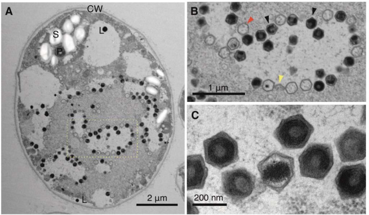
Fig. 5. Transmission electron microscopy of ultra-thin sectioned Ors 24 Chlamydomonas sp. cells.
(A) Infected Chlamydomonas sp. cell in early exponential phase (left). The nucleus and chloroplast are not clearly distinguishable. (B) Enlargement of the section delimited by a rectangle showing hexagonal viral particles. Virion production in a clearly delineated, lighter colored area with virions in later stages of completion accumulating at the edges of the production area, i.e. the virus factory/viroplasm. Red and black arrows indicate empty and full capsids, respectively. Yellow arrows indicate partially assembled capsids. (C) Enlarged picture of assembled virions. P – pyrenoid; CW – cell wall; L – lipid vesicle/plastoglobule; S – starch sheath.

