Summary:
Different surgical approaches exist for lower eyelid reconstruction. The hard palate mucosa graft stands out due to its abundance, accessibility, good tolerance, and ability to yield long-term stable results in eyelid elevation. This case report details the successful full-thickness reconstruction of the lower eyelid in an anophthalmic patient using a palatal mucosal graft, complemented by orbicularis muscle suspension. The patient presented with severe lower eyelid retraction state and instability of the ocular prosthesis. After a thorough assessment, the decision was made to address the mucosal defect using a split-thickness palatal mucosal graft, supplemented by lateral canthus suspension. Postoperatively, there were no complications, and the cosmetic result was excellent. With our method, we were able to obtain a functional and cosmetically good result of lower eyelid reconstruction in an anophthalmic socket.
Post-enucleation socket syndrome (PESS) is a set of deformities that can occur following eye removal surgery, encompassing features resulting from the loss of tissue around and within the eye socket. Common deformities associated with PESS include enophthalmos, superior sulcus depression, upper lid ptosis, lower lid sagging, socket contracture, and lower eyelid retraction.1 Lower eyelid retraction is a frequent complication of PESS, often attributed to factors like mechanical stress from frequent prosthesis insertion and removal, gravitational burden from prosthesis weight, and atrophy of structures supporting the lower lid.2,3 Addressing lower eyelid retraction is crucial not only for cosmetic reasons but also for improving prosthesis retention. Various surgical techniques, including hard palate mucosa graft, fascia lata graft, oral mucosa graft, and auricular cartilage graft, have been used for lower eyelid reconstruction.4 Among these, the hard palate mucosa graft5 stands out for its abundance, accessibility, tolerance, and long-term stability in achieving eyelid elevation. This technique, initially utilized in 1985 for eyelid reconstruction,5 has been successfully applied in correcting lower eyelid retraction resulting from various causes.6
This study presents a case where lower eyelid retraction repair was performed using a hard palate graft combined with lateral canthus suspension in a patient with an anophthalmic socket.
METHODS
Case Presentation
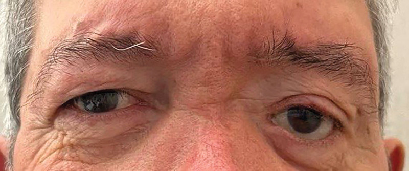
We present a case of a 71-year-old man who sustained multiple facial fractures in a car accident 56 years before his arrival at our hospital. A severe fracture of the left orbital floor resulted in significant enophthalmos, leading to vision loss that led to the enucleation of the left eye. Over the years, the patient underwent multiple reconstructive procedures to restore facial functionality and aesthetics. The left orbit required an ocular prosthesis to address the void caused by the damaged orbital floor. Despite previous interventions, the patient’s condition continued to deteriorate, primarily due to the worsening of the lower eyelid retraction state and, subsequently, due to the ocular prosthesis instability (Fig. 1). This deterioration prompted his referral to our hospital for further evaluation and management. The patient, before surgery, provided detailed and exhaustive informed consent.
Fig. 1.
Preoperative view of our patient showing severe lower eyelid retraction state.
Surgical Technique
In addressing the main concern of spontaneous ocular prosthesis dislocation, a combination of two surgical techniques was planned for complete eyelid closure and correction of lower lid laxity and retraction. The two procedures were performed in one surgical time and the patient underwent general anesthesia. The first procedure focused on harvesting a mucosal graft from the hard palate to vertically augment the posterior lamella of the lower eyelid. The second procedure involved the suspension of the lateral canthus. Throughout the surgery, the ocular prosthesis was strategically kept in place.
First Procedure
The initial surgical procedure involved three key steps. First, a transconjunctival approach was conducted to create space at the posterior lamella of the lower eyelid, avoiding the use of anesthetic solutions with adrenaline. A Desmarres retractor was strategically positioned, and a conjunctival incision was made, exposing the areolar tissue in front of the inferior orbital septum.
The second step consisted of harvesting a palatal mucosa graft from the hard palate with attention to the nervous and vascular structures. A rectangular graft, approximately 1 × 0.8 cm, was harvested, preserving the periosteum. The donor site was left to heal by second intention.
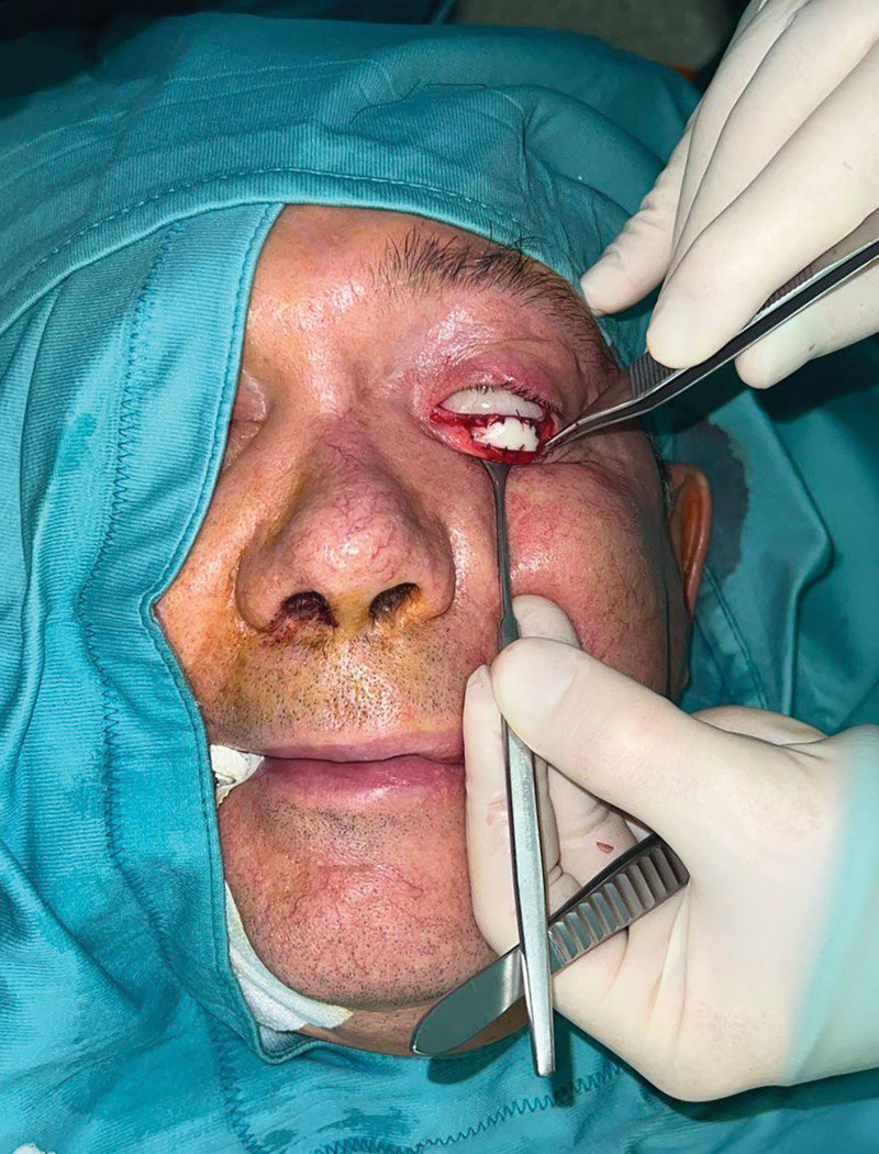
In the final step, the harvested mucosal graft was shaped, and its thickness adjusted. The graft was then placed within the prepared space and secured with absorbable and braided sutures (Fig. 2). Additional sutures were used to deepen the fornix. Postoperatively, an antibiotic gel was applied to the reconstructed area, and a chlorhexidine gel was applied to the donor site for 7 days.
Fig. 2.
Intraoperative view of the lower eyelid after the positioning of the palatal mucosal graft.
Second Procedure
Once the vertical alignment of the lower eyelid was successfully improved through mucosal grafting, the suspension of the lateral canthus was performed to provide enhanced and long-lasting support. In the second surgical procedure, an orbicularis muscle suspension was performed. An infraciliary incision was made on the lateral two-thirds of the lower eyelid, exposing the orbicularis oculi muscle beneath the lateral canthus. The distal portion of the orbicularis muscle was then rotated and engaged using a 5-0 Vicryl suture and secured to the periosteum of the orbital rim for fixation, obtaining in this way a lateral canthus elevation. We used this technique to provide better support to the lower eyelid,7 maintaining at the same time it’s flexibility.
DISCUSSION
In the management of patients with posttraumatic or reconstructive sequelae, personalized preoperative planning is essential, considering both functional and aesthetic aspects. The presented case, involving an ocular prosthesis, allowed for a more aggressive surgical approach without concerns of corneal discomfort. However, challenges related to gravitational effects and prosthesis retention emerged.
The use of an autologous graft, specifically the hard palatal mucosal graft, played a crucial role in augmenting the vertical vector as a spacer and reinforcing the posterior lamella. This approach improved the ocular prosthesis retention capacity. In comparison with alternative techniques,8 the palatal mucosa exhibits advantages such as acting as a rigid substitute for the tarsal plate, closely matching the natural curvature of the eyelid, and displaying excellent graft survival rates. The metaplasia of palatal mucosa into a nonkeratinized epithelium and its integration in the tarsoconjunctival structure contributes to stable outcomes.9 The graft retains its stiffness and experiences minimal shrinkage, reducing the risk of ectropion or entropion. However, a slight overcorrection is recommended to optimize the outcome. This can be achieved by harvesting the graft approximately 10% larger in the vertical dimension to anticipate any potential evolution.10
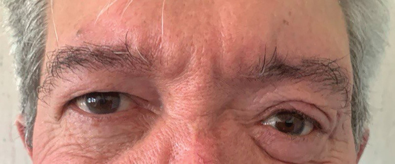
To further bolster graft support, the strategic incorporation of orbicularis muscle suspension was implemented. This additional technique provided increased stability to the ocular prosthesis, ensuring long-term functionality. Moreover, the lateral canthus suspension offers more flexibility when supporting the ocular prosthesis compared with other techniques such as canthopexy or canthotomy. The combined approach synergistically addressed both functional and aesthetic aspects, offering a comprehensive solution for the challenging case of lower eyelid reconstruction (Fig. 3).
Fig. 3.
Postoperative view of our patient at 10 months follow-up showing optimal functional and aesthetic results.
CONCLUSIONS
The combined use of orbicularis muscle suspension alongside the graft technique has proven to be a valuable and synergistic approach. By implementing their strengths, a successful outcome was achieved, a result that might have been hard to obtain with either technique alone. Although acknowledging that our 10-month follow-up duration might be considered insufficient to fully depict the long-term results, this timeframe has demonstrated positive outcomes, instilling confidence in the efficacy of this combined approach for future cases.
DISCLOSURE
The authors have no financial interest to declare in relation to the content of this article.
PATIENT CONSENT
The patient provided written consent for the use of his image.
Footnotes
Published online 13 September 2024.
Disclosure statements are at the end of this article, following the correspondence information.
REFERENCES
- 1.Shah CT, Hughes MO, Kirzhner M. Anophthalmic syndrome: a review of management. Ophthalmic Plast Reconstr Surg. 2014;30:361–365. [DOI] [PubMed] [Google Scholar]
- 2.Moon J, Choung H, Khwarg S. Correction of lower lid retraction combined with entropion using an ear cartilage graft in the anophthalmic socket. Korean J Ophtalmol. 2005;19:161–167. [DOI] [PubMed] [Google Scholar]
- 3.Ding J, Ma X, Xin Y, et al. Correction of lower eyelid retraction with hard palate graft in the anophthalmic socket. Can J Ophthalmol. 2018;53:458–461. [DOI] [PubMed] [Google Scholar]
- 4.Scruggs JT, McGwin G, Morgenstern KE. Use of noncadaveric human acellular dermal tissue (BellaDerm) in lower eyelid retraction repair. Ophthalmic Plast Reconstr Surg. 2015;31:379–384. [DOI] [PubMed] [Google Scholar]
- 5.Siegel R. Palatal grafts for eyelid reconstruction. Plast Reconstr Surg. 1985;76:411–414. [DOI] [PubMed] [Google Scholar]
- 6.Oestreicher JH, Pang NK, Liao W. Treatment of lower eyelid retraction by retractor release and posterior lamellar grafting: an analysis of 659 eyelids in 400 patients. Ophthalmic Plast Reconstr Surg. 2008;24:207–212. [DOI] [PubMed] [Google Scholar]
- 7.Miyamoto J, Nakajima T, Nagasao T, et al. Full-thickness reconstruction of the eyelid with rotation flap based on orbicularis oculi muscle and palatal mucosal graft: long-term results in 12 cases. J Plast Reconstr Aesthet Surg. 2009;62:1389–1394. [DOI] [PubMed] [Google Scholar]
- 8.Fin A, De Biasio F, Lanzetta P, et al. Posterior lamellar reconstruction: a comprehensive review of the literature. Orbit. 2018;38:51–66. [DOI] [PubMed] [Google Scholar]
- 9.Weinberg DA, Tham V, Hardin N, et al. Eyelid mucous membrane grafts: a histologic study of hard palate, nasal turbinate, and buccal mucosal grafts. Ophthalmic Plast Reconstr Surg. 2007;23:211–216. [DOI] [PubMed] [Google Scholar]
- 10.Hendriks S, Bruant-Rodier C, Lupon E, et al. The palatal mucosal graft: the adequate posterior lamellar reconstruction in extensive full-thickness eyelid reconstruction. Ann Chir Plast Esthet. 2020;65:61–69. [DOI] [PubMed] [Google Scholar]


