Abstract
Background and Objective
Breast reconstruction with microsurgical techniques allows for autologous reconstruction after mastectomy without the complications associated with alloplastic reconstruction. Autologous reconstruction has undergone significant improvement and now offers patients a variety of options depending on patient specific factors and aesthetic outcomes. This review aims to focus on the history of autologous reconstruction, operative considerations, general surgical techniques for flaps, and indications for choosing the ideal free tissue transfer for all medical specialties and not only plastic surgeons.
Methods
A comprehensive review of the literature was performed using PubMed and Embase databases. Manuscripts that provided objective data with respect to history of microsurgical options, surgical techniques, patient considerations, and contraindications were utilized for this review with the objective to simplify data for all non-plastic surgeon readers.
Key Content and Findings
In this study, we find that patient selection is critical in successful outcomes for microsurgical breast reconstruction. We find that abdominal free flaps are now considered gold standard for autologous reconstruction. However, reliable alternatives exist for patients who are not considered ideal candidates for this reconstruction. These include thigh-based flaps such as gracilis myocutaneous flaps, profunda artery perforator flaps, lateral thigh perforator flaps and trunk-based flaps such as lumbar artery perforator flap. Postoperative considerations involve clinical monitoring and enhanced recovery after surgery. The rate of reconstructive success and flap viability is greater that 95%, even in high-risk populations, and therefore risk stratification should be performed based on an individual basis. While there are no absolute contraindications to autologous reconstruction, relative contraindications do exist including obesity and elderly populations due to the increased surgical and medical complications.
Conclusions
While implant-based reconstruction remains the predominant method of breast reconstruction in the United States, there have been many exciting advancements in autologous reconstruction that offers high aesthetic outcomes and patient satisfaction.
Keywords: Breast, microsurgery, autologous, reconstruction
Introduction
Breast reconstruction with microsurgical techniques allows for autologous reconstruction after mastectomy without the inherent detriments of prosthetic implants. While still less commonly utilized than alloplastic reconstruction in the United States, autologous reconstruction has some significant advantages. By using a patient’s own tissue, numerous studies have found improved quality of life scores, patient satisfaction, increased psychosocial and sexual wellbeing, and enduring natural aesthetic and tactile outcomes when compared to implant-based reconstruction (1-3). This review aims to focus on the history of autologous reconstruction, operative considerations, surgical techniques for flaps, and indications for choosing the ideal free-tissue transfer for all non-plastic surgeon readers. We present this article in accordance with the Narrative Review reporting checklist (available at https://gs.amegroups.com/article/view/10.21037/gs-24-63/rc).
Methods
A stepwise approach was used in the design of this study. First, a comprehensive review of the literature was performed to identify landmark studies in the field of microsurgical intervention. This was then followed by the below comprehensive review forming the basis of the narrative review presented herein.
A comprehensive review of the literature was performed using PubMed and Embase databases, as seen in Table 1. Several searches were used to identify articles meeting the following criteria. Inclusion criteria were those manuscripts that provided objective data with respect to history of microsurgical options, surgical techniques, patient considerations, and contraindications in the United States. Additional secondary objectives included case series and reports of flap versatility, use cases, and outcomes. Articles that were not in English, full text not available online through the Emory University intranet, abstract only published, editorials, and letters to the editor were excluded in our review. This comprehensive review of the literature was utilized with the objective to simplify data for all non-plastic surgeon readers, as well as to provide an educational resource for new trainees and other healthcare staff.
Table 1. The search strategy summary.
| Items | Specification |
|---|---|
| Date of search | July 15, 2023 |
| Databases and other sources searched | PubMed, Embase |
| Search terms used | Breast Microsurgery [MeSH] OR Microsurgery OR Autologous Reconstruction AND Breast Surgery |
| Timeframe | 1983–2023 |
| Inclusion and exclusion criteria | Inclusion: full text, English. Exclusion: letters, commentaries |
| Selection process | Selection: author C.A.B., A.M.; ties: author G.d.P.G.N.; consensus: author A.L. |
Microsurgical breast reconstruction
History of autologous reconstruction
Although reconstruction has become a fundamental component of oncologic care after mastectomy, it was only added onto the plastic surgeon’s arsenal this past century (4). French surgeon, Dr. Aristide Verneuil, was the first one to describe in 1887 a technique where he used a pedicle tissue transfer from one breast to reconstruct another (4). Muscle flaps were soon thereafter introduced by Louis Ombrédanne with the utilization of the pectoralis minor flap (5). These evolved to myocutaneous flaps including the latissimus dorsi flap pioneered by Tansini and the pedicled transverse rectus abdominis myocutaneous flap (TRAM) introduced by Holstrom in 1979 and then popularized in the United States by Carl Hartrampf in Atlanta, GA soon after (4,6,7). In an effort to mitigate sacrificing the entire rectus muscle, Allen and Treece introduced the deep inferior epigastric perforator artery (DIEP) flap in 1994 (8), which remains the gold standard in autologous breast reconstruction. Since this time, there have been significant efforts to create durable, aesthetically pleasing results using both autologous and prosthetic options. Today, implant-based reconstruction accounts for 81% of post-mastectomy reconstruction in the United States as compared to the 19% of autologous breast reconstruction (9). Of the autologous reconstructions, approximately 60% are free-flap reconstructions compared to 40% of pedicled flap breast reconstruction, with an increasing trend towards free tissue transfer in the United States (10).
Patient selection and risk factors
Autologous breast reconstruction requires consideration of multiple risk factors. Specific patient factors have been well studied, though consensus has not been reached on the “ideal” patient for microsurgical reconstruction. When considering comorbidities such as obesity, which is understood to increase postoperative complications across surgical specialties, studies have found that increasing body mass index (BMI) have a higher morbidity in autologous and implant-based reconstruction, with higher minor and major postoperative complications (11-16). Similarly, smoking has been found to negatively influence flap-based reconstruction due to its effects on wound healing and donor site complications, although some studies have not found any relationship between nicotine use and flap loss or increased postoperative complications (13-18). While the literature demonstrates that higher age increases the risk of postoperative complications in prosthetic-based reconstruction, many studies have noted that age, when taken as an independent risk factor, does not affect postoperative complications in microsurgical reconstruction (14,16,18-20). Alternatively, patients with diabetes have been found to have an increased rate of complications with any flap-based reconstruction (21). Similarly, hypertension has been found to be an independent risk factor for autologous breast reconstruction, with increased breast and donor site morbidity (19,20,22).
Radiation therapy negatively affects the success of implant-based reconstruction, with increased rates of capsular contracture, fibrosis, and poor aesthetic outcomes (23,24). In autologous reconstruction however, there is conflicting evidence on the effects of post mastectomy radiation therapy (PMRT) (25). While some studies have found that there is significantly less morbidity when undergoing autologous reconstruction in patients who underwent PMRT when compared to alloplastic reconstruction, others have found that the radiated tissue may have poor cosmesis and increased rates of fat necrosis (20). Alternatively, radiotherapy after autologous reconstruction poses a challenge for appropriate delivery of the radiation around the autologous breast mound (20,26). Thus, some advocate that delayed autologous reconstruction after PMRT may offer the best long-term results in regards to both aesthetic and oncologic considerations (23,24,26,27). Neoadjuvant or adjuvant chemotherapy is not predictive of flap loss or microsurgical complications in autologous reconstruction. Similarly, chemotherapy did not affect rate of complications or outcomes in either autologous or implant-based reconstruction (20,28,29).
Relative contraindications include any patient that is high-risk for any lengthy operation including patients with severe lung or cardiac disease, obesity, elderly patients, current tobacco use, prior abdominal or thoracic surgery which may affect blood supply to potential flaps, or advanced breast cancer (30).
Successful breast reconstruction requires a multidisciplinary approach and team in order to optimize disease control, reconstructive outcome, and patient psychosocial wellbeing and satisfaction (31). Thus, mastectomy approach, timing and design of radiation delivery, as well as reconstructive technique require a team of physicians and providers coordinating care to provide the best patient outcome.
Operative considerations
Donor site
Microsurgical breast reconstruction involves free tissue transfer from one region of the body to another. Preoperative counseling must include donor site considerations and patient preference. While the abdominal region is the most frequently selected donor site, other regions include flaps from the thighs, buttocks, and lower back. In our current era, it is beneficial for reconstructive microsurgeons to offer flaps from various regions, as patients may prefer a given donor site based on body habitus. A common adage of reconstructive surgery remains “robbing Peter to pay Paul”, emphasizing how all microsurgical reconstructions involve some degree of morbidity. Advancements in microsurgical techniques have led to refinements that maximize aesthetics, of which will be commented on at the end of this section.
The quality of donor tissues is dependent on patient habitus—as adequate skin and subcutaneous tissues are needed to create the breast mound. Therefore, very thin patients, with minimal underlying fat, may not have sufficient tissues for microsurgical breast reconstruction. Alternatively, literature suggests that patients with elevated BMI have higher complication profiles in regard to both the donor site and flap outcomes (32). Flap types are typically named by their vascularity.
In this section, we will provide a brief overview for some of the commonly performed free flaps, along with their advantages and disadvantages.
Abdominal-based free flaps
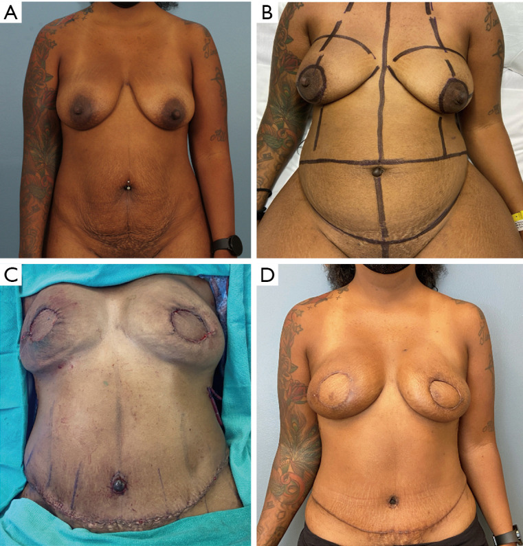
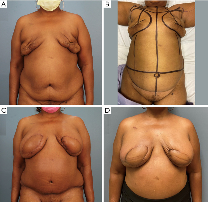
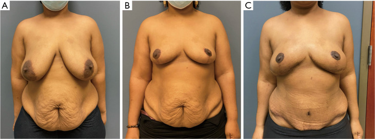
Of the microsurgical donor sites, abdominal-based flaps are the most utilized option. Advantages of the abdominal region include a favorable donor site, in which excess tissue of the lower abdomen is removed, resulting in an inconspicuous scar that is often hidden with clothes, undergarments, and bathing apparel. The blood supply of the abdominal wall has been well described—with vascularity arising from the epigastric arcades (33,34). Reconstructive trends have transitioned from a pedicled transverse rectus abdominus muscle (TRAM), perfused by the superior epigastric artery, towards microsurgical flaps, perfused by the deep-inferior system, which is more robust (35). Flaps from this region are categorized by their source blood vessel and components, of which microsurgical options include the free TRAM and its muscle-sparing variations, the deep inferior epigastric artery perforator (DIEP) flap and the superficial inferior epigastric artery perforator (SIEA) flap. The DIEP flap, though first described in 1989, was introduced as a breast reconstruction option by Robert Allen in 1994 (8). Since then, it has become the gold standard free flap for autologous breast reconstruction (36). DIEPs are reliable options for both immediate and delayed reconstruction across a large range of BMIs as seen in Figures 1-3.
Figure 1.
Before and after photos for a patient with DIEP flaps. (A) A 57-year-old female patient with PMH of obesity (BMI 35 kg/m2) and left invasive breast cancer 1 year status-post bilateral mastectomies with failed implant reconstruction and adjuvant chemotherapy. Preoperative planning for delayed reconstruction with free tissue transfer; (B) preoperative markings on table, day of surgery; (C) 3 months status-post bilateral delayed breast reconstruction with DIEPs; (D) 3 months status-post breast reconstruction revision with mastopexy. BMI, body mass index; DIEP, deep inferior epigastric perforator; PMH, past medical history.
Figure 2.

Before and after photos for a patient with autologous breast reconstruction. (A) A 44-year-old female patient with left DCIS desiring mastectomies and immediate autologous reconstruction, with grade 1 ptosis on examination; (B) preoperative markings with peri-areolar design for mastectomies; (C) on table results intraoperative status-post bilateral free DIEPs for immediate breast reconstruction; (D) 3 months status-post bilateral nipple sparing mastectomies and immediate reconstruction with DIEP flaps. DCIS, ductal carcinoma in situ; DIEP, deep inferior epigastric perforator.
Figure 3.
Before and after photos for a patient with first stage breast reduction and second stage autologous breast reconstruction. (A) A 35-year-old female patient with PMH of BRCA2 gene mutation and BMI 25 kg/m2 desiring mastectomies and autologous reconstruction, with grade 2 ptosis and macromastia on examination. Preoperative planning for breast reduction; (B) 3 months status-post breast reduction and preop for mastectomies and free flap reconstruction; (C) 3 months status-post bilateral nipple sparing mastectomies and immediate reconstruction with DIEP flap. PMH, past medical history; BRCA, breast cancer related gene; BMI, body mass index; DIEP, deep inferior epigastric perforator.
Preoperative imaging
There is robust literature regarding the various imaging modalities prior to abdominal-based free tissue transfer. In a large meta-analysis, Kiely et al. (37) suggest that cross-sectional imaging, in the form of computed tomography angiography (CTA) and magnetic resonance angiography (MRA) are superior to other modalities [ultrasound (US) and Doppler]. Literature suggests that preoperative CTA results in improved flap outcomes and decreased operative time (4,38-40). However, imaging accessibility, cost, radiation exposure (CTA) and insurance approval may limit its use based on institution and insurance type. Advances in technology suggest that color-enhanced blood flow US can accurately identify abdominal wall perforators while avoiding radiation exposure; however, limitations include availability and sonographer skill (41). Alternatively, many surgeons successfully perform perforator-free tissue transfer without preoperative imaging—of which perforators are selected based on intraoperative assessment of caliber, visible pulsations, and palpable pulse.
Considerations on donor site morbidity
With microsurgical refinements, perforator flap dissection is often performed to minimize donor site morbidity and violation of the abdominal wall. TRAM flaps and its muscle sparing variations harvest some of the abdominal wall with the flap. Perforator dissection of the deep inferior epigastric (DIE) vessels uses the same source pedicle (DIEA and DIEV)—while minimizing donor site morbidity. Literature supports that DIEP flaps reduces abdominal wall morbidity (42,43) in both hernia and bulge formations by 20% (43).
Alternatively, the SIEA flap is perfused by the SIEA, which runs superficial to the rectus fascia, and therefore flap harvest avoids abdominal wall violation—preserving abdominal wall integrity. Literature reports higher incidences of free flap failure due to vascular thrombosis when the SIEA is selected—with failure rates from 5% to 15% (34,44,45). If the SIEA is selected—authors recommend an arterial caliber of >1.5 mm, along with visible arterial pulsations to ensure pedicle adequacy (42,46-48). Some literature suggests that flap delay with ligation of the DIEA prior to SIEA flap harvest increases vessel caliber and flow (49); however, this entails an additional surgical procedure.
An advantage of evaluation and preservation of the superficial inferior epigastric vein (SIEV) includes venous supercharging to mitigate venous congestion, which can occur due to dominant superficial venous drainage (50-52). Systematic reviews (53) report significantly decreased venous congestion rates with SIEV supercharging, and a 20% reduction of fat necrosis and partial flap loss. While not every flap requires use of the SIEV, preservation can serve as a lifeboat during the index procedure or if a salvage operation is needed.
Surgical technique
❖ In the preoperative holding area, the lower abdominal tissue is marked with skin pinch. The superior breast boarder, the lateral breast border, the natural inframammary fold (IMF), and the medial breast border are also marked either by comparing with the native breast in cases of unilateral reconstruction or to provide symmetry in cases of bilateral reconstruction.
❖ Harvested tissues include skin, subcutaneous fat, and varying amounts of rectus muscle and overlying rectus fascia based on surgeon preference, patient anatomy, and technique of choice.
❖ Flap boarders include anterior superior iliac spine, inguinal crease, and umbilicus—drawn in an elliptical incision.
❖ The incision is carried down to the abdominal wall on the superior and inferior incisions, with care to evaluate the superficial inferior epigastric system vessels. As in some patients—these vessels are adequate, and may assist with venous drainage, and if used as the vascular supply, can limit abdominal wall violation. The authors prefer dissecting 5–8 cm long SIEV to assist in venous drainage if needed by anastomosing this to the internal mammary vein (IMV) or comittante vein of the DIE vessels.
❖ Flap elevation proceeds lateral to medial, then medial to lateral, identifying all perforators from the medial and lateral row, with caution once passing the linea semilunaris, to avoid iatrogenic injury to source blood supply. The senior authors prefer identifying all perforators before selecting the dominant ones and proceeding with perforator dissection.
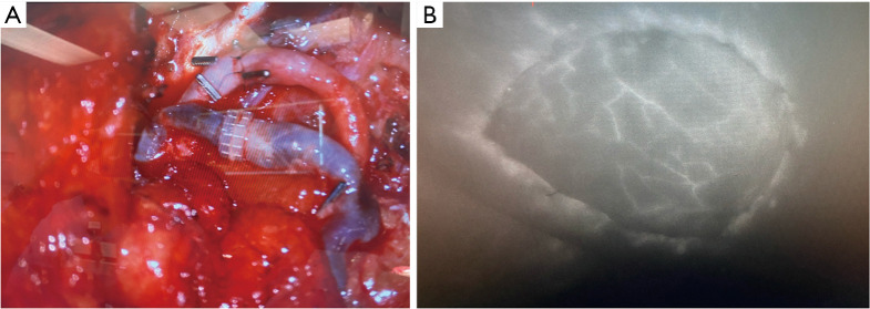
❖ The dominant perforators are then confirmed using SPY angiography to evaluate flap perfusion, after using bulldogs to clip the blood supply of the superior epigastric vessels and other non-dominant perforators as seen in Figure 4.
❖ After flap harvest, the source vessel is ligated, and the abdominal-based flap is inset to create a breast mound.
❖ Flexing of the surgical bed (beach chair position) is often necessary to allow closure of the abdominal donor site and provide an appropriate anatomical flap inset.
❖ A layered closure of the abdominal donor site is often necessary.
❖ Various techniques exist regarding umbilicoplasty, use of surgical drains, and progressive tension sutures.
Figure 4.
Intraoperative autologous reconstruction anastomosis. (A) Example right hemi-abdomen DIEP vessel anastomosis to left chest internal mammary vessels; (B) example SPY angiography using IV injection of 7.5 mg ICG after flap inset to confirm flap perfusion at 1 min after injection. DIEP, deep inferior epigastric perforator; ICG, indocyanine green; IV, intravenous.
Alternative flaps for breast reconstruction
While many conclude that the DIEP flap is the best option for microsurgical breast reconstruction, advancements have led to the development of flaps from other regions of the body. These alternative flaps can be harvested from the thighs, buttocks, and lower trunk, and can be preferred in some patients. This next section provides a brief overview on flap alternatives.
Alternative breast reconstruction
Thigh based flaps
Multiple autologous flaps can be derived from the upper thigh. In general, thigh-based flaps offer sufficient soft tissues for small to modest sized breasts. Donor site considerations include scar location, minimization of tension and lymphatic preservation.
Upper thigh options include the Gracilis myocutaneous flaps with various skin designs, the profunda artery perforator (PAP) flap and the lateral thigh perforator (LTP) flap.
Gracilis myocutaneous flaps
Gracilis myocutaneous flaps harvest tissues from the upper, inner thigh, based off medial femoral circumflex artery (MFCA). Benefits of this flap include is reliable pedicle location, which enters the gracilis muscle in the proximal third of the muscle, about 10 cm distal to the pubic tubercle (54). The skin design can be in a transverse, diagonal or vertical dimensions, based on surgeon desire. Traditionally, the transverse orientation has been used, of which the scar in hidden with the medial aspect of the thigh. In 49 patients, Craggs et al. (55) report 4% total flap loss with 59% of donor site complications, albeit high patient satisfaction.
PAP flap
The PAP flap derives tissue from the posterior-medial thigh, of which when carefully executed, results in a scar hidden within the inferior gluteal fold. This flap is derived off of perforators that penetrate the abductor magnus, typically within 7 cm from the inferior gluteal fold with a pedicle length of nearly 10 cm (56,57). While large studies are limited—success rates of 98–99% have been reported in 2 of the largest series (56-58). In fact, many, including Haddock, have posited that the PAP flap may be the second-choice flap following the DIEP if an abdominal-based flap is not an option (59). Disadvantages of this flap include sacrifice of the posterior cutaneous nerve of the thigh (60).
LTP flap
The region referred to as “saddle bags” in layman’s terms provides the donor tissues for the LTP. This flap is supplied by the septocutaneous perforators from the ascending branch of the lateral femoral circumflex artery, of which superficializes between the tensor facia lata and the gluteus medius (61). The septocutaneous course facilities quick dissection with high flap viability (61). Preoperative discussion should include that removal of this tissue can masculinize the thigh contour.
Trunk based flaps
Lumbar artery perforator (LAP) flap
The LAP flap—proposed in 2003 by de Weed and colleagues (62), derives its donor tissue from posterior excess, often described as the “love handle” (63). Flap boundaries include the ASIS and the posterior midline, with superior and inferior aspects based on sufficient skin pinch to allow for donor site closure. In most cases, the flap’s dominant LAP penetrates the thoracolumbar fascia about 7–10 cm lateral to the midline, between the erector spine and quadratus lumbar muscles, and is then traced to the transverse process of the spine (56,62-64).
Benefits of this flap include is an inconspicuous donor site with limited morbidity, as the harvested tissue corresponds to the tissue excised from a posterior body lift. Sultan and Greenspun (65) advocate that the LAP flap is the ideal alternative flap for breast reconstruction if an abdominal site is unavailable, as it avoids the disfigurement inherent to buttock or thigh-based flaps. While the donor site is aesthetically favorable, this flap is not without shortcomings. Due to its short pedicle length (2–4 cm) (65,66), this flap requires interposition arterial and venous grafting, most often from the DIEA or the thoracodorsal vessels. Like all perforator flaps, meticulous dissection is required, and continues to the transverse process of the spine, with the potential for serious bleeding. Further, due to flap location, a position change is required, which can be burdensome for operative room personnel (67). Additionally, literature reports a flap loss rate of 3–9% (66,68,69), which is higher than other flap types, acute revision rates of 17–24% (68-70) for attempted flap salvage, and symptomatic seroma rates of 31% that require intervention (69). In summary, this flap provides an aesthetic donor site with that can be utilized in patients with lower BMI’s (65,66,71), although is perhaps more technically demanding than other autologous sources.
Thoracodorsal artery perforator flap (TDAP)
In attempts to minimize donor site morbidity from the latissimus dorsi flap, TDAP flaps were first introduced in 1995. This allows for reduced functional deficits from muscle-sparing as well as improved aesthetic effects without contour defects (72). While originally it was vascularly pedicled based, TDAP flaps now offer a simpler approach utilizing a propeller TDAP where the flap is rotated 180 degrees over the pedicle, thereby covering mastectomy site (72). This adjustment eliminates the need for the dissection of the intramuscular pedicle (72). TDAPs offer multiple muscle-sparing techniques now including the propeller, flip-over, and conventional perforator (73). By including the superior and inferior fat compartments, the TDAP can also be extended for successful reconstruction in large body mass indexes or in medium to large defect cases, although these have been found to have the greatest morbidity of the TDAP armamentarium (73,74).
Minimally invasive autologous reconstruction
Laparoscopic-assisted autologous reconstruction
Total extraperitoneal laparoscopic DIEP flaps allowed for a decreased in myofascial dissection and incision size, thereby reducing donor site morbidity (75-77). The operative technique includes placing a supraumbilical camera port at the medial edge of the rectus, developing an extraperitoneal plane using insufflation and a balloon dissector. Two ports for dissection instruments are placed below the umbilicus in the linear alba, then dissecting out the DIE vessels from the underside of the rectus. The vessels are then ligated and delivered through a minimal fascial incision (76). While more expensive when compared to open, the reduced donor site morbidity makes minimally invasive autologous reconstruction a viable alternative (75). As a far more readily available technique when compared to robotic, as well as cheaper, faster, and an easier learning curve, laparoscopic-assisted autologous reconstruction may be an improved alternative when it relates to patient’s outcomes of autologous reconstruction and surgeon’s ease of performing (75).
Robotic-assisted autologous reconstruction
As robotic surgery becomes a standard option in a surgeon’s toolbox, plastic surgeons have expanded their techniques for breast reconstruction to include robotic surgery with promising preoperative and postoperative results (75,78). Robotic reconstruction has been found to be an alternative for mastectomies with immediate reconstruction including flaps utilizing latissimus dorsi and the DIE perforators (78-80). Results comparing robotic versus conventional open autologous reconstruction found decreased postoperative pain in those undergoing robotic operations, equivocal complication rates, and increased operative time (78,80). This indicates the feasibility, safety, and effectiveness of robotic autologous reconstruction, with increased efficiency with more comfort and training.
Neurotization during autologous reconstruction
While autologous breast reconstruction is becoming the gold standard for breast reconstruction following mastectomies and has been found to improve psychosocial wellbeing postoperatively, an ongoing challenge is that of loss of sensation following the procedure (67,81). This sensation is through feedback from the anterior and lateral branches of the second through sixth intercostal nerve, with the primary sensation to the nipple areolar complex being the fourth and fifth ICN (67). Breast sensation involves the many components of touch including pressure, temperature, and pain (67). While some patients have been found to regain some form of sensation, few have been able to standardize or predict who will recover and the speed at which it will happen. Thus, while neurotization is still outside the scope of standard of care and has not yet been widely adopted, the significant positive psychosocial impact has led to more research and clinical expertise in those who are utilizing it. Yet, there is still variable technique, including recipient nerve choices, decision to use conduits, and the extent of ICN dissection (81). Studies including those by Momeni et al. [2021] demonstrate significant improvement in 1 year sensation for those who underwent abdominal flap neurotization with allograft compared to those who didn’t (81). This demonstrates an area of continued research and refinement of clinical acumen.
Pre-operative perforator marking
As autologous breast reconstruction has become standard of care and more accessible to both patients and surgeons, efforts have been made to improve the efficiency in selection of a vascular pedicle preoperatively and intra-operative visual evaluation of the perforators (82). Options for perforator mapping preoperatively included abdominal MRA, CTA, contrast-enhanced US, and color Doppler US as safe reliable methodologies to do so (42,82-84). However, CTA is currently considered gold standard in this regard (84). Intraoperatively, the use of fluorescent angiography, utilizing indocyanine green (ICG), allows for assessing blood flow and patency in the anastomosis when insetting the autologous flap (85,86). The use of ICG during autologous breast reconstruction has been found to reduce fat necrosis and be a more accurate predictor of patent anastomoses than clinical assessment alone (86). Lastly, the use of smartphone thermal imagining technology has been found to be a valuable, reliable and cost-effective preoperative modality to design perforator flaps in identifying the most dominant perforator as well as intraoperatively to assess perfusion pattern, decision to discard least perfused areas, and in evaluating patency of the microvascular anastomosis (87).
Post operative considerations
Based on surgeon preference, post operative protocols vary widely. Considerations include intensive care unit admission versus trained nursing unit, flap monitoring, duration of hospital stay, analgesia, and mobility. Multiple studies looking at free tissue transfer to both the lower extremity and head and neck have found that there was no reduction in complications or flap failure with ICU admission, and thus limiting ICU admission to those requiring it may reduce duration and cost of stay (88-90). However, others posit that due to the highest risk of thrombotic events and hematomas within the first 24–72 hours, close monitoring every 1–2 hours in the ICU or dedicated flap units are still warranted (91). This decision may rely on surgeon’s clinical acumen regarding patient risk to limit overutilization of ICU resources, yet more research still needs to be done in this area.
Monitoring
Clinical examination remains a valuable component of flap monitoring; however, it relies on education and experience to identify flap compromise. Flap color and capillary refill can aid in clinical examination; although in darker skin individuals, skin color may disguise congestion based on these parameters alone. Based on the location of cutaneous perforators and flap inset, arterial and venous flow can often be identified by Doppler US; however, this technology is often user dependent and is less reliable in buried flaps. Halani et al. (92) thoroughly discuss various modalities for flap monitoring.
Literature suggests that tissue oximetry can detect flap compromise before changes in clinical exam are appreciated (93), and is a widely used modality. Early recognition of flap perfusion can allow for timely exploration and salvage attempts, preventing irreversible ischemia. At our institution, we commonly utilize tissue oximetry in addition to clinical and doppler examination—and have had great success with this technology.
Enhanced recover after surgery (ERAS)
Implementation of ERAS pathways have demonstrated improved outcomes, decreased complications, decreased hospital LOS, and decreased overall costs across nearly every surgical specialty. The literature suggests similar benefits in microvascular breast reconstruction as well (94-98). While ERAS pathway specifics vary, most emphasize narcotic reduction with multi-modal pain analgesia, early ambulation/mobility, early removal of foley, and early enteral nutrition. Based on institutional protocols and perioperative educations, we believe ERAS pathways are essential in microvascular breast free flap reconstruction.
Drains
While majority of abdominal autologous breast reconstruction still leaves two abdominal drains post-operatively, in abdominoplasty, progression tension sutures have been found comparable to use of abdominal drains. Studies have been looking at utilizing barbed progressive tension sutures in donor-site closure of DIEPs (99). Findings indicated that complication rates were not significantly different between closure using progressive tension suture and abdominal drain placement (99). Elimination of abdominal drains increases patient satisfaction, mobility, independence upon discharge, and many of the postoperative questions and concerns (99).
Risk stratification/complication profile
In general, when compared to implant-based breast reconstruction (IBR) and oncoplastic or breast conservation techniques, microsurgical reconstruction is a longer reconstruction with an increased duration of anesthetic exposure. Therefore, appropriate preoperative risk stratification is necessary to mitigate complication development.
In a meta-analysis, Schaverien and Mcculley (100) compared complication profiles in abdominal-based autologous reconstruction between obese (BMI >30 kg/m2) and non-obese (BMI <30 kg/m2) cohorts. In non-obese patients, the overall complication rate was 31%, with 12% donor site complications and 17% recipient site complications. The incidence of bulge/hernia and donor site infection was 3% each. Further, partial flap loss was 1.3% and total flap loss was 1%. Alternatively, obese patients had significantly increased complications with an overall complication rate of 60%, donor site rate of 32%, recipient site rate of 52%, 8% donor site infection, 5.5% hernia/bulge, 4% partial flap loss and 2.2% complete flap loss. When patients were sub-stratified to BMI >40 kg/m2, the complication profile approached 65%, with 5% total flap loss. The authors suggest a BMI of 40 kg/m2 as a cutoff to which the risks of surgery may surpass the benefits.
While this analysis suggests a relatively high complication profile, the rate of reconstructive success and flap viability is greater that 95%, even in high-risk populations, and therefore risk stratification should be performed based on an individual basis.
Contraindications
In microsurgical breast reconstruction, the overall health of the patient should be considered as these procedures are generally lengthy in nature. It is well known that obesity increases complications in free flap breast reconstruction (100-102). Panayi et al. conclude that obesity (BMI >30 kg/m2) results in greater than 2-fold risk of both surgical and medical complications (101). However, it is important to consider that while obesity increases the complication profile, reconstructive success is still very high in obese patients, and therefore an individualized approach to risk stratification should be implemented. It is also known that radiotherapy and chemotherapy may impact the overall tissue health, autologous reconstruction has been found to be safe in patients who have had radiotherapy (103).
In regards to age, there is a paucity of literature on older patients undergoing free tissue transfer; however, success rates of 96% have been reported in patients >70 years of age (104). Overall, patient preference, habitus, physiologic fitness and comorbidities should all be considered when determining candidacy for flap reconstruction.
Aesthetic refinements
We are now at an era in microsurgery where breast reconstruction should not only be viable, but also cosmetically pleasing. Aesthetic considerations apply not only to the breast mound, but also to the donor site. Autologous reconstruction allows the removal of tissue excess, of which body contouring principles can and should be emphasized.
The similarities between an abdominal free flap donor site and abdominal contouring procedure, such as an abdominoplasty, are numerous, including removal of excess adipose tissue from the lower abdominal region. However, the scar location for abdominal-based free flaps tends to be higher than a traditional abdominoplasty scar. To place the abdominal scar in a more inconspicuous location, the “Low DIEP flap” (105) has been described, in which the scar is placed 4 to 6 cm inferior to the umbilicus. However, this technique tends to have high rates of venous congestion (up to 30%), requiring supercharging of venous drainage with the SIEV (105). While the scar is better hidden, the flaps harvested are smaller in size and authors of this technique recommend preoperative imaging to ensure adequate perforator incorporation within the proposed flap area (105). Therefore, while this technique offers a solution for improved abdominal scar location—it has limitations and selection should be dependent on patient habitus, perforator location and patient expectations. Additionally, other techniques include the use of progressive tension sutures to eliminate the need for drains in the abdominal donor site. Mohan et al. (106) demonstrated a significantly decreased complication rate and decreased seroma rate in patients with the drainless technique compared to traditional methods (2.4% vs. 7.8% respectively).
Thigh-based options allow for removal of excess thigh tissues, with a scar that is relatively well hidden. Removal of medial thigh tissues pose the opportunity to create a “thigh gap” in patients. There should be caution in the usage of lateral thigh donor sites, such as in the lateral thigh flap—which can masculinize the thigh—and should be discussed preoperatively. Furthermore, aesthetic principles from a lower body lift can be applied to the LAP flap, as this effectively removes “love handles”.
Other aesthetic considerations include flap size and projection of the newly reconstructed breast. Blondeel et al. (107) propose an algorithm for shaping of reconstructive breasts, with emphasis on the breast footprint, projection and skin components. In patients undergoing unilateral reconstruction, a bi-pedicled DIEP flap can be used to increase flap size. Adjuncts to increasing projection include utilization of mastectomy skin flaps, autologous fat graft and placement of a prosthesis. Residual mastectomy skin flaps can be de-epithelialized to provide additional bulk. Hamdi et al. (108) propose a technique in which the inferior skin flap is de-epithelialized, with the medial and lateral components folded centrally to increase projection, analogous to a “Hug”. Results from the “Hug” flap include significantly decreased rates of secondary fat grafting, thus decreasing additional surgical procedure (108). Further, breast augmentation can be performed with the use of an implant either at the time of free flap reconstruction or in a staged fashion. Literature suggests that this can improve size and symmetry between breasts, with good aesthetic outcomes and without flap compromise (109-112). Systematic reviews suggest high rates (97.5%) of successful reconstruction in patients with free tissue transfer and implant placement; albeit significantly reduced complications when the prosthesis is placed in a staged fashion (32% vs. 18%) (112). As plastic surgeons, we are familiar and adept with breast implant placement for both aesthetic and reconstructive procedures, placement of a foreign device entails implant-related complication, that are avoided in purely autologous methods. Other symmetrizing procedures include flap debulking and flap advancement, or combinations of the aforementioned techniques. Like many aesthetic and reconstructive procedures, the patient’s adipose distribution, BMI, and skin quality affect the outcome, and it is important to set patient expectations preoperatively.
Secondary surgeries
The ideal breast reconstruction would limit the need for additional surgeries. In the short term, most secondary surgeries are tributary to complications. However, revisionary surgeries for aesthetic refinement are common in all methods of breast reconstruction. In a 25-year experience, encompassing 888 reconstructed breasts (including both implant-based and autologous), Losken et al. report that 3.99 (in unilateral reconstructions) and 5.54 (in bilateral reconstructions) additional surgeries were required to achieve reconstruction completion (113). A complete reconstruction was defined as the performance of nipple reconstruction—and risk factors for additional procedures included radiation therapy, patient comorbidities, post operative complication, and bilateral reconstructions (113). Recent microsurgical literature (114-116) suggests that the majority of patients following abdominal-based breast reconstruction require at least one additional procedure, and this is crucial to include in the preoperative setting.
Conclusions
While implant-based reconstruction remains the predominant method of breast reconstruction in the US, there have been many exciting advancements in autologous reconstruction that offers high aesthetic outcomes and patient satisfaction. An obvious benefit of total autologous reconstruction is the avoidance of implants and their associated sequalae. Although the most common donor site remains the abdomen, alternative flaps can be preferred depending on patient habitus, patient preference, and surgeon experience. Microsurgical refinements, such as perforator dissection, limit donor site morbidity with reliable outcomes. The literature suggests high rates of reconstruction success in multiple systematic reviews. Risk stratification and setting patient expectations are a crucial component of the preoperative experience.
Supplementary
The article’s supplementary files as
Acknowledgments
Funding: None.
Ethical Statement: The authors are accountable for all aspects of the work in ensuring that questions related to the accuracy or integrity of any part of the work are appropriately investigated and resolved. All clinical procedures described in this study were performed in accordance with the ethical standards of the institutional and/or national research committee(s) and with the Helsinki Declaration (as revised in 2013). Written informed consent was obtained from the patients for the publication of this article and accompanying images.
Footnotes
Provenance and Peer Review: This article was commissioned by the Guest Editors (Tine Engberg Damsgaard, C. Andrew Salzberg and Jørn Bo Thomsen) for the series “Hot Topics in Breast Reconstruction World Wide” published in Gland Surgery. The article has undergone external peer review.
Reporting Checklist: The authors have completed the Narrative Review reporting checklist. Available at https://gs.amegroups.org/article/view/10.21037/gs-24-63/rc
Conflicts of Interest: All authors have completed the ICMJE uniform disclosure form (available at https://gs.amegroups.org/article/view/10.21037/gs-24-63/coif). The series “Hot Topics in Breast Reconstruction World Wide” was commissioned by the editorial office without any funding or sponsorship. The authors have no other conflicts of interest to declare.
References
- 1.Garza R, 3rd, Ochoa O, Chrysopoulo M. Post-mastectomy Breast Reconstruction with Autologous Tissue: Current Methods and Techniques. Plast Reconstr Surg Glob Open 2021;9:e3433. 10.1097/GOX.0000000000003433 [DOI] [PMC free article] [PubMed] [Google Scholar]
- 2.Ochoa O, Garza R, 3rd, Pisano S, et al. Prospective Longitudinal Patient-Reported Satisfaction and Health-Related Quality of Life following DIEP Flap Breast Reconstruction: Relationship with Body Mass Index. Plast Reconstr Surg 2019;143:1589-600. 10.1097/PRS.0000000000005616 [DOI] [PubMed] [Google Scholar]
- 3.Coriddi M, Shenaq D, Kenworthy E, et al. Autologous Breast Reconstruction after Failed Implant-Based Reconstruction: Evaluation of Surgical and Patient-Reported Outcomes and Quality of Life. Plast Reconstr Surg 2019;143:373-9. 10.1097/PRS.0000000000005197 [DOI] [PMC free article] [PubMed] [Google Scholar]
- 4.Rozen WM, Rajkomar AK, Anavekar NS, et al. Post-mastectomy breast reconstruction: a history in evolution. Clin Breast Cancer 2009;9:145-54. 10.3816/CBC.2009.n.024 [DOI] [PubMed] [Google Scholar]
- 5.Teimourian B, Adham MN. Louis Ombredanne and the origin of muscle flap use for immediate breast mound reconstruction. Plast Reconstr Surg 1983;72:905-10. 10.1097/00006534-198312000-00037 [DOI] [PubMed] [Google Scholar]
- 6.Hartrampf CR, Scheflan M, Black PW. Breast reconstruction with a transverse abdominal island flap. Plast Reconstr Surg 1982;69:216-25. 10.1097/00006534-198202000-00006 [DOI] [PubMed] [Google Scholar]
- 7.Hartrampf CR, Jr. The transverse abdominal island flap for breast reconstruction. A 7-year experience. Clin Plast Surg 1988;15:703-16. [PubMed] [Google Scholar]
- 8.Allen RJ, Treece P. Deep inferior epigastric perforator flap for breast reconstruction. Ann Plast Surg 1994;32:32-8. 10.1097/00000637-199401000-00007 [DOI] [PubMed] [Google Scholar]
- 9.Broyles JM, Balk EM, Adam GP, et al. Implant-based versus Autologous Reconstruction after Mastectomy for Breast Cancer: A Systematic Review and Meta-analysis. Plast Reconstr Surg Glob Open 2022;10:e4180. 10.1097/GOX.0000000000004180 [DOI] [PMC free article] [PubMed] [Google Scholar]
- 10.Masoomi H, Hanson SE, Clemens MW, et al. Autologous Breast Reconstruction Trends in the United States: Using the Nationwide Inpatient Sample Database. Ann Plast Surg 2021;87:242-7. 10.1097/SAP.0000000000002664 [DOI] [PubMed] [Google Scholar]
- 11.Hanwright PJ, Davila AA, Hirsch EM, et al. The differential effect of BMI on prosthetic versus autogenous breast reconstruction: a multivariate analysis of 12,986 patients. Breast 2013;22:938-45. 10.1016/j.breast.2013.05.009 [DOI] [PubMed] [Google Scholar]
- 12.Momeni A, Ahdoot MA, Kim RY, et al. Should we continue to consider obesity a relative contraindication for autologous microsurgical breast reconstruction? J Plast Reconstr Aesthet Surg 2012;65:420-5. 10.1016/j.bjps.2011.10.005 [DOI] [PubMed] [Google Scholar]
- 13.Selber JC, Kurichi JE, Vega SJ, et al. Risk factors and complications in free TRAM flap breast reconstruction. Ann Plast Surg 2006;56:492-7. 10.1097/01.sap.0000210180.72721.4a [DOI] [PubMed] [Google Scholar]
- 14.Seidenstuecker K, Munder B, Mahajan AL, et al. Morbidity of microsurgical breast reconstruction in patients with comorbid conditions. Plast Reconstr Surg 2011;127:1086-92. 10.1097/PRS.0b013e318205f255 [DOI] [PubMed] [Google Scholar]
- 15.Booi DI, Debats IB, Boeckx WD, et al. Risk factors and blood flow in the free transverse rectus abdominis (TRAM) flap: smoking and high flap weight impair the free TRAM flap microcirculation. Ann Plast Surg 2007;59:364-71. 10.1097/SAP.0b013e318030b083 [DOI] [PubMed] [Google Scholar]
- 16.Fischer JP, Nelson JA, Serletti JM, et al. Peri-operative risk factors associated with early tissue expander (TE) loss following immediate breast reconstruction (IBR): a review of 9305 patients from the 2005-2010 ACS-NSQIP datasets. J Plast Reconstr Aesthet Surg 2013;66:1504-12. 10.1016/j.bjps.2013.06.030 [DOI] [PubMed] [Google Scholar]
- 17.Nahabedian MY. Symmetrical breast reconstruction: analysis of secondary procedures after reconstruction with implants and autologous tissue. Plast Reconstr Surg 2005;115:257-60. [PubMed] [Google Scholar]
- 18.Nahabedian MY, Momen B, Galdino G, et al. Breast Reconstruction with the free TRAM or DIEP flap: patient selection, choice of flap, and outcome. Plast Reconstr Surg 2002;110:466-75; discussion 476-7. 10.1097/00006534-200208000-00015 [DOI] [PubMed] [Google Scholar]
- 19.Gill PS, Hunt JP, Guerra AB, et al. A 10-year retrospective review of 758 DIEP flaps for breast reconstruction. Plast Reconstr Surg 2004;113:1153-60. 10.1097/01.prs.0000110328.47206.50 [DOI] [PubMed] [Google Scholar]
- 20.Voineskos SH, Frank SG, Cordeiro PG. Breast reconstruction following conservative mastectomies: predictors of complications and outcomes. Gland Surg 2015;4:484-96. 10.3978/j.issn.2227-684X.2015.04.13 [DOI] [PMC free article] [PubMed] [Google Scholar]
- 21.Wang XL, Liu LB, Song FM, et al. Meta-analysis of the safety and factors contributing to complications of MS-TRAM, DIEP, and SIEA flaps for breast reconstruction. Aesthetic Plast Surg 2014;38:681-91. 10.1007/s00266-014-0333-3 [DOI] [PubMed] [Google Scholar]
- 22.Fischer JP, Sieber B, Nelson JA, et al. Comprehensive outcome and cost analysis of free tissue transfer for breast reconstruction: an experience with 1303 flaps. Plast Reconstr Surg 2013;131:195-203. 10.1097/PRS.0b013e318277856f [DOI] [PubMed] [Google Scholar]
- 23.Javaid M, Song F, Leinster S, et al. Radiation effects on the cosmetic outcomes of immediate and delayed autologous breast reconstruction: an argument about timing. J Plast Reconstr Aesthet Surg 2006;59:16-26. 10.1016/j.bjps.2005.07.007 [DOI] [PubMed] [Google Scholar]
- 24.Barry M, Kell MR. Radiotherapy and breast reconstruction: a meta-analysis. Breast Cancer Res Treat 2011;127:15-22. 10.1007/s10549-011-1401-x [DOI] [PubMed] [Google Scholar]
- 25.Thorarinsson A, Fröjd V, Kölby L, et al. Patient determinants as independent risk factors for postoperative complications of breast reconstruction. Gland Surg 2017;6:355-67. 10.21037/gs.2017.04.04 [DOI] [PMC free article] [PubMed] [Google Scholar]
- 26.Motwani SB, Strom EA, Schechter NR, et al. The impact of immediate breast reconstruction on the technical delivery of postmastectomy radiotherapy. Int J Radiat Oncol Biol Phys 2006;66:76-82. 10.1016/j.ijrobp.2006.03.040 [DOI] [PubMed] [Google Scholar]
- 27.Williams JK, Bostwick J, 3rd, Bried JT, et al. TRAM flap breast reconstruction after radiation treatment. Ann Surg 1995;221:756-64; discussion 764-6. 10.1097/00000658-199506000-00014 [DOI] [PMC free article] [PubMed] [Google Scholar]
- 28.Mehrara BJ, Santoro TD, Arcilla E, et al. Complications after microvascular breast reconstruction: experience with 1195 flaps. Plast Reconstr Surg 2006;118:1100-9. 10.1097/01.prs.0000236898.87398.d6 [DOI] [PubMed] [Google Scholar]
- 29.Hart SE, Brown DL, Kim HM, et al. Association of Clinical Complications of Chemotherapy and Patient-Reported Outcomes After Immediate Breast Reconstruction. JAMA Surg 2021;156:847-55. 10.1001/jamasurg.2021.2239 [DOI] [PMC free article] [PubMed] [Google Scholar]
- 30.Regan JP, Casaubon JT. Breast Reconstruction. Treasure Island (FL): StatPearls Publishing; 2024. [PubMed] [Google Scholar]
- 31.Kaidar-Person O, Hermann N, Poortmans P, et al. A multidisciplinary approach for autologous breast reconstruction: A narrative (re)view for better management. Radiother Oncol 2021;157:263-71. 10.1016/j.radonc.2021.01.036 [DOI] [PubMed] [Google Scholar]
- 32.Lee KT, Mun GH. Effects of Obesity on Postoperative Complications After Breast Reconstruction Using Free Muscle-Sparing Transverse Rectus Abdominis Myocutaneous, Deep Inferior Epigastric Perforator, and Superficial Inferior Epigastric Artery Flap: A Systematic Review and Meta-analysis. Ann Plast Surg 2016;76:576-84. 10.1097/SAP.0000000000000400 [DOI] [PubMed] [Google Scholar]
- 33.Saint-Cyr M. Assessing perforator architecture. Clin Plast Surg 2011;38:175-202. 10.1016/j.cps.2011.03.015 [DOI] [PubMed] [Google Scholar]
- 34.Avraham T, Clavin N, Mehrara BJ. Microsurgical breast reconstruction. Cancer J 2008;14:241-7. 10.1097/PPO.0b013e31817fb7e3 [DOI] [PubMed] [Google Scholar]
- 35.Grotting JC, Urist MM, Maddox WA, et al. Conventional TRAM flap versus free microsurgical TRAM flap for immediate breast reconstruction. Plast Reconstr Surg 1989;83:828-41; discussion 842-4. 10.1097/00006534-198905000-00009 [DOI] [PubMed] [Google Scholar]
- 36.Varnava C, Wiebringhaus P, Hirsch T, et al. Breast Reconstruction with DIEP Flap: The Learning Curve at a Breast Reconstruction Center and a Single-Surgeon Study. J Clin Med 2023;12:2894. 10.3390/jcm12082894 [DOI] [PMC free article] [PubMed] [Google Scholar]
- 37.Kiely J, Kumar M, Wade RG. The accuracy of different modalities of perforator mapping for unilateral DIEP flap breast reconstruction: A systematic review and meta-analysis. J Plast Reconstr Aesthet Surg 2021;74:945-56. 10.1016/j.bjps.2020.12.005 [DOI] [PubMed] [Google Scholar]
- 38.Teunis T, Heerma van Voss MR, Kon M, et al. CT-angiography prior to DIEP flap breast reconstruction: a systematic review and meta-analysis. Microsurgery 2013;33:496-502. 10.1002/micr.22119 [DOI] [PubMed] [Google Scholar]
- 39.Wade RG, Watford J, Wormald JCR, et al. Perforator mapping reduces the operative time of DIEP flap breast reconstruction: A systematic review and meta-analysis of preoperative ultrasound, computed tomography and magnetic resonance angiography. J Plast Reconstr Aesthet Surg 2018;71:468-77. 10.1016/j.bjps.2017.12.012 [DOI] [PubMed] [Google Scholar]
- 40.Rozen WM, Anavekar NS, Ashton MW, et al. Does the preoperative imaging of perforators with CT angiography improve operative outcomes in breast reconstruction? Microsurgery 2008;28:516-23. 10.1002/micr.20526 [DOI] [PubMed] [Google Scholar]
- 41.Zinser MJ, Kröger N, Malter W, et al. Preoperative Perforator Mapping in DIEP Flaps for Breast Reconstruction. The Impact of New Contrast-Enhanced Ultrasound Techniques. J Pers Med 2022;13:64. 10.3390/jpm13010064 [DOI] [PMC free article] [PubMed] [Google Scholar]
- 42.Lipa JE. Breast reconstruction with free flaps from the abdominal donor site: TRAM, DIEAP, and SIEA flaps. Clin Plast Surg 2007;34:105-21; abstract vii. 10.1016/j.cps.2006.11.015 [DOI] [PubMed] [Google Scholar]
- 43.Egeberg A, Rasmussen MK, Sørensen JA. Comparing the donor-site morbidity using DIEP, SIEA or MS-TRAM flaps for breast reconstructive surgery: a meta-analysis. J Plast Reconstr Aesthet Surg 2012;65:1474-80. 10.1016/j.bjps.2012.07.001 [DOI] [PubMed] [Google Scholar]
- 44.Grünherz L, Wolter A, Andree C, et al. Autologous Breast Reconstruction with SIEA Flaps: An Alternative in Selected Cases. Aesthetic Plast Surg 2020;44:299-306. 10.1007/s00266-019-01554-8 [DOI] [PubMed] [Google Scholar]
- 45.Park JE, Shenaq DS, Silva AK, et al. Breast Reconstruction with SIEA Flaps: A Single-Institution Experience with 145 Free Flaps. Plast Reconstr Surg 2016;137:1682-9. 10.1097/PRS.0000000000002158 [DOI] [PubMed] [Google Scholar]
- 46.Chevray PM. Breast reconstruction with superficial inferior epigastric artery flaps: a prospective comparison with TRAM and DIEP flaps. Plast Reconstr Surg 2004;114:1077-83; discussion 1084-5. 10.1097/01.prs.0000135328.88101.53 [DOI] [PubMed] [Google Scholar]
- 47.Ulusal BG, Cheng MH, Wei FC, et al. Breast reconstruction using the entire transverse abdominal adipocutaneous flap based on unilateral superficial or deep inferior epigastric vessels. Plast Reconstr Surg 2006;117:1395-403; discussion 1404-6. 10.1097/01.prs.0000207401.78491.43 [DOI] [PubMed] [Google Scholar]
- 48.Spiegel AJ, Khan FN. An Intraoperative algorithm for use of the SIEA flap for breast reconstruction. Plast Reconstr Surg 2007;120:1450-9. 10.1097/01.prs.0000270282.92038.3f [DOI] [PubMed] [Google Scholar]
- 49.Gregorič M, Flis V, Milotić F, et al. Delaying the superficial inferior epigastric artery flap: a solution to the problem of the small calibre of the donor artery. J Plast Reconstr Aesthet Surg 2011;64:1181-6. 10.1016/j.bjps.2011.04.009 [DOI] [PubMed] [Google Scholar]
- 50.Park SO, Imanishi N, Chang H. The Anatomic Features and Role of Superficial Inferior Epigastric Vein in Abdominal Flap. Arch Plast Surg 2022;49:482-7. 10.1055/s-0042-1748645 [DOI] [PMC free article] [PubMed] [Google Scholar]
- 51.Rozen WM, Pan WR, Le Roux CM, et al. The venous anatomy of the anterior abdominal wall: an anatomical and clinical study. Plast Reconstr Surg 2009;124:848-53. 10.1097/PRS.0b013e3181b037a2 [DOI] [PubMed] [Google Scholar]
- 52.Carramenha e Costa MA, Carriquiry C, Vasconez LO, et al. An anatomic study of the venous drainage of the transverse rectus abdominis musculocutaneous flap. Plast Reconstr Surg 1987;79:208-17. 10.1097/00006534-198702000-00010 [DOI] [PubMed] [Google Scholar]
- 53.Lee KT, Mun GH. Benefits of superdrainage using SIEV in DIEP flap breast reconstruction: A systematic review and meta-analysis. Microsurgery 2017;37:75-83. 10.1002/micr.22420 [DOI] [PubMed] [Google Scholar]
- 54.Bains RMZaRD. Chapter 49: Gracilis Flap. In: Mardini FCWaS, editor. Flaps and reconstructive Surgery, 2nd Edition Elsevier Inc.; 2017. [Google Scholar]
- 55.Craggs B, Vanmierlo B, Zeltzer A, et al. Donor-site morbidity following harvest of the transverse myocutaneous gracilis flap for breast reconstruction. Plast Reconstr Surg 2014;134:682e-691e. 10.1097/PRS.0000000000000612 [DOI] [PubMed] [Google Scholar]
- 56.Myers PL, Nelson JA, Allen RJ, Jr. Alternative flaps in autologous breast reconstruction. Gland Surg 2021;10:444-59. 10.21037/gs.2020.03.16 [DOI] [PMC free article] [PubMed] [Google Scholar]
- 57.Allen RJ, Jr, Lee ZH, Mayo JL, et al. The Profunda Artery Perforator Flap Experience for Breast Reconstruction. Plast Reconstr Surg 2016;138:968-75. 10.1097/PRS.0000000000002619 [DOI] [PubMed] [Google Scholar]
- 58.Haddock NT, Gassman A, Cho MJ, et al. 101 Consecutive Profunda Artery Perforator Flaps in Breast Reconstruction: Lessons Learned with Our Early Experience. Plast Reconstr Surg 2017;140:229-39. 10.1097/PRS.0000000000003553 [DOI] [PubMed] [Google Scholar]
- 59.Haddock NT, Teotia SS. Consecutive 265 Profunda Artery Perforator Flaps: Refinements, Satisfaction, and Functional Outcomes. Plast Reconstr Surg Glob Open 2020;8:e2682. 10.1097/GOX.0000000000002682 [DOI] [PMC free article] [PubMed] [Google Scholar]
- 60.Cohen Z, Azoury SC, Matros E, et al. Modern Approaches to Alternative Flap-Based Breast Reconstruction: Profunda Artery Perforator Flap. Clin Plast Surg 2023;50:289-99. 10.1016/j.cps.2022.10.001 [DOI] [PMC free article] [PubMed] [Google Scholar]
- 61.Tuinder SMH, Beugels J, Lataster A, et al. The Lateral Thigh Perforator Flap for Autologous Breast Reconstruction: A Prospective Analysis of 138 Flaps. Plast Reconstr Surg 2018;141:257-68. 10.1097/PRS.0000000000004072 [DOI] [PubMed] [Google Scholar]
- 62.de Weerd L, Elvenes OP, Strandenes E, et al. Autologous breast reconstruction with a free lumbar artery perforator flap. Br J Plast Surg 2003;56:180-3. 10.1016/s0007-1226(03)00039-0 [DOI] [PubMed] [Google Scholar]
- 63.Stillaert FBJL, Opsomer D, Blondeel PN, et al. The Lumbar Artery Perforator Flap in Breast Reconstruction. Plast Reconstr Surg 2023;151:41-4. 10.1097/PRS.0000000000009782 [DOI] [PubMed] [Google Scholar]
- 64.Thomas J, Waxweiler C, Leveque R, et al. Anatomical Study of Lumbar Artery Perforators in Male Subjects. Plast Reconstr Surg Glob Open 2020;8:e2628. 10.1097/GOX.0000000000002628 [DOI] [PMC free article] [PubMed] [Google Scholar]
- 65.Sultan SM, Greenspun DT. Lumbar Artery Perforator Flaps in Autologous Breast Reconstruction. Clin Plast Surg 2023;50:301-12. 10.1016/j.cps.2022.11.005 [DOI] [PubMed] [Google Scholar]
- 66.Haddock NT, Teotia SS. Lumbar Artery Perforator Flap: Initial Experience with Simultaneous Bilateral Flaps for Breast Reconstruction. Plast Reconstr Surg Glob Open 2020;8:e2800. 10.1097/GOX.0000000000002800 [DOI] [PMC free article] [PubMed] [Google Scholar]
- 67.Vartanian ED, Lo AY, Hershenhouse KS, et al. The role of neurotization in autologous breast reconstruction: Can reconstruction restore breast sensation? J Surg Oncol 2021;123:1215-31. 10.1002/jso.26422 [DOI] [PubMed] [Google Scholar]
- 68.Peters KT, Blondeel PN, Lobo F, et al. Early experience with the free lumbar artery perforator flap for breast reconstruction. J Plast Reconstr Aesthet Surg 2015;68:1112-9. 10.1016/j.bjps.2015.03.031 [DOI] [PubMed] [Google Scholar]
- 69.Opsomer D, Stillaert F, Blondeel P, et al. The Lumbar Artery Perforator Flap in Autologous Breast Reconstruction: Initial Experience with 100 Cases. Plast Reconstr Surg 2018;142:1e-8e. 10.1097/PRS.0000000000004522 [DOI] [PubMed] [Google Scholar]
- 70.Vonu PM, Chopan M, Sayadi L, et al. Lumbar Artery Perforator Flaps: A Systematic Review of Free Tissue Transfers and Anatomical Characteristics. Ann Plast Surg 2022;89:465-71. 10.1097/SAP.0000000000003216 [DOI] [PubMed] [Google Scholar]
- 71.Haddock NT, Mejia Martinez V, Teotia SS. Surgical Outcomes of Autologous Breast Reconstruction in Low BMI Patients; Beyond the Gold Standard DIEP Flap. Plast Reconstr Surg 2023;152:3S-15S. 10.1097/PRS.0000000000010490 [DOI] [PMC free article] [PubMed] [Google Scholar]
- 72.Angrigiani C, Rancati A, Escudero E, et al. Propeller thoracodorsal artery perforator flap for breast reconstruction. Gland Surg 2014;3:174-80. 10.3978/j.issn.2227-684X.2014.06.04 [DOI] [PMC free article] [PubMed] [Google Scholar]
- 73.Angrigiani C, Rancati A, Escudero E, et al. Extended thoracodorsal artery perforator flap for breast reconstruction. Gland Surg 2015;4:519-27. 10.3978/j.issn.2227-684X.2015.04.20 [DOI] [PMC free article] [PubMed] [Google Scholar]
- 74.Thomsen JB, Rindom MB, Rancati A, et al. Thoracodorsal artery flaps for breast reconstruction-the variants and its approach. Arch Plast Surg 2021;48:15-25. 10.5999/aps.2020.01410 [DOI] [PMC free article] [PubMed] [Google Scholar]
- 75.Nores GDPG, Kanchwala S. Minimally Invasive Approaches to DIEP flap Harvest: A review of current data. Handchir Mikrochir Plast Chir 2023;55:120-5. 10.1055/a-1936-3464 [DOI] [PubMed] [Google Scholar]
- 76.Shakir S, Spencer AB, Kozak GM, et al. Laparoscopically Assisted DIEP Flap Harvest Minimizes Fascial Incision in Autologous Breast Reconstruction. Plast Reconstr Surg 2020;146:265e-75e. 10.1097/PRS.0000000000007048 [DOI] [PubMed] [Google Scholar]
- 77.Shakir S, Spencer AB, Piper M, et al. Laparoscopy allows the harvest of the DIEP flap with shorter fascial incisions as compared to endoscopic harvest: A single surgeon retrospective cohort study. J Plast Reconstr Aesthet Surg 2021;74:1203-12. 10.1016/j.bjps.2020.10.098 [DOI] [PubMed] [Google Scholar]
- 78.Roy N, Alessandro CJ, Ibelli TJ, et al. The Expanding Utility of Robotic-Assisted Flap Harvest in Autologous Breast Reconstruction: A Systematic Review. J Clin Med 2023;12:4951. 10.3390/jcm12154951 [DOI] [PMC free article] [PubMed] [Google Scholar]
- 79.Donnely E, Griffin MF, Butler PE. Robotic Surgery: A Novel Approach for Breast Surgery and Reconstruction. Plast Reconstr Surg Glob Open 2020;8:e2578. 10.1097/GOX.0000000000002578 [DOI] [PMC free article] [PubMed] [Google Scholar]
- 80.Elameen AM, Dahy AA. Surgical outcomes of robotic versus conventional autologous breast reconstruction: a systematic review and meta-analysis. J Robot Surg 2024;18:189. 10.1007/s11701-024-01913-x [DOI] [PMC free article] [PubMed] [Google Scholar]
- 81.Momeni A, Meyer S, Shefren K, et al. Flap Neurotization in Breast Reconstruction with Nerve Allografts: 1-year Clinical Outcomes. Plast Reconstr Surg Glob Open 2021;9:e3328. 10.1097/GOX.0000000000003328 [DOI] [PMC free article] [PubMed] [Google Scholar]
- 82.Agrawal MD, Thimmappa ND, Vasile JV, et al. Autologous breast reconstruction: preoperative magnetic resonance angiography for perforator flap vessel mapping. J Reconstr Microsurg 2015;31:1-11. 10.1055/s-0034-1372475 [DOI] [PubMed] [Google Scholar]
- 83.Fong A, Park HS, Ross DA, et al. Preoperative planning of unilateral breast reconstruction with pedicled transverse rectus abdominis myocutaneous (TRAM) flaps: a pilot study of perforator mapping. Gland Surg 2023;12:366-73. 10.21037/gs-22-529 [DOI] [PMC free article] [PubMed] [Google Scholar]
- 84.Mijuskovic B, Tremp M, Heimer MM, et al. Color Doppler ultrasound and computed tomographic angiography for perforator mapping in DIEP flap breast reconstruction revisited: A cohort study. J Plast Reconstr Aesthet Surg 2019;72:1632-9. 10.1016/j.bjps.2019.06.008 [DOI] [PubMed] [Google Scholar]
- 85.Griffiths M, Chae MP, Rozen WM. Indocyanine green-based fluorescent angiography in breast reconstruction. Gland Surg 2016;5:133-49. 10.3978/j.issn.2227-684X.2016.02.01 [DOI] [PMC free article] [PubMed] [Google Scholar]
- 86.Parmeshwar N, Sultan SM, Kim EA, et al. A Systematic Review of the Utility of Indocyanine Angiography in Autologous Breast Reconstruction. Ann Plast Surg 2021;86:601-6. 10.1097/SAP.0000000000002576 [DOI] [PubMed] [Google Scholar]
- 87.Yassin AM, Kanapathy M, Khater AME, et al. Uses of Smartphone Thermal Imaging in Perforator Flaps as a Versatile Intraoperative Tool: The Microsurgeon's Third Eye. JPRAS Open 2023;38:98-108. 10.1016/j.jpra.2023.08.004 [DOI] [PMC free article] [PubMed] [Google Scholar]
- 88.Deldar R, Abu El Hawa AA, Gupta N, et al. Intensive care unit versus floor admission following lower extremity free flap surgery: Is there a difference in outcomes? Microsurgery 2022;42:696-702. 10.1002/micr.30935 [DOI] [PubMed] [Google Scholar]
- 89.Mashrah MA, Aldhohrah T, Abdelrehem A, et al. Postoperative care in ICU versus non-ICU after head and neck free-flap surgery: a systematic review and meta-analysis. BMJ Open 2022;12:e053667. 10.1136/bmjopen-2021-053667 [DOI] [PMC free article] [PubMed] [Google Scholar]
- 90.Stevens MN, Prasad K, Sharma RK, et al. Comparative Outcomes for Microvascular Free Flap Monitoring Outside the Intensive Care Unit. Otolaryngol Head Neck Surg 2024;171:381-6. 10.1002/ohn.780 [DOI] [PubMed] [Google Scholar]
- 91.Cornejo A, Ivatury S, Crane CN, et al. Analysis of free flap complications and utilization of intensive care unit monitoring. J Reconstr Microsurg 2013;29:473-79. 10.1055/s-0033-1345434 [DOI] [PubMed] [Google Scholar]
- 92.Halani SH, Hembd AS, Li X, et al. Flap Monitoring Using Transcutaneous Oxygen or Carbon Dioxide Measurements. J Hand Microsurg 2022;14:10-8. 10.1055/s-0040-1718862 [DOI] [PMC free article] [PubMed] [Google Scholar]
- 93.Smith AR, Sonneveld GJ, Kort WJ, et al. Clinical application of transcutaneous oxygen measurements in replantation surgery and free tissue transfer. J Hand Surg Am 1983;8:139-45. 10.1016/s0363-5023(83)80004-5 [DOI] [PubMed] [Google Scholar]
- 94.Kaoutzanis C, Ganesh Kumar N, O'Neill D, et al. Enhanced Recovery Pathway in Microvascular Autologous Tissue-Based Breast Reconstruction: Should It Become the Standard of Care? Plast Reconstr Surg 2018;141:841-51. 10.1097/PRS.0000000000004197 [DOI] [PMC free article] [PubMed] [Google Scholar]
- 95.Afonso A, Oskar S, Tan KS, et al. Is Enhanced Recovery the New Standard of Care in Microsurgical Breast Reconstruction? Plast Reconstr Surg 2017;139:1053-61. 10.1097/PRS.0000000000003235 [DOI] [PMC free article] [PubMed] [Google Scholar]
- 96.Oh C, Moriarty J, Borah BJ, et al. Cost analysis of enhanced recovery after surgery in microvascular breast reconstruction. J Plast Reconstr Aesthet Surg 2018;71:819-26. 10.1016/j.bjps.2018.02.018 [DOI] [PubMed] [Google Scholar]
- 97.Rochlin DH, Leon DS, Yu C, et al. The Power of Patient Norms: Postoperative Pathway Associated With Shorter Hospital Stay After Free Autologous Breast Reconstruction. Ann Plast Surg 2019;82:S320-4. 10.1097/SAP.0000000000001767 [DOI] [PubMed] [Google Scholar]
- 98.Sharif-Askary B, Hompe E, Broadwater G, et al. The Effect of Enhanced Recovery after Surgery Pathway Implementation on Abdominal-Based Microvascular Breast Reconstruction. J Surg Res 2019;242:276-85. 10.1016/j.jss.2019.04.062 [DOI] [PubMed] [Google Scholar]
- 99.Nagarkar P, Lakhiani C, Cheng A, et al. No-drain DIEP Flap Donor-site Closure Using Barbed Progressive Tension Sutures. Plast Reconstr Surg Glob Open 2016;4:e672. 10.1097/GOX.0000000000000049 [DOI] [PMC free article] [PubMed] [Google Scholar]
- 100.Schaverien MV, Mcculley SJ. Effect of obesity on outcomes of free autologous breast reconstruction: a meta-analysis. Microsurgery 2014;34:484-97. 10.1002/micr.22244 [DOI] [PubMed] [Google Scholar]
- 101.Panayi AC, Agha RA, Sieber BA, et al. Impact of Obesity on Outcomes in Breast Reconstruction: A Systematic Review and Meta-Analysis. J Reconstr Microsurg 2018;34:363-75. 10.1055/s-0038-1627449 [DOI] [PubMed] [Google Scholar]
- 102.Fischer JP, Nelson JA, Kovach SJ, et al. Impact of obesity on outcomes in breast reconstruction: analysis of 15,937 patients from the ACS-NSQIP datasets. J Am Coll Surg 2013;217:656-64. 10.1016/j.jamcollsurg.2013.03.031 [DOI] [PubMed] [Google Scholar]
- 103.Thiruchelvam PTR, Leff DR, Godden AR, et al. Primary radiotherapy and deep inferior epigastric perforator flap reconstruction for patients with breast cancer (PRADA): a multicentre, prospective, non-randomised, feasibility study. Lancet Oncol 2022;23:682-90. 10.1016/S1470-2045(22)00145-0 [DOI] [PMC free article] [PubMed] [Google Scholar]
- 104.Coskunfirat OK, Chen HC, Spanio S, et al. The safety of microvascular free tissue transfer in the elderly population. Plast Reconstr Surg 2005;115:771-5. 10.1097/01.prs.0000152424.91250.a5 [DOI] [PubMed] [Google Scholar]
- 105.Eom JS, Kim DY, Kim EK, et al. The Low DIEP Flap: An Enhancement to the Abdominal Donor Site. Plast Reconstr Surg 2016;137:7e-13e. 10.1097/PRS.0000000000001867 [DOI] [PubMed] [Google Scholar]
- 106.Mohan AT, Rammos CK, Gaba P, et al. Modified aesthetic abdominoplasty approach in perforator free-flap breast reconstruction: Impact of drain free donor site on patient outcomes. J Plast Reconstr Aesthet Surg 2015;68:800-9. 10.1016/j.bjps.2015.03.008 [DOI] [PubMed] [Google Scholar]
- 107.Blondeel PN, Hijjawi J, Depypere H, et al. Shaping the breast in aesthetic and reconstructive breast surgery: an easy three-step principle. Part IV--aesthetic breast surgery. Plast Reconstr Surg 2009;124:372-82. 10.1097/PRS.0b013e3181aeeb21 [DOI] [PubMed] [Google Scholar]
- 108.Hamdi M, Al Harami S, Chahine F, et al. The "Hug Flap": Surgical Technique to Enhance the Aesthetic Breast Projection in Autologous Breast Reconstruction. Aesthet Surg J 2021;41:NP1462-70. 10.1093/asj/sjab030 [DOI] [PubMed] [Google Scholar]
- 109.Figus A, Canu V, Iwuagwu FC, et al. DIEP flap with implant: a further option in optimising breast reconstruction. J Plast Reconstr Aesthet Surg 2009;62:1118-26. 10.1016/j.bjps.2007.12.089 [DOI] [PubMed] [Google Scholar]
- 110.Alhefzi M, Voineskos SH, Coroneos CJ, et al. Secondary Implant Augmentation in the Subpectoral Plane following Abdominal-based Perforator Flaps for Breast Reconstruction. Plast Reconstr Surg Glob Open 2020;8:e3180. 10.1097/GOX.0000000000003180 [DOI] [PMC free article] [PubMed] [Google Scholar]
- 111.Roehl KR, Baumann DP, Chevray PM, et al. Evaluation of outcomes in breast reconstructions combining lower abdominal free flaps and permanent implants. Plast Reconstr Surg 2010;126:349-57. 10.1097/PRS.0b013e3181de1b67 [DOI] [PubMed] [Google Scholar]
- 112.Black CK, Graziano FD, Fan KL, et al. Combining Abdominal Flaps and Implants in the Breast Reconstruction Patient: A Systematic and Retrospective Review of Complications and Outcomes. Plast Reconstr Surg 2019;143:495e-503e. 10.1097/PRS.0000000000005373 [DOI] [PubMed] [Google Scholar]
- 113.Losken A, Carlson GW, Schoemann MB, et al. Factors that influence the completion of breast reconstruction. Ann Plast Surg 2004;52:258-61; discussion 262. 10.1097/01.sap.0000110560.03010.7c [DOI] [PubMed] [Google Scholar]
- 114.Smith JM, Boukovalas S, Chang EI, et al. Analysis of Breast Aesthetic Revision Procedures after Unilateral Abdominal-based Free-flap Breast Reconstruction: A Single-center Experience with 1251 Patients. Plast Reconstr Surg Glob Open 2023;11:e4861. 10.1097/GOX.0000000000004861 [DOI] [PMC free article] [PubMed] [Google Scholar]
- 115.Enajat M, Smit JM, Rozen WM, et al. Aesthetic refinements and reoperative procedures following 370 consecutive DIEP and SIEA flap breast reconstructions: important considerations for patient consent. Aesthetic Plast Surg 2010;34:306-12. 10.1007/s00266-009-9424-y [DOI] [PubMed] [Google Scholar]
- 116.Damen TH, Mureau MA, Timman R, et al. The pleasing end result after DIEP flap breast reconstruction: a review of additional operations. J Plast Reconstr Aesthet Surg 2009;62:71-6. 10.1016/j.bjps.2007.09.045 [DOI] [PubMed] [Google Scholar]


