Abstract
Objectives
A systematic review and meta-analysis was performed to evaluate the preventive effectiveness of Helicobacter pylori eradication against metachronous gastric cancer (MGC) or dysplasia following endoscopic resection (ER) for early gastric cancer (EGC) or dysplasia.
Methods
PubMed, Cochrane Library, MEDLINE, and EMBASE were searched until 31 October 2023, and randomized controlled trials or cohort studies were peer-reviewed. The incidence of metachronous gastric lesions (MGLs) including MGC or dysplasia was compared between Helicobacter pylori persistent and negative groups, eradicated and negative groups, and eradicated and persistent groups.
Results
Totally, 21 eligible studies including 82,256 observations were analyzed. Compared to those never infected, Helicobacter pylori persistent group (RR = 1.58, 95% CI = 0.98–2.53) trended to have a higher risk of MGLs and significantly in partial subgroups, while the post-ER eradicated group (RR = 0.79, 95% CI = 0.43–1.45) did not increase the risk of MGLs. Moreover, successful post-ER eradication could significantly decrease the risk of MGLs (RR = 0.54, 95% CI = 0.44–0.65) compared to those persistently infected. Sensitivity analysis obtained generally consistent results, and no significant publication bias was found.
Conclusion
The persistent Helicobacter pylori infection trends to increase the post-ER incidence of MGC or dysplasia, but post-ER eradication can decrease the risk correspondingly. Post-ER screening and eradication of Helicobacter pylori have preventive effectiveness on MGC, and the protocol should be recommended to all the post-ER patients.
Systematic review registration: The PROSPERO registration identification was CRD42024512101.
Keywords: early gastric cancer, metachronous gastric cancer, Helicobacter pylori, endoscopic resection, eradication
Introduction
Gastric cancer is the fifth most common cancer and the fourth most common cause of cancer death globally, according to 768,793 deaths in 2020 (1, 2). Epidemiologic and clinical studies indicate that the hotspot of incidence and mortality events of gastric cancer exists in East Asia, probably due to the differences in population-specific genetic risk factors and infectious agents such as Helicobacter pylori (H. pylori) (3–7).
Gastrectomy with lymphadenectomy was regarded as the standard treatment for gastric cancer (8–10). In recent decades, endoscopic resection (ER) including endoscopic mucosal resection (EMR) and endoscopic submucosal dissection (ESD) for early gastric cancer (EGC) has been widely accepted as curative therapy (8). Current clinical evidence suggests that long-term survival after ER for EGC is comparable to surgical resection (11–13). Additionally, patients who underwent ER might have a higher risk of metachronous gastric cancer (MGC) compared with those who underwent gastrectomy, but MGC after ER was successfully re-treated without affecting overall survival (14).
In 1975, Correa et al. first reported the astute observation that intestinal-type gastric adenocarcinoma was associated with an inflammatory process in the stomach, namely Correa’s cascade: normal gastric mucosa, non-atrophic gastritis, atrophic gastritis, intestinal metaplasia, intraepithelial neoplasia (dysplasia), and then gastric cancer (15). Later reports showed that H. pylori, as a particular bacterial species, could colonize the stomach and initiate an inflammatory response or atrophic gastritis, and therefore H. pylori infection was the most well-described risk factor for non-cardia gastric cancer (16–19).
However, the exact role of H. pylori infection in the development of post-ER metachronous gastric lesions (MGL) including late-stage precancerous lesion (dysplasia) or MGC has not been clearly elucidated. Some studies indicated that post-ER H. pylori eradication could reduce the risk of MGC, but a few suggested it was not worthy instead (20, 21). Thus, we aimed to conduct a systematic review and meta-analysis to evaluate the association between H. pylori status and the risk of MGLs. We hypothesized that (A) post-ER persistent infection of H. pylori might increase the risk of MGLs and then (B) successful post-ER eradication of H. pylori might decrease the risk of MGLs.
Methods
Literature search
PubMed, Cochrane Library, MEDLINE, and EMBASE were searched until 31 October 2023. The search strategy combined the following MESH items: Helicobacter pylori; Endoscopy; Gastrointestinal, Neoplasms, Second Primary. The synonyms of these items were also included in the search strategy, such as Helicobacter nemestrinae, Campylobacter pylori, Endoscopic Gastrointestinal Surgery, Neoplasms, Metachronous, Second Malignancy. The search link in PubMed was shown in Supplementary materials. We mainly used PubMed, and the same search strategy was used in the Cochrane Library, MEDLINE, and EMBASE databases as supplements.
Eligibility
Either randomized controlled trials (RCTs) or cohort studies were potentially eligible. ERs were performed for EGC or dysplasia. The outcome of post-ER H. pylori eradication on the prevention of MGLs was compared to negative controls. The status of H. pylori infection was examined in all patients by any possible test, and they were classified into three categories: (A) H. pylori–negative group, (B) H. pylori-eradicated group, and (C) H. pylori-persistent group. The H. pylori-negative group consisted of patients who was never detected before the ER and were negative during follow-up. The H. pylori-eradicated group consisted of patients who were diagnosed with H. pylori infection at or before the time of ER and received eradication therapy, with no evidence of H. pylori infection after the re-examination during follow-up. The H. pylori-persistent group consisted of patients who remained H. pylori positive during follow-up regardless of eradication or not. Endoscopic follow-up was performed at the post-ER 1-year visit or later. The outcome measure was defined as the incidence of MGLs, including the subsets (A) MGC and dysplasia or (B) dysplasia only. There was no limitation on publication date, language, or country. If the outcome data were unextractable, the studies were excluded.
Selection, assessment, and data extraction
The eligibility of literature was peer-reviewed, and any disagreement was resolved through discussion between peer-reviewers or arbitration by a third party. Quality assessment was carried out using the Cochrane risk-of-bias tool for RCT and Newcastle–Ottawa scale for observational studies (22, 23). The general information of eligible studies was extracted including the first author, publication year, country, primary disease, endoscopic intervention, H. pylori test, eradication regimen, and follow-up duration. Besides, the number of observed participants and the number of MGL events were extracted or estimated in each group.
Statistics
This meta-analysis was conducted using the R Studio software with the R package “meta.” The risk ratio (RR) and 95% confidence interval (CI) were estimated as the effect size. Between-study heterogeneity was assessed by I-square and Cochran’s Q. A fixed-effects model was used for those with I-square value of <50%, or a random-effects model was used instead. The forest plots were presented to display the meta-analysis. Subgroup analysis was conducted with regard to the study design and primary outcome. Publication bias was first estimated using the funnel plot and then confirmed using the Egger’s test, the AS-Thompson test, the Duval and Tweedie trim-and-fill method, the contour-enhanced meta-analysis funnel plot, and the Baujat plot, where applicable (24–26). A p-value of <0.05 was considered statistically significant.
Comparisons were conducted between persistent and negative groups for hypothesis A, as well as between eradicated and negative groups as secondary analyses. Comparisons were conducted between persistent and eradicated groups for hypothesis B. Subgroup analyses were carried out regarding the subsets of study designs, countries, primary diseases, and outcome measures. Sensitive analysis by the leave-one-out method was also performed to evaluate whether any study had excessive influence on the results of the pooled analysis.
Reporting
This systematic review and meta-analysis was conducted according to the Meta-analysis Of Observational Studies in Epidemiology (MOOSE) 2000 statements (27), and a flow diagram was drawn in accordance with the Preferred Reporting Items for Systematic Reviews and Meta-Analysis (PRISMA) guidelines (28).
Registration
The present meta-analysis was registered in the PROSPERO International Prospective Register of Systematic Reviews supported by the National Institute for Health Research of the National Health Service (NHS), UK (ID: CRD42024512101) (29).
Ethics
Ethical approval was not required due to the nature of literature-based research.
Results
Study information
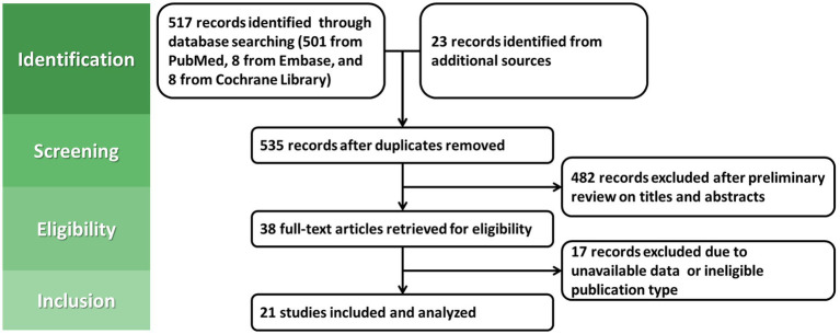
The flow diagram of study selection is presented in Figure 1. Finally, 21 studies were included for the meta-analysis, including 3 RCTs (30–32) and 18 cohort studies (20, 21, 33–48). The brief characteristics of the included studies are summarized in Supplementary Table S1. Due to the lack of relevant research in European and American countries, all the included studies were from Japan and Korea. All the included studies were of high or moderate quality (Supplementary Table S2, S3). Among the 21 studies, MGLs developed in 1,233 out of 38,931 eradicated patients and 2,982 out of 41,969 persistent patients, compared with 92 out of 1,356 negative patients. Additionally, among those with dysplasia only receiving post-ER eradication therapy, 917 out of 34,494 eradicated patients and 2,469 out of 36,059 persistent patients found MGLs.
Figure 1.
The PRISMA flow diagram of the meta-analysis.
Helicobacter pylori infection and MGL risk
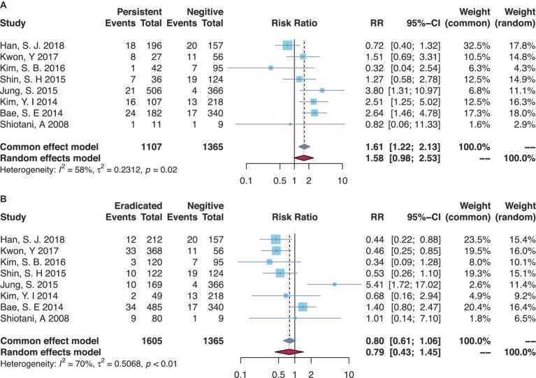
There were eight studies that compared the MGL risk between H. pylori-persistent and negative statuses, while the risk trended to be increased in the persistent group but not significantly yet (RR = 1.58, 95% CI = 0.98–2.53, I-square = 58%) (Table 1, Figure 2A). Subgroup analyses are shown in Table 1 and Supplementary Figures S1–S8. The majority of Korean studies obtained consistent results (RR = 1.61, 95% CI = 0.99–2.61, I-square = 63%). In the primary disease subsets of EGC combining dysplasia or not, higher risks of MGLs were found in the persistent group than those in the negative group (RR = 3.80, 95% CI = 1.31–10.97). Similarly, in the MGL outcome subsets of MGC combining dysplasia or not, the persistent group had a higher risk of MGL events (RR = 1.93, 95% CI = 1.17–3.17). Besides, the secondary analyses by comparing the eradicated group to the negative group demonstrated generally comparable risks of post-ER MGLs (Table 1, Figure 2B).
Table 1.
Meta-analyses on associations between H. pylori infection and MGL risk.
| Subsets | Persistent vs. negative | Eradicated vs. negative | ||||
|---|---|---|---|---|---|---|
| Study count | RR (95% CI) | p-value | Study count | RR (95% CI) | p-value | |
| Total | 8 | 1.58 (0.98–2.53) a | 0.06 | 8 | 0.79 (0.43–1.45) a | 0.44 |
| Study design | ||||||
| RCT only | 0 | N/A | N/A | 0 | N/A | N/A |
| Cohort only | 8 | 1.58 (0.98–2.53) a | 0.06 | 8 | 0.79 (0.43–1.45) a | 0.21 |
| Country | ||||||
| Japan | 1 | 0.82 (0.06–11.33) | 0.88 | 1 | 1.01 (0.14–7.10) | 0.99 |
| Korea | 7 | 1.61 (0.99–2.61) a | 0.06 | 7 | 0.78 (0.40–1.50) a | 0.45 |
| Primary disease | ||||||
| EGC or dysplasia | 1 | 3.80 (1.31–10.97) | 0.01 | 1 | 5.41 (1.72–17.01) | <0.01 |
| EGC only | 6 | 1.42 (0.79–2.57) a | 0.24 | 6 | 0.64 (0.37–1.10) a | 0.10 |
| Dysplasia only | 1 | 1.27 (0.58–2.78) | 0.55 | 1 | 0.53 (0.26–1.10) | 0.09 |
| MGL outcome | ||||||
| MGC or dysplasia | 3 | 1.93 (1.17–3.17) | 0.01 | 3 | 1.03 (0.23–4.61) a | 0.97 |
| MGC only | 5 | 1.35 (0.63–2.92) a | 0.44 | 5 | 0.71 (0.37–1.35) a | 0.29 |
| Dysplasia only | 0 | N/A | N/A | 0 | N/A | N/A |
CI, confidence interval; EGC, early gastric cancer; MGC, metachronous gastric cancer; MGL, metachronous gastric lesion; N/A, not applicable; RCT, randomized controlled trial; RR, risk ratio.
Random-effects model was used due to heterogeneity.
Figure 2.
Forest plots of overall comparisons (A) between H. pylori-persistent and negative patients and (B) between eradicated and negative patients.
Preventive effectiveness of post-ER eradication
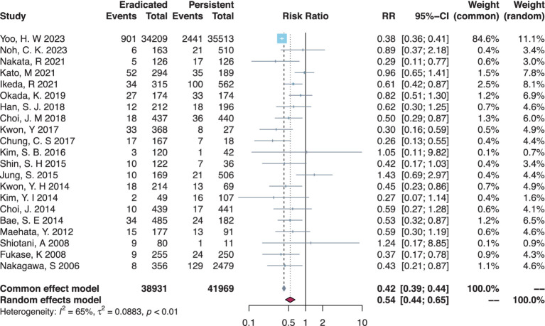
There were 21 studies that compared the MGL risk between H. pylori-eradicated and persistent statuses, and post-ER eradication had a significant protective effect by lowering the MGL risk (RR = 0.54, 95% CI = 0.44–0.65, I-square = 65%) (Table 2, Figure 3). In subgroup analysis (Table 2, Supplementary Figures S9–S12), meta-analyses on RCTs or cohort studies did not differ in results. Either Japanese or Korean studies obtained consistent results, respectively. In the primary disease subset of either EGC only or dysplasia only, similar protective effects against MGLs were found in the eradicated group compared to the persistent group. Additionally, in the MGL outcome subsets of MGC combining dysplasia or not, the eradicated group had a significantly lower risk of MGLs.
Table 2.
Meta-analyses on the preventive effectiveness of post-ER H. pylori eradication.
| Subsets | Eradicated vs. Persistent | ||
|---|---|---|---|
| Study count | RR (95% CI) | p-value | |
| Total | 21 | 0.54 (0.44–0.65) a | <0.01 |
| Study design | |||
| RCT only | 3 | 0.48 (0.33–0.70) | <0.01 |
| Cohort only | 18 | 0.55 (0.44–0.68) a | <0.01 |
| Country | |||
| Japan | 8 | 0.63 (0.52–0.77) | <0.01 |
| Korea | 13 | 0.40 (0.37–0.42) | <0.01 |
| Primary disease | |||
| EGC or dysplasia | 3 | 0.73 (0.39–1.38) a | 0.33 |
| EGC only | 15 | 0.57 (0.49–0.68) | <0.01 |
| Dysplasia only | 3 | 0.39 (0.36–0.42) | <0.01 |
| MGL outcome | |||
| MGC or dysplasia | 8 | 0.49 (0.33–0.70) a | <0.01 |
| MGC only | 13 | 0.60 (0.51–0.71) | <0.01 |
| Dysplasia only | 0 | N/A | N/A |
CI, confidence interval; EGC, early gastric cancer; MGC, metachronous gastric cancer; MGL, metachronous gastric lesion; N/A, not applicable; RCT, randomized controlled trial; RR, risk ratio.
Random-effects model was used due to heterogeneity.
Figure 3.
Forest plot of the overall comparison between H. pylori-eradicated and persistent patients.
Sensitivity analysis
Through leave-one-out analyses, consistent results were found in all the three principal meta-analyses, including the comparisons between H. pylori-persistent and negative groups, eradicated and negative groups, and eradicated and persistent groups (data not shown).
Publication bias
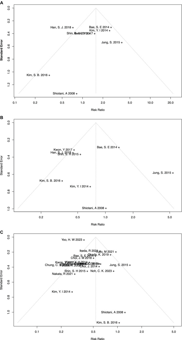
In the meta-analysis of H. pylori-persistent and negative groups, no significant publication bias was found using the funnel plot (Figure 4A) or Egger’s test (p = 0.63). Similarly, no significant publication bias was found in the meta-analysis of eradicated and negative groups using the funnel plot (Figure 4B) or Egger’s test (p = 0.78).
Figure 4.
Funnel plots of meta-analyses on overall comparisons (A) between persistent and negative patients, (B) between eradicated and negative patients, and (C) between eradicated and persistent patients.
In particular, the meta-analysis of eradicated and persistent groups included more than 10 studies, and therefore the AS-Thompson test was used to test the potential sources of high heterogeneity. No significant publication bias was tested (p = 0.24), along with the funnel plot (Figure 4C). Additionally, the Duval and Tweedie trim-and-fill method and contour-enhanced meta-analysis funnel plots were also applied to visually depict publication bias. By using the Duval and Tweedie trim-and-fill method, seven more studies were included and all the added studies were located in the p < 0.05 area of the contour-enhanced meta-analysis funnel plots, namely, null publication bias (Supplementary Figure S13). Moreover, the Duval and Tweedie trim-and-fill method was also used as part of sensitivity analysis, which suggested consistent and reliable results (RR = 0.41, 95% CI = 0.32–0.53, I-square = 75%). Finally, the Baujat plot indicated that the study by Yoo et al. had the greatest impact on the overall result (33), and the study by Kato et al. had the greatest impact on heterogeneity (35).
Discussion
This system review and meta-analysis analyzed 21 studies with 82,256 observations. H. pylori-persistent group trended to have a higher risk of MGLs compared to those never infected, but not in the post-ER eradicated group. Moreover, successful post-ER eradication could decrease the risk of MGLs compared to those persistently infected. Sensitivity analysis obtained generally consistent results, and no significant publication bias was found. However, heterogeneity commonly existed among meta-analyses, and a random-effects model was used accordingly. In the bibliographic view, the present report might be a most updated meta-analysis.
Gastric cancer is still one of the top malignancies in China, with a heavy burden of high population incidence (19, 49). H. pylori infection is one of the most important risk factors for gastric cancer in East Asia, particularly in China (50, 51). A multicenter prospective cohort study including 512,715 Chinese indicates that H. pylori infection accounted for 78.5% of non-cardia and 62.1% of cardia cancers, and up to 339,955 incident gastric cancers could be attributable to H. pylori infection (52). Due to consistent advocacy, health education, improved sanitary condition, and drinking water quality in China, the H. pylori infection rate among Chinese has slowly declined over the past 30 years, especially among the urban health check-up population (50, 53). Active H. pylori eradication within organized massive screening might simultaneously lower the incidence and improve population survival of gastric cancer (54).
Currently, a low proportion of EGC, usually no more than 20%, is the principal difficulty in improving the population survival of gastric cancer in China (55). In contrast, the proportions of EGC are fairly high in both Japan and Korea due to national organized massive screening for gastric cancer. Therefore, ER as a minimally invasive procedure is well-practiced for the early treatment of EGC, with a negligible risk of lymph node metastasis among high-selected candidates. According to Japanese Gastric Cancer Treatment Guidelines 2021 (6th edition), the EMR or ESD absolute indications for EGC include a differentiated-type adenocarcinoma without ulcerative findings in which the depth of invasion is clinically diagnosed as T1a and the maximal size is ≤ 2 cm (56). The experiences of post-ER management in Japan and Korea should be much substantial, and thus the present meta-analysis pooled studies just available from Japan and Korea.
At the 1-year or later endoscopic follow-up after ER or gastrectomy for EGC, the MGC has been more prone to occur after ER instead of gastrectomy (57). It may be due to the fact that the stomach can be preserved after ER, while most of the stomach is removed postgastrectomy with a limited area of mucosa left. Meanwhile, some studies have shown that conventional mucosal resection has a higher incidence of post-ER MGC than expanded mucosal resection (58). However, providing repeated ER treatment, there is no significant difference in long-term survival outcomes between the expanded and conventional groups (58). Besides, the other potential risk factors for MGC after ER for EGC included male, older age, severe or corpus intestinal metaplasia, family history of gastric cancer, synchronous adenoma, and H. pylori infection (59–61). It is the rationale why the present meta-analysis hypothesizes H. pylori infection could increase MGC risk and post-ER eradication might benefit in preventing MGC.
Regarding H. pylori infection, some consider eradication is no longer meaningful since gastric cancer has already developed, but investigation has shown that after eradication, the richness and uniformity of the gastric microbiota could be restored to a state similar to that of negative subjects. From bench to bedside, our updated findings evidence the capacity of reducing MGL incidence by post-ER eradication with plausible reporting quality, consistent with previous studies (62–64). Some studies suggested that the effectiveness of eradication could be observed by decreasing MGC incidence after the 6th post-ER year, but not at 3-year follow-up (31). Specifically, adequate length of endoscopic surveillance must be quite important in common practice, and diverse follow-up situations might introduce the source of heterogeneity in the present meta-analysis. Additionally, the time interval between the first ER and H. pylori eradication needs to be taken into account. As reported in the study by Kato et al. (35), all the patients were required to undergo H. pylori eradication within no more than 1 year after ER. Moreover, the regimens of H. pylori eradication are unspecific to post-ER patients. Actually, the risk factors of MGLs and the best follow-up strategy are still underestimated up to the known knowledge.
Limitation
Some limitations need attention yet. First, all the included studies came from Japan and Korea, and the extrapolation of results kept unclear to other high-risk areas. Second, although the quality of the included cohort studies was high or moderate, only three available RCTs indicated that selection and performance biases were inevitable. Third, in many studies, follow-up work was not long enough and without regular endoscopic surveillance, which could introduce between-study heterogeneity. Fourth, the time interval between H. pylori eradication and the first ER had not been detailed in several studies, and thus the prolonged infection status might potentially influence the preventive effectiveness. Finally, other confounders for gastric cancer risk such as infection-attributable oncoviruses, family history of gastric cancer, and ethnicity had not been considered in the present investigation (65–68).
Conclusion
In summary, the present meta-analysis can further evidence that the H. pylori infection might potentially increase the post-ER incidence of MGC or dysplasia, but post-ER eradication can decrease the risk of MGC or dysplasia. Specifically, post-ER screening and eradication of Helicobacter pylori have preventive effectiveness on MGC, and the protocol should be recommended to all the post-ER patients. Moreover, larger-scale RCTs with longer follow-up duration are still warranted to define the best cost-effective interval and length for endoscopic surveillance among H. pylori-infected individuals after ER.
Data availability statement
The original contributions presented in the study are included in the article/Supplementary material, further inquiries can be directed to the corresponding author.
Author contributions
T-HY: Conceptualization, Data curation, Formal analysis, Investigation, Methodology, Software, Writing – original draft, Writing – review & editing. DB: Data curation, Investigation, Methodology, Writing – review & editing. KL: Data curation, Methodology, Writing – review & editing. W-HZ: Data curation, Investigation, Methodology, Supervision, Writing – review & editing. X-ZC: Conceptualization, Data curation, Funding acquisition, Investigation, Methodology, Project administration, Resources, Software, Supervision, Validation, Writing – original draft, Writing – review & editing. J-KH: Funding acquisition, Resources, Supervision, Writing – review & editing.
Acknowledgments
This study was a part of series of studies from the Sichuan Gastric Cancer Early Detection and Screening (SIGES) project. The authors thank the substantial work of the Volunteer Team of Gastric Cancer Surgery (VOLTGA), West China Hospital, Sichuan University, China. Additionally, Yibin Cancer Prevention and Control Center, Second People’s Hospital of Yibin—West China Yibin Hospital, Sichuan University, Yibin, China, supported the SIGES research project.
Funding Statement
The author(s) declare financial support was received for the research, authorship, and/or publication of this article. Foundation of Science and Technology Department of Sichuan Province, China (No. 23ZDYF0839); The 1.3.5 Project for Disciplines of Excellence, West China Hospital, Sichuan University, China (No. ZY2017304).
Abbreviations
CI, Confidence interval; EGC, Early gastric cancer; EMR, Endoscopic mucosal resection; ER, Endoscopic resection; ESD, Endoscopic submucosal dissection; H. pylori, Helicobacter pylori; MGC, Metachronous gastric cancer; MGL, Metachronous gastric lesion; MGLs, Metachronous gastric lesions; MOOSE, Meta-analysis Of Observational Studies in Epidemiology; PRISMA, Preferred Reporting Items for Systematic Reviews and Meta-Analysis; RCTs, Randomized controlled trials; RR, Risk ratio.
Conflict of interest
The authors declare that the research was conducted in the absence of any commercial or financial relationships that could be construed as a potential conflict of interest.
Publisher’s note
All claims expressed in this article are solely those of the authors and do not necessarily represent those of their affiliated organizations, or those of the publisher, the editors and the reviewers. Any product that may be evaluated in this article, or claim that may be made by its manufacturer, is not guaranteed or endorsed by the publisher.
Supplementary material
The Supplementary material for this article can be found online at: https://www.frontiersin.org/articles/10.3389/fmed.2024.1393498/full#supplementary-material
References
- 1.Smyth EC, Nilsson M, Grabsch HI, van Grieken NCT, Lordick F. Gastric cancer. Lancet. (2020) 396:635–48. doi: 10.1016/S0140-6736(20)31288-5 [DOI] [PubMed] [Google Scholar]
- 2.Sung H, Ferlay J, Siegel RL, Laversanne M, Soerjomataram I, Jemal A, et al. Global Cancer Statistics 2020: GLOBOCAN estimates of incidence and mortality worldwide for 36 cancers in 185 countries. CA Cancer J Clin. (2021) 71:209–49. doi: 10.3322/caac.21660, PMID: [DOI] [PubMed] [Google Scholar]
- 3.Li Y, Choi H, Leung K, Jiang F, Graham DY, Leung WK. Global prevalence of Helicobacter pylori infection between 1980 and 2022: a systematic review and meta-analysis. Lancet Gastroenterol Hepatol. (2023) 8:553–64. doi: 10.1016/S2468-1253(23)00070-5, PMID: [DOI] [PubMed] [Google Scholar]
- 4.Arnold M, Abnet CC, Neale RE, Vignat J, Giovannucci EL, McGlynn KA, et al. Global burden of 5 major types of gastrointestinal Cancer. Gastroenterology. (2020) 159:335–49.e15. doi: 10.1053/j.gastro.2020.02.068, PMID: [DOI] [PMC free article] [PubMed] [Google Scholar]
- 5.Bray F, Ferlay J, Laversanne M, Brewster DH, Gombe Mbalawa C, Kohler B, et al. Cancer incidence in five continents: inclusion criteria, highlights from volume X and the global status of cancer registration. Int J Cancer. (2015) 137:2060–71. doi: 10.1002/ijc.29670, PMID: [DOI] [PubMed] [Google Scholar]
- 6.Chen XZ, Schöttker B, Castro FA, Chen H, Zhang Y, Holleczek B, et al. Association of helicobacter pylori infection and chronic atrophic gastritis with risk of colonic, pancreatic and gastric cancer: a ten-year follow-up of the ESTHER cohort study. Oncotarget. (2016) 7:17182–93. doi: 10.18632/oncotarget.7946, PMID: [DOI] [PMC free article] [PubMed] [Google Scholar]
- 7.Wang R, Zhang MG, Chen XZ, Wu H. Risk population of Helicobacter pylori infection among Han and Tibetan ethnicities in western China: a cross-sectional, longitudinal epidemiological study. Lancet. (2016) 388:S17. doi: 10.1016/S0140-6736(16)31944-4 [DOI] [Google Scholar]
- 8.Japanese Gastric Cancer Association . Japanese gastric cancer treatment guidelines 2018 (5th edition). Gastric Cancer. (2020) 24:1–21. doi: 10.1007/s10120-020-01042-y [DOI] [PMC free article] [PubMed] [Google Scholar]
- 9.Chen XZ, Hu JK, Zhou ZG, Rui YY, Yang K, Wang L, et al. Meta-analysis of effectiveness and safety of D2 plus Para-aortic lymphadenectomy for resectable gastric cancer. J Am Coll Surg. (2010) 210:100–5. doi: 10.1016/j.jamcollsurg.2009.09.033, PMID: [DOI] [PubMed] [Google Scholar]
- 10.Zhang WH, Chen XZ, Liu K, Chen XL, Yang K, Zhang B, et al. Outcomes of surgical treatment for gastric cancer patients: 11-year experience of a Chinese high-volume hospital. Med Oncol. (2014) 31:150. doi: 10.1007/s12032-014-0150-1, PMID: [DOI] [PubMed] [Google Scholar]
- 11.Gotoda T, Ono H. Stomach: endoscopic resection for early gastric cancer. Dig Endosc. (2022) 34:58–60. doi: 10.1111/den.14167 [DOI] [PubMed] [Google Scholar]
- 12.Suzuki H, Ono H, Hirasawa T, Takeuchi Y, Ishido K, Hoteya S, et al. Long-term survival after endoscopic resection for gastric Cancer: real-world evidence from a multicenter prospective cohort. Clin Gastroenterol Hepatol. (2023) 21:307–18.e2. doi: 10.1016/j.cgh.2022.07.029 [DOI] [PubMed] [Google Scholar]
- 13.Ono H, Yao K, Fujishiro M, Oda I, Uedo N, Nimura S, et al. Guidelines for endoscopic submucosal dissection and endoscopic mucosal resection for early gastric cancer (second edition). Dig Endosc. (2021) 33:4–20. doi: 10.1111/den.13883, PMID: [DOI] [PubMed] [Google Scholar]
- 14.Choi KS, Jung HY, Choi KD, Lee GH, Song HJ, Kim DH, et al. EMR versus gastrectomy for intramucosal gastric cancer: comparison of long-term outcomes. Gastrointest Endosc. (2011) 73:942–8. doi: 10.1016/j.gie.2010.12.032, PMID: [DOI] [PubMed] [Google Scholar]
- 15.Correa P, Haenszel W, Cuello C, Tannenbaum S, Archer M. A model for gastric cancer epidemiology. Lancet. (1975) 306:58–60. doi: 10.1016/S0140-6736(75)90498-5 [DOI] [PubMed] [Google Scholar]
- 16.Marshall BJ, Warren JR. Unidentified curved bacilli in the stomach of patients with gastritis and peptic ulceration. Lancet. (1984) 1:1311–5. PMID: [DOI] [PubMed] [Google Scholar]
- 17.Correa P. Human gastric carcinogenesis: a multistep and multifactorial process--first American Cancer Society award lecture on cancer epidemiology and prevention. Cancer Res. (1992) 52:6735–40. PMID: [PubMed] [Google Scholar]
- 18.Zavros Y, Merchant JL. The immune microenvironment in gastric adenocarcinoma. Nat Rev Gastroenterol Hepatol. (2022) 19:451–67. doi: 10.1038/s41575-022-00591-0, PMID: [DOI] [PMC free article] [PubMed] [Google Scholar]
- 19.Wang R, Chen XZ. High mortality from hepatic, gastric and esophageal cancers in mainland China: 40 years of experience and development. Clin Res Hepatol Gastroenterol. (2014) 38:751–6. doi: 10.1016/j.clinre.2014.04.014, PMID: [DOI] [PubMed] [Google Scholar]
- 20.Kim SB, Lee SH, Bae SI, Jeong YH, Sohn SH, Kim KO, et al. Association between Helicobacter pylori status and metachronous gastric cancer after endoscopic resection. World J Gastroenterol. (2016) 22:9794–802. doi: 10.3748/wjg.v22.i44.9794, PMID: [DOI] [PMC free article] [PubMed] [Google Scholar]
- 21.Noh CK, Lee E, Park B, Lim SG, Shin SJ, Lee KM, et al. Effect of Helicobacter pylori eradication treatment on metachronous gastric neoplasm prevention following endoscopic submucosal dissection for gastric adenoma. J Clin Med. (2023) 12:1512. doi: 10.3390/jcm12041512, PMID: [DOI] [PMC free article] [PubMed] [Google Scholar]
- 22.Jüni P, Altman DG, Egger M. Systematic reviews in health care: assessing the quality of controlled clinical trials. BMJ. (2001) 323:42–6. doi: 10.1136/bmj.323.7303.42, PMID: [DOI] [PMC free article] [PubMed] [Google Scholar]
- 23.Kim SY, Park JE, Lee YJ, Seo HJ, Sheen SS, Hahn S, et al. Testing a tool for assessing the risk of bias for nonrandomized studies showed moderate reliability and promising validity. J Clin Epidemiol. (2013) 66:408–14. doi: 10.1016/j.jclinepi.2012.09.016, PMID: [DOI] [PubMed] [Google Scholar]
- 24.Bai D, Liu K, Wang R, Zhang WH, Chen XZ, Hu JK. Prevalence difference of Helicobacter pylori infection between Tibetan and Han ethnics in China: a meta-analysis on epidemiologic studies (SIGES). Asia Pac J Public Health. (2023) 35:103–11. doi: 10.1177/10105395221134651, PMID: [DOI] [PubMed] [Google Scholar]
- 25.Sterne JA, Sutton AJ, Ioannidis JP, Terrin N, Jones DR, Lau J, et al. Recommendations for examining and interpreting funnel plot asymmetry in meta-analyses of randomised controlled trials. BMJ. (2011) 343:d4002. doi: 10.1136/bmj.d4002, PMID: [DOI] [PubMed] [Google Scholar]
- 26.Peters JL, Sutton AJ, Jones DR, Abrams KR, Rushton L. Contour-enhanced meta-analysis funnel plots help distinguish publication bias from other causes of asymmetry. J Clin Epidemiol. (2008) 61:991–6. doi: 10.1016/j.jclinepi.2007.11.010, PMID: [DOI] [PubMed] [Google Scholar]
- 27.Stroup DF, Berlin JA, Morton SC, Olkin I, Williamson GD, Rennie D, et al. Meta-analysis of observational studies in epidemiology: a proposal for reporting. Meta-analysis of observational studies in epidemiology (MOOSE) group. JAMA. (2000) 283:2008–12. doi: 10.1001/jama.283.15.2008, PMID: [DOI] [PubMed] [Google Scholar]
- 28.McInnes MDF, Moher D, Thombs BD, McGrath TA, Bossuyt PM, and the PRISMA-DTA Group et al. Preferred reporting items for a systematic review and Meta-analysis of diagnostic test accuracy studies: the PRISMA-DTA statement. JAMA. (2018) 319:388–96. doi: 10.1001/jama.2017.19163 [DOI] [PubMed] [Google Scholar]
- 29.Yu TH, Chen XZ. Helicobacter pylori eradication following endoscopic resection and metachronous gastric cancer: a systematic review and meta-analysis. PROSPERO (CRD42024512101) (2024). Available at: https://www.crd.york.ac.uk/prospero/display_record.php?ID=CRD42024512101
- 30.Choi JM, Kim SG, Choi J, Park JY, Oh S, Yang HJ, et al. Effects of Helicobacter pylori eradication for metachronous gastric cancer prevention: a randomized controlled trial. Gastrointest Endosc. (2018) 88:475–85.e2. doi: 10.1016/j.gie.2018.05.009 [DOI] [PubMed] [Google Scholar]
- 31.Choi J, Kim SG, Yoon H, Im JP, Kim JS, Kim WH, et al. Eradication of Helicobacter pylori after endoscopic resection of gastric tumors does not reduce incidence of metachronous gastric carcinoma. Clin Gastroenterol Hepatol. (2014) 12:793–800.e1. doi: 10.1016/j.cgh.2013.09.057, PMID: [DOI] [PubMed] [Google Scholar]
- 32.Fukase K, Kato M, Kikuchi S, Inoue K, Uemura N, Okamoto S, et al. Effect of eradication of Helicobacter pylori on incidence of metachronous gastric carcinoma after endoscopic resection of early gastric cancer: an open-label, randomised controlled trial. Lancet. (2008) 372:392–7. doi: 10.1016/S0140-6736(08)61159-9, PMID: [DOI] [PubMed] [Google Scholar]
- 33.Yoo HW, Hong SJ, Kim SH. Helicobacter pylori treatment and gastric cancer risk after endoscopic resection of dysplasia: a Nationwide Cohort Study. Gastroenterology. (2024) 166:313–22.e3. doi: 10.1053/j.gastro.2023.10.013, PMID: [DOI] [PubMed] [Google Scholar]
- 34.Nakata R, Nagami Y, Hashimoto A, Sakai T, Ominami M, Fukunaga S, et al. Successful eradication of Helicobacter pylori could prevent metachronous gastric cancer: a propensity matching analysis. Digestion. (2021) 102:236–45. doi: 10.1159/000504132, PMID: [DOI] [PubMed] [Google Scholar]
- 35.Kato M, Hayashi Y, Nishida T, Oshita M, Nakanishi F, Yamaguchi S, et al. Helicobacter pylori eradication prevents secondary gastric cancer in patients with mild-to-moderate atrophic gastritis. J Gastroenterol Hepatol. (2021) 36:2083–90. doi: 10.1111/jgh.15396, PMID: [DOI] [PubMed] [Google Scholar]
- 36.Ikeda R, Hirasawa K, Sato C, Sawada A, Nishio M, Fukuchi T, et al. Incidence of metachronous gastric cancer after endoscopic submucosal dissection associated with eradication status of Helicobacter pylori. Eur J Gastroenterol Hepatol. (2021) 33:17–24. doi: 10.1097/MEG.0000000000001788, PMID: [DOI] [PubMed] [Google Scholar]
- 37.Okada K, Suzuki S, Naito S, Yamada Y, Haruki S, Kubota M, et al. Incidence of metachronous gastric cancer in patients whose primary gastric neoplasms were discovered after Helicobacter pylori eradication. Gastrointest Endosc. (2019) 89:1152–9.e1. doi: 10.1016/j.gie.2019.02.026, PMID: [DOI] [PubMed] [Google Scholar]
- 38.Han SJ, Kim SG, Lim JH, Choi JM, Oh S, Park JY, et al. Long-term effects of Helicobacter pylori eradication on metachronous gastric cancer development. Gut Liver. (2018) 12:133–41. doi: 10.5009/gnl17073, PMID: [DOI] [PMC free article] [PubMed] [Google Scholar]
- 39.Kwon Y, Jeon S, Nam S, Shin I. Helicobacter pylori infection and serum level of pepsinogen are associated with the risk of metachronous gastric neoplasm after endoscopic resection. Aliment Pharmacol Ther. (2017) 46:758–67. doi: 10.1111/apt.14263 [DOI] [PubMed] [Google Scholar]
- 40.Chung CS, Woo HS, Chung JW, Jeong SH, Kwon KA, Kim YJ, et al. Risk factors for metachronous recurrence after endoscopic submucosal dissection of early gastric cancer. J Korean Med Sci. (2017) 32:421–6. doi: 10.3346/jkms.2017.32.3.421, PMID: [DOI] [PMC free article] [PubMed] [Google Scholar]
- 41.Shin SH, Jung DH, Kim JH, Chung HS, Park JC, Shin SK, et al. Helicobacter pylori eradication prevents metachronous gastric neoplasms after endoscopic resection of gastric dysplasia. PLoS One. (2015) 10:e0143257. doi: 10.1371/journal.pone.0143257, PMID: [DOI] [PMC free article] [PubMed] [Google Scholar]
- 42.Jung S, Park CH, Kim EH, Shin SJ, Chung H, Lee H, et al. Preventing metachronous gastric lesions after endoscopic submucosal dissection through Helicobacter pylori eradication. J Gastroenterol Hepatol. (2015) 30:75–81. doi: 10.1111/jgh.12687, PMID: [DOI] [PubMed] [Google Scholar]
- 43.Kwon YH, Heo J, Lee HS, Cho CM, Jeon SW. Failure of Helicobacter pylori eradication and age are independent risk factors for recurrent neoplasia after endoscopic resection of early gastric cancer in 283 patients. Aliment Pharmacol Ther. (2014) 39:609–18. doi: 10.1111/apt.12633 [DOI] [PubMed] [Google Scholar]
- 44.Kim YI, Choi IJ, Kook MC, Cho SJ, Lee JY, Kim CG, et al. The association between Helicobacter pylori status and incidence of metachronous gastric cancer after endoscopic resection of early gastric cancer. Helicobacter. (2014) 19:194–201. doi: 10.1111/hel.12116, PMID: [DOI] [PubMed] [Google Scholar]
- 45.Bae SE, Jung HY, Kang J, Park YS, Baek S, Jung JH, et al. Effect of Helicobacter pylori eradication on metachronous recurrence after endoscopic resection of gastric neoplasm. Am J Gastroenterol. (2014) 109:60–7. doi: 10.1038/ajg.2013.404, PMID: [DOI] [PubMed] [Google Scholar]
- 46.Maehata Y, Nakamura S, Fujisawa K, Esaki M, Moriyama T, Asano K, et al. Long-term effect of Helicobacter pylori eradication on the development of metachronous gastric cancer after endoscopic resection of early gastric cancer. Gastrointest Endosc. (2012) 75:39–46. doi: 10.1016/j.gie.2011.08.030, PMID: [DOI] [PubMed] [Google Scholar]
- 47.Shiotani A, Uedo N, Iishi H, Yoshiyuki Y, Ishii M, Manabe N, et al. Predictive factors for metachronous gastric cancer in high-risk patients after successful Helicobacter pylori eradication. Digestion. (2008) 78:113–9. doi: 10.1159/000173719, PMID: [DOI] [PubMed] [Google Scholar]
- 48.Nakagawa S, Asaka M, Kato M, Nakamura T, Kato C, Fujioka T, et al. Helicobacter pylori eradication and metachronous gastric cancer after endoscopic mucosal resection of early gastric cancer. Aliment Pharmacol Ther. (2006) 24:214–8. doi: 10.1111/j.1365-2036.2006.00048.x [DOI] [Google Scholar]
- 49.Chen XZ, Liu Y, Wang R, Zhang WH, Hu JK. Improvement of cancer control in mainland China: epidemiological profiles during the 2004–10 National Cancer Prevention and Control Program. Lancet. (2016) 388:S40. doi: 10.1016/S0140-6736(16)31967-5 [DOI] [Google Scholar]
- 50.Ding SZ, Du YQ, Lu H, Wang WH, Cheng H, Chen SY, et al. Chinese consensus report on family-based Helicobacter pylori infection control and management (2021 Edition). Gut. (2022) 71:238–53. doi: 10.1136/gutjnl-2021-325630, PMID: [DOI] [PMC free article] [PubMed] [Google Scholar]
- 51.Huang J, Lucero-Prisno DE, 3rd, Zhang L, Xu W, Wong SH, Ng SC, et al. Updated epidemiology of gastrointestinal cancers in East Asia. Nat Rev Gastroenterol Hepatol. (2023) 20:271–87. doi: 10.1038/s41575-022-00726-3, PMID: [DOI] [PubMed] [Google Scholar]
- 52.Yang L, Kartsonaki C, Yao P, de Martel C, Plummer M, Chapman D, et al. The relative and attributable risks of cardia and non-cardia gastric cancer associated with Helicobacter pylori infection in China: a case-cohort study. Lancet Public Health. (2021) 6:e888–96. doi: 10.1016/S2468-2667(21)00164-X, PMID: [DOI] [PMC free article] [PubMed] [Google Scholar]
- 53.Zou JC, Wen MY, Huang Y, Chen XZ, Hu JK. Helicobacter pylori infection prevalence declined among an urban health check-up population in Chengdu, China: a longitudinal analysis of multiple cross-sectional studies. Front Public Health. (2023) 11:1128765. doi: 10.3389/fpubh.2023.1128765, PMID: [DOI] [PMC free article] [PubMed] [Google Scholar]
- 54.Zou JC, Yang Y, Chen XZ, Sichuan Gastric Cancer Early Detection and Screening Research Group . Active eradication of within organized massive screening might improve survival of gastric cancer patients. Gastroenterology. (2023) 164:162–3. doi: 10.1053/j.gastro.2022.05.009, PMID: [DOI] [PubMed] [Google Scholar]
- 55.Chen XZ, Zhang WH, Hu JK. A difficulty in improving population survival outcome of gastric cancer in mainland China: low proportion of early diseases. Med Oncol. (2014) 31:315. doi: 10.1007/s12032-014-0315-y, PMID: [DOI] [PubMed] [Google Scholar]
- 56.Japanese Gastric Cancer Association . Japanese gastric cancer treatment guidelines 2021 (6th edition). Gastric Cancer. (2023) 26:1–25. doi: 10.1007/s10120-022-01331-8, PMID: [DOI] [PMC free article] [PubMed] [Google Scholar]
- 57.Ortigão R, Figueirôa G, Frazzoni L, Pimentel-Nunes P, Hassan C, Dinis-Ribeiro M, et al. Risk factors for gastric metachronous lesions after endoscopic or surgical resection: a systematic review and meta-analysis. Endoscopy. (2022) 54:892–901. doi: 10.1055/a-1724-7378, PMID: [DOI] [PubMed] [Google Scholar]
- 58.Probst A, Schneider A, Schaller T, Anthuber M, Ebigbo A, Messmann H. Endoscopic submucosal dissection for early gastric cancer: are expanded resection criteria safe for Western patients? Endoscopy. (2017) 49:855–65. doi: 10.1055/s-0043-110672 [DOI] [PubMed] [Google Scholar]
- 59.Lee E, Kim SG, Kim B, Kim JL, Kim J, Chung H, et al. Metachronous gastric neoplasm beyond 5 years after endoscopic resection for early gastric cancer. Surg Endosc. (2023) 37:3901–10. doi: 10.1007/s00464-023-09889-9, PMID: [DOI] [PubMed] [Google Scholar]
- 60.Rei A, Ortigão R, Pais M, Afonso LP, Pimentel-Nunes P, Dinis-Ribeiro M, et al. Metachronous lesions after gastric endoscopic submucosal dissection: first assessment of the FAMISH prediction score. Endoscopy. (2023) 55:909–17. doi: 10.1055/a-2089-6849, PMID: [DOI] [PubMed] [Google Scholar]
- 61.Na YS, Kim SG, Cho SJ. Risk assessment of metachronous gastric cancer development using OLGA and OLGIM systems after endoscopic submucosal dissection for early gastric cancer: a long-term follow-up study. Gastric Cancer. (2023) 26:298–306. doi: 10.1007/s10120-022-01361-2, PMID: [DOI] [PubMed] [Google Scholar]
- 62.Yoon SB, Park JM, Lim CH, Cho YK, Choi MG. Effect of Helicobacter pylori eradication on metachronous gastric cancer after endoscopic resection of gastric tumors: a meta-analysis. Helicobacter. (2014) 19:243–8. doi: 10.1111/hel.12146, PMID: [DOI] [PubMed] [Google Scholar]
- 63.Jung DH, Kim JH, Chung HS, Park JC, Shin SK, Lee SK, et al. Helicobacter pylori eradication on the prevention of metachronous lesions after endoscopic resection of gastric neoplasm: a meta-analysis. PLoS One. (2015) 10:e0124725. doi: 10.1371/journal.pone.0124725, PMID: [DOI] [PMC free article] [PubMed] [Google Scholar]
- 64.Xiao S, Li S, Zhou L, Jiang W, Liu J. Helicobacter pylori status and risks of metachronous recurrence after endoscopic resection of early gastric cancer: a systematic review and meta-analysis. J Gastroenterol. (2019) 54:226–37. doi: 10.1007/s00535-018-1513-8, PMID: [DOI] [PubMed] [Google Scholar]
- 65.Chen XZ, Chen H, Castro FA, Hu JK, Brenner H. Epstein-Barr virus infection and gastric cancer: a systematic review. Medicine. (2015) 94:e792. doi: 10.1097/MD.0000000000000792, PMID: [DOI] [PMC free article] [PubMed] [Google Scholar]
- 66.Wang H, Chen XL, Liu K, Bai D, Zhang WH, Chen XZ, et al. Associations between gastric cancer risk and virus infection other than Epstein-Barr virus: a systematic review and meta-analysis based on epidemiological studies. Clin Transl Gastroenterol. (2020) 11:e00201. doi: 10.14309/ctg.0000000000000201, PMID: [DOI] [PMC free article] [PubMed] [Google Scholar]
- 67.Wang R, Bai D, Xiang W, Chen XZ. Tibetan ethnicity, birthplace, Helicobacter pylori infection, and gastric cancer risk. Am J Gastroenterol. (2022) 117:1010. doi: 10.14309/ajg.0000000000001757, PMID: [DOI] [PubMed] [Google Scholar]
- 68.Yu TH, Wang R, Chen XZ. Multifactorial co-exposure within family of gastric cancer: increased risk of familial gastric cancer and tailored prevention. Med Oncol. (2022) 40:19. doi: 10.1007/s12032-022-01882-x, PMID: [DOI] [PubMed] [Google Scholar]
Associated Data
This section collects any data citations, data availability statements, or supplementary materials included in this article.
Supplementary Materials
Data Availability Statement
The original contributions presented in the study are included in the article/Supplementary material, further inquiries can be directed to the corresponding author.


