



Abstract
Restoring somatosensory feedback in individuals with lower-limb amputations would reduce the risk of falls and alleviate phantom limb pain. Here we show, in three individuals with transtibial amputation (one traumatic and two owing to diabetic peripheral neuropathy), that sensations from the missing foot, with control over their location and intensity, can be evoked via lateral lumbosacral spinal cord stimulation with commercially available electrodes and by modulating the intensity of stimulation in real time on the basis of signals from a wireless pressure-sensitive shoe insole. The restored somatosensation via closed-loop stimulation improved balance control (with a 19-point improvement in the composite score of the Sensory Organization Test in one individual) and gait stability (with a 5-point improvement in the Functional Gait Assessment in one individual). And over the implantation period of the stimulation leads, the three individuals experienced a clinically meaningful decrease in phantom limb pain (with an average reduction of nearly 70% on a visual analogue scale). Our findings support the further clinical assessment of lower-limb neuropr ostheses providing somatosensory feedback.
Every year, approximately 150,000 people in the United States undergo amputation of a lower limb1. Loss of a lower limb leads to chronic challenges including major mobility impairments and emergence of chronic pain that appears to emanate from the missing limb (that is, phantom limb pain, PLP). Current clinical practice involves prescribing a prosthetic limb to improve functional mobility, along with neuroleptic and opiate pharmaceuticals to treat PLP. Even with these interventions, people with lower-limb amputation exhibit a high rate of falls, a lack of confidence during gait, abnormal gait patterns and persistent PLP. All these problems have been associated with the disruption of somatosensory feedback from the missing limb. Tactile feedback from the sole of the foot is critical for maintaining balance and postural stability2, and the loss of somatosensory feedback after an amputation causes a sensorimotor mismatch between attempted movements and expected sensory feedback. This mismatch and the persistent absence of somatosensory input to the brain have been implicated in the development and maintenance of PLP3,4. One potential way to address the sequelae of lower-limb amputation is to restore somatosensation in the missing limb, thereby improving functional outcomes and reducing PLP.
Previous studies have demonstrated that electrical stimulation of peripheral nerves in the residual limb can evoke sensations in the missing hand or foot5-7. Tactile feedback via peripheral nerve stimulation has been shown to enhance control of the prosthesis and improve balance and gait8-12. In addition, anecdotal evidence suggests that chronic peripheral nerve stimulation reduces PLP8,13-15. To date, most studies to restore somatosensory feedback from the missing limb have relied on complex surgical techniques to implant devices inside or around peripheral nerves or to reroute those nerves to other regions of the body7,8,10,12,16,17. While these approaches clearly showed the promise of electrical stimulation, their surgical complexity remains a barrier to widespread clinical adoption. Evoking sensations via peripheral nerve stimulation may also be challenging in individuals with severe peripheral neuropathy, a common co-morbidity for people with amputations related to vascular disease and diabetes, which account for up to 82% of lower-limb amputations18. To our knowledge, no study to date has demonstrated restored somatosensation in the amputated foot for people with diabetic amputation.
In this Article, we aimed to address these challenges by leveraging spinal cord stimulation (SCS) as an alternative to peripheral nerve stimulation, to restore somatosensory feedback from the missing lower limb. SCS is an existing clinical technology that is implanted in as many as 50,000 people each year to treat chronic pain19. The surgical procedures involved in the implantation of these devices and the associated risks are well understood, and most major medical centres throughout developed countries have physicians that routinely perform SCS implants20. Recently, we have shown that cervical SCS can be used to restore somatosensation from the missing hand in people with upper-limb amputation21. Our goal in this study was to demonstrate that lumbosacral SCS could evoke sensations in the missing foot and that the restored somatosensory feedback could improve functional use of the prosthesis and reduce PLP. Importantly, we aimed to demonstrate that we could achieve these effects regardless of whether the amputation was traumatic or secondary to diabetic peripheral neuropathy, which substantially increases the pool of people that might benefit from these devices. To that end, we included participants both with and without sensory neuropathies in the residual and contralateral limb (Extended Data Fig. 1 and Supplementary Table 1).
In three people with below-knee amputation (Table 1), we implanted commercially available SCS leads in the thoracolumbar epidural space to stimulate the lateral lumbosacral spinal cord. Participants attended multiple testing sessions each week for the duration of the implantation period. Testing sessions typically lasted 4–6 h. During those sessions, we identified electrode contacts that evoked sensation experienced on the missing foot and performed psychophysical assessments to characterize those sensations. We developed a closed-loop system (Fig. 1 and Supplementary Video 1) where SCS was modulated by pressure signals wirelessly recorded from an insole in the shoe under the prosthetic limb. Using this system to deliver real-time somatosensory feedback, we assessed balance and gait, as well as changes in PLP over the duration of the multi-week implantation period. Our results indicate that lumbosacral SCS is a promising intervention to restore sensations, improve function and reduce PLP in lower-limb amputees.
Table 1 ∣.
Study participant demographics and amputation data
| Participant | Age (years) |
Gender | Ambulation level | Years since amputation |
Side of amputation |
Nature of amputation |
Time with prosthesis (months) |
Prosthesis wear time (h per day) |
Implant duration (days) |
|---|---|---|---|---|---|---|---|---|---|
| 1 | 56 | M | Limited community | 3.5 | Left | Diabetic | 42 | 8 | 28 |
| 2 | 56 | M | Active | 7 | Left | Traumatic | 84 | 8 | 28 |
| 3 | 65 | W | Limited community | 5 | Left | Diabetic | 52 | 20 | 84 |
Fig. 1 ∣. Schematic of the closed-loop SCS system used in this study.

Electrical stimulation was delivered to the spinal cord via two or three 8- or 16-contact leads implanted percutaneously near the lateral lumbosacral spinal cord. The leads were tunnelled through the skin and connected to an external stimulation system. A sensorized insole was inserted into the shoe to measure pressure under the prosthetic foot, the signals from this insole were used to modulate stimulation amplitude for SCS electrodes implanted in the lateral thoracolumbar epidural space, and the stimulation evoked sensations that appeared to emanate from the missing limb. The purple region of the leg and foot shows the location of evoked sensation in one participant, and the red dotted line represents the end of the residual limb for this participant with left trans-tibial amputation.
Results
SCS evokes sensations in the missing foot
The first goal of this study was to characterize the location and perceptual qualities of sensations evoked by lumbosacral SCS. To map the location of evoked sensations, we delivered 1-s-long stimulation trains and asked the participants to draw the location of the perceived sensations on a graphic representation of the foot and legs. For all three participants, SCS evoked sensations in the missing limb, including the toes and heel (Fig. 2a). The sensations were absent during the first 1–2 weeks of the study and gradually stabilized in the missing foot over the following weeks (Fig. 2c and Extended Data Fig. 2). During experiments to characterize the location of evoked sensations, the choice of stimulation electrodes and parameters was randomized, and participants were blinded to those choices. The sensations in the missing limb were always accompanied by sensations in the residual limb, and higher stimulation amplitudes were required to evoke sensations in the missing limb than in the residual limb alone (Extended Data Fig. 3). The rostral–caudal arrangement of the electrodes across different levels of the spinal cord elicited sensations that corresponded to the dermatomal distribution22 (Fig. 2b and Extended Data Fig. 4).
Fig. 2 ∣. SCS evokes percepts in the missing limb.
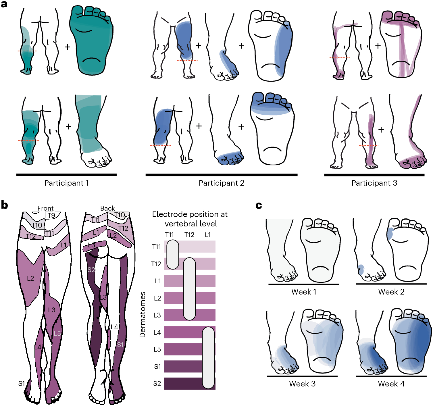
a, Examples of percepts evoked in the missing and residual limbs from one session for each participant. Two different sensations (corresponding to stimulation through two different electrodes) are shown for each participant (top and bottom). The red dashed line indicates the level of the amputation. The coloured area represents the location of the perceived sensation, with darker colours representing more frequent reports of sensation at that location across trials, normalized within each participant. b, Dermatome activation by electrodes located at different vertebral levels for participant 3. Left: expected dermatomal innervation in the leg, adapted from ref. 22. Right: horizontal bars indicate different dermatomes, and the white ovals indicate the approximate electrode position that evoked sensations in those dermatomes, with respect to vertebral level. c, Rate of occurrence of sensations in the missing limb across weeks from one electrode in participant 2. Darker shades indicate more frequent reports of evoked sensations in the foot.
The participants also reported the perceived quality of the sensations using a list of descriptors compiled from previous literature23. For analytical purposes, we grouped these descriptors as sensations that participants might experience commonly in their daily life (naturalistic) or rare, less familiar sensations (paresthetic). All participants reported a combination of naturalistic and paresthetic descriptors in different proportions (Extended Data Fig. 5). We explored the effect of stimulation frequency on perceived sensations but did not find a consistent relationship between frequency and properties such as intensity or quality of the evoked sensations across participants.
Sensory magnitude can be systematically manipulated by varying stimulation amplitude
A key step in designing a sensory prosthesis is to assess the dependence of the sensation on stimulation parameters. With this in mind, we first established the stimulation intensity required to evoke a conscious percept. To this end, we had the participants perform a detection task in a two-alternative forced choice paradigm. In brief, a 1 s stimulation train at 1 of 5 to 10 amplitudes, determined in preliminary experiments to be peri-liminal, was presented in 1 of 2 visually cued stimulus intervals, and the participant’s task was to report which interval contained the stimulus. Each stimulus was presented at least four times, and we tallied the proportion of times the participant correctly identified the interval containing the stimulus for each amplitude (Fig. 3a). The detection threshold was the amplitude (estimated from the fitted logistic psychometric function) at which the participant would correctly identify the stimulus interval 75% of the time. Detection thresholds varied across electrodes and participants from 0.6 to 4 mA, but there were no large and systematic differences across participants (Fig. 3b). We also measured detection thresholds for a small set of multipolar configurations of cathodes and anodes to determine whether there were any differences compared to monopolar stimulation. The detection thresholds for multipolar stimulation were slightly higher than for monopolar stimulation (mean monopolar detection threshold across all participants, 1.27 ± 0.43 mA; multipolar detection thresholds for participants 1 and 2, respectively, 1.81 and 3.95 mA).
Fig. 3 ∣. Psychophysical assessment of evoked sensations.

a, Performance of participant 1 on the detection task for one electrode, showing the proportion of times the stimulus interval was correctly selected as a function of stimulus amplitude. The bold line shows a cumulative-normal curve fit to the data. b, Psychometric functions for the subset of electrodes (N = 7 across all participants) in which psychophysical assessment of detection threshold was assessed, colour-coded by participant. Solid lines correspond to monopolar electrode configurations, and dashed lines correspond to multipolar configurations. The vertical black dashed lines indicate the detection threshold for each electrode. c, Performance of participant 3 on the amplitude discrimination task with a standard amplitude of 2 mA for one electrode. The bold line shows the fitted psychometric function, and the dashed lines indicate the range used to compute the JND (in this case 0.08 mA), given by half the distance between the dotted lines. d, Distribution of JNDs across the three participants on a subset of electrodes (N = 10 sessions from 1 electrode for participant 1, N = 14 sessions from 3 electrodes for participant 2 and N = 2 sessions from 2 electrodes for participant 3). Filled circles correspond to monopolar electrode configurations, and filled diamonds correspond to multipolar configurations. e, Average normalized magnitude ratings as a function of stimulus amplitude for one electrode for participant 2 with 6 repetitions of each stimulus amplitude. The bold line indicates the linear fit to the data. The error bar denotes the mean ± standard deviation across repeated presentations of the same stimulus. f, Scatter plot showing predicted magnitude estimated by a linear model for each electrode (N = 3 sessions from 1 electrode for participant 1, N = 11 sessions from 4 electrodes for participant 2 and N = 4 sessions from 3 electrodes for participant 3) and participant versus the actual stimulation magnitude. Points show average magnitude for each presented amplitude for each electrode, colour-coded by participant. Filled circles correspond to monopolar electrode configurations, and filled diamonds correspond to multipolar configurations. The dashed line represents the unity line.
Next, we measured the participants’ sensitivity to changes in stimulation amplitude. To this end, we had them perform an amplitude discrimination task. On each trial, the participant was presented with two stimuli: (1) a standard, the amplitude of which was fixed within the block, and (2) a comparison, the value of which varied from trial to trial. After both presentations, the participant reported which of the two felt stronger (Fig. 3c). For each electrode and participant, we fitted a logistic psychometric function and computed the just-noticeable difference (JND), the change in amplitude required for the participant to correctly identify the more intense stimulus 75% of the time. JNDs varied from 0.05 to 0.3 mA across participants and electrodes (Fig. 3d and Extended Data Fig. 6). The range of JNDs overlapped across participants, although participant 2 tended to have higher JNDs than the other two participants. We also measured the JND for a single multipolar configuration in participant 3. The JND for multipolar stimulation was slightly higher than for monopolar stimulation, although this difference was within the interparticipant variability of JNDs (mean monopolar JND across all participants, 0.15 ± 0.08 mA; multipolar JND for Participant 3, 0.14 mA).
Finally, we wished to explicitly measure the relationship between stimulation amplitude and perceived magnitude. To this end, we delivered stimuli that spanned a range of intensities and had the participant report how intense the stimulus felt with the following instructions: (1) If they did not feel the stimulus, they ascribed to it a rating of 0; (2) if one stimulus felt twice as strong as another, it was to be ascribed a number that was twice as high (other examples were also provided); and (3) they could use any scale they wanted and were encouraged to use decimals, if necessary. Perceived magnitude increased nearly linearly with stimulation amplitude for all participants and electrodes (R2 of the linear regression for participant 1 was 0.978, participant 2 was 0.854 and participant 3 was 0.952; Fig. 3e,f), as has been previously found with stimulation of the peripheral nerves9,24-26 and of the somatosensory cortex27. We also measured this relationship for one multipolar configuration each in participants 2 and 3 and found the relationship to be similarly linear to that of monopolar stimulation. Because of this linear relationship between the perceived intensity and stimulation amplitude, we used linear modulation of stimulation amplitude in subsequent experiments assessing functional outcomes (see below).
SCS improves functional use of a prosthesis
The second goal of this study was to demonstrate that restored somatosensation can improve functional use of a prosthetic limb. To restore somatosensation during functional tasks, such as standing and walking, we placed a wireless pressure-sensing insole (Moticon Insole 3) under the prosthetic foot and used the output from that insole to control stimulation in real time. For participants 2 and 3, we selected an SCS electrode that reliably evoked sensation on the plantar surface of the missing foot and used the pressure signal from the same location under the prosthetic foot to control stimulation amplitude (Fig. 1a). Due to time constraints, we could not perform these experiments in participant 1. We used clinical measures of balance and gait to compare postural stability with and without restored somatosensory feedback.
To assess standing balance with and without sensory feedback, we used the Sensory Organization Test (SOT), a clinical outcome measure that quantifies reliance on visual, vestibular or somatosensory feedback to maintain balance control. The SOT requires the participant to maintain balance (Fig. 4a) despite erroneous visual information from a visual surround that can sway and/or erroneous somatosensory information from a support surface that can also sway. To characterize reliance on vision, vestibular sense and somatosensory feedback, the SOT comprises six different conditions, with each condition obscuring different combinations of the relevant sensory feedback. With somatosensory feedback restored via SCS, both participants 2 and 3 achieved improvements in SOT scores (Fig. 4c; participant 2, +4.6 points; participant 3, +19.0 points), with greater improvements in the most challenging conditions (platform sway with eyes closed and platform sway with visual surround sway; Extended Data Fig. 7). Notably, both participants experienced at least one ‘fall’ without stimulation (that is, one fall for participant 2, three falls for participant 3), but neither participant fell with stimulation (Fig. 4c). A ‘fall’ denotes a failure to complete the trial due to taking a step, falling in the harness or grabbing the walls for support. Performance was slightly worse with stimulation during the least challenging conditions (that is, no visual or support surface sway) with eyes open and eyes closed (Extended Data Fig. 7), although this difference was negligible (that is, smaller than the minimum clinically important difference). In participant 2, we implemented a sham stimulation condition, in which stimulation evoked sensation only in the residual limb and not in the missing foot. In this case, we saw decreased performance from baseline for condition 2 (static platform with eyes closed, −5.3 points), condition 5 (platform sway with eyes closed, −8.0 points) and condition 6 (platform sway with visual surround sway, −10.9 points), and the decrease for condition 6 was larger than a minimum clinically important difference (that is, >8.0 points; Extended Data Fig. 7). Biomechanical analyses of centre of gravity traces (Fig. 4d) revealed that both participants exhibited decreases in sway area (indicating greater stability) with eyes closed condition and an unstable support surface during stimulation (mean sway area decreased for participant 2 by 13.72 cm2 and decreased for participant 3 by 41.83 cm2).
Fig. 4 ∣. Closed-loop sensory feedback improves postural stability.

a, The SOT comprises six conditions, defined by whether the visual surround (middle) and/or platform (right) are swaying while participants have their eyes open or closed. b, For analysis, the centre of gravity (COG) is a projection of the pressure trace onto the force plate to indicate their centre of mass (COM) movement throughout a trial. The equilibrium score is an indication of how well participants maintain their COM within a normative 12.5° limit of anteroposterior sway. Beyond these limits, a fall can occur (red). c, Falls occurred only during conditions without stimulation for both participants (left), and both participants exhibited an improvement in composite equilibrium score (right, N = 1 per condition). This improvement was above the MDC for participant 2 (*MDC = 3.98) and above the threshold for a clinically meaningful difference in participant 3 (**clinically meaningful difference = 8.0). d, Both participants showed a decrease in sway area, indicating greater stability, with stimulation.
To assess stability during gait, participants performed the Functional Gait Assessment (FGA), a clinical measure of dynamic balance, commonly applied to detect reliance on visual and somatosensory systems for maintaining balance during walking28,29. This task consists of ten items, including walking with eyes closed, walking with a narrow base of support and walking over obstacles. Restored somatosensation led to a clinically meaningful improvement (>4 points) in FGA score for participant 3 (5-point improvement) but not participant 2 (1-point improvement; Fig. 5b). Notably, participant 2 demonstrated baseline performance 3.9 points below age-matched able-bodied controls, whereas the baseline score for participant 3 was 13.5 points below age-matched normative data28.
Fig. 5 ∣. Closed-loop sensory feedback improves gait stability.

a, Example of amplitude modulation with plantar pressure throughout the gait cycle. Stimulation was triggered above a threshold for the metatarsals (purple shading), and either it was maintained at a constant amplitude (for participant 2) or amplitude was modulated linearly with pressure signals (for participant 3). b, The FGA score increased in both participants with stimulation and increased beyond the MDC (**MDC > 4 points) for participant 3, who had a lower baseline score. For bar plot, N = 1 for each participant per condition.
SCS reduces PLP
To assess the impact of stimulation on PLP, we examined participants’ reports of their current pain level on a visual analogue scale (VAS). Participants were instructed to report the pain level perceived specifically in the missing limb for this assessment. As the study progressed, we observed a clinically meaningful decrease in PLP score (defined as a 50% reduction from the baseline pain score) for participants 1 and 3 (Fig. 6). While the PLP score for participant 2 also decreased to 0.48 from 1.2 points, this improvement is considered sub-clinical because it is less than 1 point. For both participants 1 and 3, the first clinically meaningful decrease in PLP coincided with the emergence of electrically evoked sensations in the missing limb (that is, week 3 and week 2, respectively). For participant 3, experiments were suspended over a 1 week holiday (week 11), at which time pain scores increased sharply (3.65 times greater than week 10), consistent with the hypothesis that SCS relieves PLP.
Fig. 6 ∣. SCS reduces PLP.

PLP intensity as reported weekly on a VAS for participants 1 and 2 (top row, implanted for 4 weeks) and participant 3 (bottom row, implanted for 12 weeks). The dashed line indicates a clinically meaningful decrease in the pain score. For participant 3, no experiments were conducted during week 11 (marked with a grey box).
We also conducted the McGill Pain Questionnaire30 (MPQ) once per week to characterize the sensory and affective dimensions of the participants’ pain. This questionnaire provides a holistic measure of a patients’ pain experience and can be used to infer overall patient well-being. All participants were instructed to rate their pain in the missing limb over the most recent week of the study (Extended Data Fig. 8). We observed a clinically meaningful decrease in the MPQ scores (defined as a >5-point decrease) for participant 1 (28 points) and participant 2 (10 points) across the 4 week implant phase. For participant 3, across the 12 week implant, there was a reduction in the pain scores until week 8 (15 points) followed by an increase from week 9 onwards, including a 24-point increase that coincided with the break in testing during week 11. However, participant 3 anecdotally reported a substantial reduction in PLP episodes.
Discussion
In this study, we provide preliminary evidence that lateral lumbosacral SCS can evoke sensations in the missing limb in people with transtibial amputation and that this restored somatosensation can improve balance control and gait stability and reduce PLP. Importantly, we showed these effects in a heterogenous cohort of three participants, including persons with amputations that spanned 3–7 years before enrolment in the study, and with different levels of mobility. Among this cohort, we included one participant with traumatic amputation and two others with diabetic peripheral neuropathy and associated distal sensory impairments. Critically, the implantable electrodes used in this study were commercially available devices that are currently implanted in more than 50,000 people each year for the treatment of pain19. The devices were implanted via a percutaneous approach in an outpatient surgical procedure, and future development and translation of our approach can leverage the vast infrastructure of clinicians and surgical techniques that already exist for SCS. While we cannot demonstrate broad claims of safety of efficacy from a study with three participants with devices percutaneously implanted for fewer than 29 or 90 days, and translation will still require substantial technical and clinical development, this study shows the feasibility of using SCS to restore somatosensation from the missing foot with the potential to improve quality of life for people with lower-limb amputation.
In all participants, we found that multiple SCS electrodes evoked sensations in the missing limb, and each participant reported more than one sensation in different locations on the missing limb. However, we also found that the sensations evoked in the missing limb always co-occurred with sensations in the residual limb. Participants 1 and 2 reported simultaneous sensations in distinct areas of the residual and missing limb, whereas participant 3 reported contiguous sensations spanning the residual and missing limb. This may reflect a difference in the electrodes used in participants 1 and 2 versus participant 3 (that is, devices manufactured by Boston Scientific versus Abbott Laboratories), although the differences in electrode geometry were small (for example, lead diameters of 1.3 mm and 1.4 mm, respectively; a ~7% difference in electrode size) and there were not clear differences in the size of the percepts evoked in the foot. In a previous study that focused on people with upper-limb amputation, we observed similar coincidence of SCS-evoked sensations in the residual and missing limbs in only two out of four participants, and the sensations on the residual limb tended to be more focal in the arm than in the leg21. This difference may reflect anatomical differences between the cervical and lumbosacral regions of the spinal cord. Indeed, sensory neurons enter the cervical spinal cord at a shallow angle, nearly perpendicular to the rostrocaudal axis, whereas they travel parallel to the rostrocaudal axis for several spinal segments before entering the lumbosacral cord31. Accordingly, afferents that innervate multiple regions of the legs are more densely packed in the lumbosacral region than in the cervical region. Delivering charge in the epidural space using the large commercially available SCS electrodes likely recruits more sensory afferents in the lumbosacral cord, increasing the likelihood of activating neurons projecting from both missing and residual limbs. While there were small differences in detection threshold and JNDs within a participant for monopolar versus multipolar stimulation, there was not a clear improvement in focality when using multipolar configurations of electrodes. Moving forward, designing SCS leads with smaller and more densely packed electrodes may allow us to achieve more selective stimulation and, consequently, more focal sensations in the missing limb. Future work should examine the relationship between the size of epidural SCS electrodes and the focality of stimulation to understand the trade-offs between electrode size, interelectrode spacing and the added complexity of tuning stimulation delivered by a device with higher channel count and a higher density of electrodes.
We also found that participants did not report sensations in the missing limb until the second or third week of the study. Throughout the study, participants attended testing sessions multiple times per week, and during the first 1–2 weeks, a majority of reported sensations were diffuse and limited to the residual limb. Following this period, participants consistently reported sensations in the missing limb. Other studies using peripheral nerve stimulation to evoke sensation in the missing foot have also reported that the incidence of sensations in the missing limb increases with time7. This delayed emergence of sensations in the missing foot stands in contrast to our previous study, in which participants frequently reported sensations in the missing upper limb during intraoperative testing of cervical SCS21. In future studies, functional imaging of the brain and spinal cord pre- and post-implantation could shed light on the possible reorganization of neural circuitry following amputation and sensory restoration32,33. Understanding this reorganization may help guide future therapeutic development of systems to restore somatosensation.
We found that the magnitude of electrically evoked sensations can be systematically manipulated by modulating the stimulation amplitude, as has been previously reported across a variety of stimulation modalities and neural targets21,24. JNDs ranged from around 0.05 to 0.3 mA across participants and leads (mean = 0.15 mA). The JNDs were independent of the reference amplitude (Extended Data Fig. 6), violating Weber’s law, which states that the JND should be directly proportional to reference amplitude34. This is similar to results reported for both peripheral nerve stimulation24 and intracortical microstimulation35. Together, these results suggest that approximately 20 discriminate steps can be achieved from threshold (typically less than 2 mA) to maximum amplitude (4–6 mA). The dynamic range of SCS-based tactile feedback is thus comparable to or wider than its counterparts in the peripheral nerve24.
One of the goals of this study was to demonstrate that restored somatosensation could improve standing balance and gait stability. We found that SCS-evoked somatosensory feedback improved standing balance, particularly in the more challenging conditions (in which visual and somatosensory feedback were altered), consistent with previous results using peripheral nerve stimulation to restore sensation in the foot11. Furthermore, the stimulation-induced reduction of falls in the SOT constitutes a critical improvement in balance control. Note that these substantial and clinically meaningful improvements in balance were observed even though evoked sensations extended from the missing limb onto the residual limb. While evoking focal sensations in the missing limb is likely to further improve balance, our results suggest even non-focal sensations projecting from both the missing and the residual limbs may be sufficient to improve function.
During gait, we saw a clinically meaningful improvement in the FGA for participant 3. Notably, participant 3 had a lower baseline score than participant 2, allowing the possibility for greater improvements with stimulation. Because this clinical assessment serves as a relatively crude measure of dynamic balance control during ambulation, it may not be sensitive to changes with sensory feedback and, like many other clinical measures, is subject to ceiling effects. In addition, we have recently demonstrated that spatiotemporal analysis of level walking is insensitive to large differences in somatosensory ability across individuals with an amputation and is similarly unlikely to be able to reveal subtle improvements with restored sensation36. These findings indicate that, while we see improvements with stimulation, we should identify more sensitive and more challenging outcome measures to detect improvements in function with restored somatosensory feedback. In addition, evaluating fall risk itself over a longer time period will be critical in future studies to demonstrate the clinical importance of restored somatosensation after amputation.
In addition to functional improvements, we also found evidence that stimulation reduced PLP. Participants 1 and 3 showed a clinically meaningful decrease in PLP during the week in which they first reported experiencing evoked sensations in the missing limb. This observation is similar to the gradual decrease in PLP reported in other studies with lower-limb amputees and suggests neuroplastic changes in the brain may follow evoked sensations in the missing limb9. Participant 3 also reported that PLP increased when testing was paused for a week. The recurrence of PLP aligns with anecdotal evidence from traditional SCS studies that report that the wash-in and wash-out period of SCS can be 3–7 days37. Our observations build on growing evidence that somatosensory neuroprosthetic systems are associated with a decrease in PLP8,12-15.
Several important limitations remain to be addressed in future studies. First, the participant pool in this study was small and heterogenous, including two people with diabetic neuropathy and substantial mobility limitations and a third person with a traumatic amputation and a high degree of active mobility. Further, many of the experiments in the study were not blinded to either the participant or the investigators. It is possible that participants were motivated to better performance when they knew that we were delivering stimulation. However, the use of the sham stimulation in participant 2 suggests that providing sensory feedback in the residual limb was insufficient to improve balance, and instead sensation in the missing limb was important for improving postural stability. In addition, a blinded reviewer confirmed the improvements in FGA that occurred when SCS was used to provide sensory feedback. Still, future studies should use additional sham stimulation conditions (for example, tonic stimulation, sub-threshold stimulation, stimulation at incongruent time periods) and assessor blinding to further demonstrate efficacy of restored sensations for improving balance and gait. While we demonstrated initial feasibility of our approach, larger randomized controlled trials will be critical for demonstrating that SCS can improve function and reduce PLP after lower-limb amputation38. Second, the intervention in this study involved a percutaneously implanted device tested over 29 or 90 days in a laboratory setting. Future studies should include a fully implanted system, including an implantable pulse generator wirelessly communicating with external sensors on the prosthesis, as well as long-term testing of performance in real-world settings. Third, the stimulation delivered during this study involved simple, constant frequency trains in which amplitude was modulated based on pressure signals from an insole under the prosthetic foot. More complex trains of stimuli, such as biomimetic patterns that more closely match the naturalistic patterns of activity across the population of somatosensory afferents, may produce more naturalistic sensations, yielding greater functional gains and possibly stronger pain relief39-43. While epidural SCS is unlikely to achieve the fibre-type selectivity that would likely be required to evoke fully naturalistic percepts, biomimetic patterns (for example, that include higher stimulation amplitude and/or frequency at the onset of ground contact with the foot) may produce more salient sensations that better convey differences in limb state than simple amplitude-modulated trains. Fourth, we administered the MPQ to measure PLP during laboratory sessions only. Several studies38,44-48 have shown that retrospective assessment of pain can yield significantly higher pain scores than diary assessment. Future studies should include a computerized diary or at-home questionnaire to assess changes in pain intensity, location, medications and activities that improved or intensified pain. In addition, we only report VAS PLP ratings for the time period during implantation and do not compare them to pre-implantation levels. Pre-implant MPQ scores were highly elevated for participants 1 and 3 (Extended Data Fig. 8), and we instructed participants to specifically report their PLP levels, but elevated pain in other regions of the body (for example, at the surgical incision site) could produce an associated elevation in perceived PLP during the first week after implantation and this could have affected our results. Future studies should include extensive pre-intervention assessment of PLP.
This study represents a step forward towards the clinical translation of somatosensory neuroprosthetics for people with lower-limb amputation. We demonstrated that SCS delivered via commercially available electrodes and implanted through a common clinical procedure could evoke sensations in the missing foot in people with transtibial amputation. Importantly, this includes two people with amputations related to diabetic peripheral neuropathy and associated distal somatosensory impairments. We also demonstrated in two participants that these sensations, when controlled by a wireless insole in the shoe, improved balance control and gait stability. Finally, we measured decreases in PLP in all three participants during their participation in the study. Building on these promising results, we believe that SCS may be a clinically viable approach to restore sensation and improve quality of life for people with transtibial amputation.
Methods
Participants
Three participants with transtibial amputation (Table 1) were recruited for this study. Two participants had diabetes and peripheral neuropathy associated with the amputations (Supplementary Table 1 and Extended Data Fig. 1), while one participant had a traumatic amputation. All participants were active users of a non-motorized prosthetic limb before beginning the study. The two participants with diabetes (participants 1 and 3) were limited-community ambulators, and the participant with traumatic amputation was an active ambulator (exceeding community ambulation skills, participant 2). All three participants reported normal or corrected-to-normal vision. The study was approved by the University of Pittsburgh Institutional Review Board, and the extended-duration implant in participant 3 was performed under an Investigational Device Exemption from the US Food and Drug Administration (FDA). Both studies (that is, 29 day and 90 day implant periods) are registered at ClinicalTrials.gov (NCT03027947 and NCT04547582). Participants provided informed consent before participation.
Inclusion/exclusion criteria
Participants 21–70 years old were included in the study if they had a unilateral transtibial amputation and were not excluded for partial amputation (for example, one or more toes) on the contralateral limb. Participants were at least 6 months post-amputation at the time of SCS lead implantation, with no serious co-morbidities that could increase risk of participation. Women who were pregnant or breast feeding, people taking anticoagulant drugs and people with implanted metal not cleared for magnetic resonance imaging or implanted medical devices such as pacemakers, defibrillators and infusion pumps were excluded. Participants were also excluded from the 90 day implant study if they had a glycated haemoglobin level above 8.0 %, because of the increased infection risk associated with this condition.
Electrode implant
SCS leads were implanted percutaneously via a minimally invasive procedure, under local and/or twilight anaesthesia. Participants were in the prone position while leads were inserted into the dorsal epidural space using a 14-guage 4-inch epidural Tuohy needle, and the leads were steered posterior laterally using a stylet under live fluoroscopic guidance. The connector from each lead was externalized so that we could connect it to an external stimulator. From the fluoroscopic images, we used the pedicles of each vertebra to mark the boundaries between spine levels21. These boundaries provided an anatomical marker to establish the rostrocaudal location of each electrode. In the thoracic and lumbosacral region of the spinal cord, the difference between the vertebral level and spinal cord level (that is, the level at which the nerve roots exit the spinal canal) is approximately three to four segments49. As such, to target the L4–S1 spinal cord, the electrodes were typically placed near the T12–L2 spine. We performed intraoperative stimulation, and participants verbally reported the location of evoked sensations so we could iteratively adjust the placement of the leads to evoke sensations in the missing limb or as close to the end of the residual limb as possible.
In participant 1, two 16-contact leads (Infinion, Boston Scientific) were implanted near the T12–L2 vertebral levels, and a third 16-contact lead was inserted through the sacral hiatus to target the cauda equina. The third lead did not produce useful sensations in the missing limb, so this type of insertion was not repeated in subsequent participants. In participant 2, two 16-contact leads (Infinion, Boston Scientific) were inserted near the T12–L2 vertebral levels. Before enrolment of participant 3, we sought FDA approval of an investigational device exemption to allow for lead implantation for up to 90 days. For this portion of the study, we used leads manufactured by Abbott Laboratories. As such, in participant 3, three 8-contact leads (Octrode, Abbott Laboratories) were inserted near the T12–L2 levels. For participants 1 and 2, contacts were 1.3 mm in diameter and 3 mm long, with 1 mm inter-contact spacing. For participant 3, contacts were 1.4 mm in diameter and 3 mm long, with 3 mm inter-contact spacing. Lead migration was monitored by comparing intraoperative fluoroscopic images to weekly X-rays for the first 4 weeks and then bi-monthly X-rays for the following weeks for participant 3. In participant 1, leads were anchored with sutures to the superficial layers of skin at the exit sites, and all three leads showed substantial caudal migration across weeks during the implant. Therefore, to better stabilize the electrode placements, in participants 2 and 3 the leads were anchored to subcutaneous fascia via a small incision. With this anchoring procedure, we saw minimal lead migration across weeks. Participants attended testing sessions (typically 4–6 h per session) in our lab multiple days per week (average weekly testing days, 4 ± 0.8, 4 ± 2.1 and 1.8 ± 1, for participants 1, 2 and 3, respectively) for the duration of the implantation period, with the exception of a 1 week gap in testing for participant 3 at week 11. During these testing sessions, we mapped the location of percepts evoked by SCS; performed psychophysical experiments to characterize the threshold, JND, and intensity of evoked sensations; performed functional assessments of balance and gait; and measured PLP. At the end of the study, all leads were removed from the body using a procedure similar to implantation.
Stimulus pulse parameters and electrode configurations
Stimulation was delivered using a custom-built circuit board and three 32-channel stimulators (Nano 2+Stim; Ripple)21. Stimulus pulses were always charge balanced, biphasic and rectangular with symmetric anodic and cathodic phases. Each phase was 200 μs in duration with a 60 μs interphase interval. Except where otherwise noted, all stimuli were monopolar with a transcutaneous patch electrode at the scapula used as the return. For participants 1 and 2, all monopolar stimuli were anodic-first. Due to a change in the software that controls our stimulator, which we identified after completing testing in participants 1 and 2, monopolar stimulation in participant 3 was cathodic-first. This change in polarity did not drive any obvious changes in the response to stimulation (for example, a change in threshold or the focality of evoked sensations), likely because of the biphasic symmetric shape of the stimulus waveforms.
For a subset of psychophysical experiments, we explored multipolar configurations of cathodes and anodes to determine whether there was any advantage in terms of the focality or psychophysical properties of evoked sensations. To select each of these multipolar configurations, we used a single primary electrode contact that evoked sensations in the missing limb or near the end of the residual limb and added one or more neighbouring contacts to serve as the return path. We tested multiple neighbouring return electrodes to find a combination that produced the most focal sensation in the missing limb. When more than one neighbouring return electrode was selected, current was evenly divided between them. The specific configurations of cathodes and anodes are described in relevant sections below.
Mapping evoked sensations
To map the location of evoked sensations, we delivered stimulation through each electrode contact using a 1-s-long pulse train. We stimulated with amplitudes from 0.5 to 6 mA and with frequencies from 1 to 1,000 Hz. After each stimulation train, the participant reported the location of the evoked sensation on the residual and/or missing portions of the limb as well as the quality of the sensation using our previously developed touchscreen interface50. The quality of the sensation was described using a predefined list of descriptors developed specifically for characterizing sensations evoked by electrical stimulation23, including mechanical, movement, tingle and temperature sensations. The presentation of stimulation trains through different electrodes and with different parameters was randomized, and the participant was blinded to these parameters. For analytical purposes, we grouped these descriptors as sensations that participants might experience commonly in their daily life (naturalistic) or rare, less familiar sensations (paresthetic). In total, 13 descriptors were used for naturalistic modalities (pulsing, pressure, touch, sharp, tap, urge to move, vibration, flutter, itch, tickle, prick, cool and warm), and 5 descriptors were used for paresthetic modalities (electric current, tingle, buzz, shock and numb).
Psychophysical analysis: detection threshold estimation
We used a two-alternative forced choice task in which the participants were presented with two visually cued 1-s-long blocks with a variable delay period: one with stimulation and one without stimulation, assigned randomly. The participants were instructed to select the block in which they felt a sensation. The stimulus amplitudes were centred around the rough detection threshold we observed from the mapping trials on that day. Overall, stimulus amplitudes ranged from 0.5 to 6 mA, and each amplitude was repeated 4–10 times. The stimulation frequency remained constant at 50 Hz for all trains. For each stimulus amplitude, we calculated the number of times the participant responded correctly (accuracy rate). For electrodes with densely sampled stimulation amplitudes (that differ by less than 0.1 mA), the values were re-binned with 0.1 mA steps, and the amplitudes that were in the same interval bin were replaced by their mean. A logistic curve constrained between 0.5 and 1 was fit to the accuracy rate for each participant and electrode, and the stimulus amplitude corresponding to 75% accuracy rate was selected as the detection threshold. Electrodes with insufficient repetitions per condition (<5) or poor logistic fit (goodness of fit of the model is insignificant at 10%) were excluded from analysis. Overall, we tested two electrodes for participant 1, two electrodes for participant 2 and three electrodes for participant 3. One electrode each in participants 1 and 3 was used to deliver stimulation in a multipolar configuration. For the multipolar configuration in participant 1, the most rostral contact on one lead was the anode, and the third and fourth most rostral contacts on the same lead were the return path. In participant 3, the multipolar configuration included a single cathode with the neighbouring rostral contact on the same lead acting as the return path.
Psychophysical analysis: JNDs
We used a similar two-alternative forced choice task to determine the JND for the evoked sensations (that is, the minimum detectable change (MDC) in stimulation amplitude). On each trial, the participant was presented with two stimuli: (1) a standard, the amplitude of which was fixed within the set, and (2) a comparison, the value of which varied from trial to trial. The participant was asked to report which of the two stimuli felt more intense. Standard amplitudes across different sets ranged 1–3.5 mA for participant 1, 1.2–4.55 mA for participant 2 and 2–4.74 mA for participant 3. Comparison amplitudes ranged from 50% to 150% of the standard amplitude in that set. The frequency and pulse width of both standard and comparison stimuli remained constant (50 Hz, 0.2 ms). In each set, each stimulus pair was presented at least five times, and both the order of stimuli within the pair and the order of the pairs were varied pseudo randomly.
A logistic function was fit to the percentage of times the comparison interval was selected by the participant to obtain psychometric curves for each standard amplitude. Then, the JND was calculated as the change in amplitude that led to 75% accuracy according to the psychometric curve. We tested for 10 sessions from 1 electrode for participant 1, 14 sessions from 3 electrodes for participant 2 and 2 sessions from 2 electrodes for participant 3. One electrode in participant 3 was used to deliver stimulation in a multipolar configuration using the same configuration described above for the threshold detection experiment. Specifically, a single cathode was used with the neighbouring rostral contact on the same lead serving as the return path. Sets with poor psychometric fits (goodness of fit of the model is insignificant at 10%) were omitted from the analysis.
Psychophysical analysis: perceived stimulation intensity
To understand the relationship between the stimulus amplitude and the perceived intensity of the sensation, we conducted a magnitude estimation experiment. On each trial, a 1-s-long pulse train was delivered, and the participant was asked to state a number whose magnitude corresponded to the magnitude of the evoked sensation. Participants were instructed to use their own scale including decimals. If a stimulus was imperceptible, it was ascribed to the number 0. If one stimulus felt twice as intense as another, it was given a number that was twice as large. The tested amplitudes ranged from 0.5 to 6 mA and were restricted to amplitudes above detection threshold for each channel. The maximum amplitude delivered was below the pain/discomfort threshold for each participant. Each test amplitude was presented at least five times.
Ratings of sensation magnitude were normalized by the mean rating of their respective set, and linear regression was fit to the observed data for each channel separately. The residuals of regression models were tested for normality with Kolmogorov–Smirnov test to justify linear fit. We tested for 3 sessions from 1 electrode for participant 1, 11 sessions from 4 electrodes for participant 2, and 4 sessions from 3 electrodes for participant 3. One electrode each in participants 2 and 3 was used to deliver stimulation in a multipolar configuration. For the multipolar configuration in participant 2, the most caudal contact on one lead was the anode, and the neighbouring rostral contacts on the same lead was the return path. In participant 3, the multipolar configuration included a single cathode with the neighbouring rostral contact on the same lead acting as the return path.
Closed-loop stimulation for functional tasks
To use these evoked sensations for real-time feedback in a functional task, such as gait or balance, data from wireless plantar pressure sensing insoles (Moticon Insole 3) was used to trigger stimulation of the spinal cord in real time. For participants 2 and 3, an electrode that evoked sensations in the missing limb (based on perceptual mapping experiments described above) was selected to provide real-time feedback of plantar pressure. We delivered biphasic, symmetric, charge-balanced monopolar pulses, with a transcutaneous patch electrode at the scapula as the stimulation return. In both participants, the evoked sensations were in the toes and metatarsals (Fig. 1). As participant 1 experienced substantial lead migration, we spent the majority of the implant duration mapping new locations of percepts and quantifying psychophysical parameters of those evoked percepts. Due to these time constraints, participant 1 did not participate in this portion of the study. For participants 2 and 3, plantar pressure above a minimum threshold triggered stimulation in the same region as the mapped sensation. Pressure was processed and encoded linearly using custom-built software in Python (v.2.7) and MATLAB (v.2019a). Participant 2 experienced sustained quadriceps contractions for stimuli above 2.5 mA. Because of the small range of stimulation amplitudes available between threshold and these contractions (2.25–2.5 mA), for participant 2 we utilized constant amplitude stimulation, in which stimulation turned on when insole pressure was above a threshold and turned off when below that threshold. For participant 3, plantar pressure linearly modulated stimulation amplitude; as she put more weight on her metatarsals, she felt a more intense sensation (Fig. 5a). Plantar pressure was normalized and scaled within the range of stimulation amplitudes (2–3 mA). Stimulation frequency (50 Hz for participant 2, 90 Hz for participant 3) and pulse width (200 μs for both participants) were kept constant, and stimulation amplitude was updated every 20 ms. Stimulation parameters were selected empirically for each participant based on perceptual mapping experiments with the goal of evoking comfortable and salient sensations in the missing limb.
Functional assessments
The SOT was used to determine changes in balance ability using the NeuroCom Equitest system (Fig. 4b). The SOT is a clinical measure of reliance on visual, vestibular and somatosensory systems for balance using six conditions in which either the surround or platform can sway. Three 20 s trials were completed per condition. The SOT was completed pre-implant without stimulation and repeated later with stimulation. Centre of pressure traces were recorded from the support surface at 100 Hz, filtered with a low-pass fourth-order Butterworth filter and analysed for biomechanical and clinical measures of posturography. Standard posturography measures were calculated, including excursion, sway velocity, 95% confidence interval ellipse of sway area, sample and approximate entropy51. The primary clinical outcome measure for each condition is the equilibrium score, a measure of the participant’s ability to stay within a normative 12.5° of anteroposterior sway (Fig. 4b).
During walking, the FGA and kinematics of walking on a level surface were evaluated. The FGA is a ten-item test of dynamic balance, including challenging items such as walking with eyes closed, walking with a narrow base of support and walking backwards28. Each item is scored by a trained physical therapist on a scale from 0 to 3 points, where 3 indicates no impairment and 0 indicates an inability to complete the task. Videos of the FGA for participant 3 were also assessed by an additional blinded expert reviewer (also a physical therapist) and showed excellent reliability. Gait kinematics were recorded with a 16-camera OptiTrack motion analysis system (Natural Point). Sixteen reflective markers were placed on anatomical landmarks according to the OptiTrack ‘Conventional Lower Body’ model. Motion capture data were collected at 100 Hz and filtered using a fourth-order low-pass Butterworth filter at 12 Hz. Participants were instructed to walk at their self-selected speed across a 6 m walkway. For participant 2, 14 trials of walking without stimulation and with stimulation were analysed. For participant 3, 28 trials without stimulation and with stimulation were compared.
For clinical measures, published standards for clinically meaningful difference or MDC were used to compare baseline and stimulation trials52-54. For biomechanical measures of gait, comparisons between outcomes were performed using permutation testing, a non-parametric method often used for smaller sample sizes55. We completed 10,000 permutations of six 20 s plantar pressure traces for each condition at both baseline and with-stimulation groups to estimate the means of the underlying Gaussian distributions, and the difference in means of biomechanical data across trials was determined for each participant56. The P value in permutation testing is the count of permutations in which the observed difference in means is above the actual difference in means. An α of 0.05 was used for all statistical analyses.
PLP
To quantify PLP, we asked the participants to report their current pain level on a VAS at the beginning of each testing day. The scale ranged from 0 to 10 where 0 indicated no pain and 10 indicated the worst pain imaginable. VAS scores were averaged over each week. Typically, a 50% decrease (and at least a 1-point decrease) in VAS score is considered clinically meaningful57,58.
Participants also completed the MPQ once per week to describe their pain over the previous week. The MPQ is a multi-dimensional survey of the affective, evaluative and other experiences of pain and requires the participant to select from ranked lists of descriptor words (such as dull, sore, hurting, aching, heavy) about their pain. Participants also select a value ranging 0–5 to describe the intensity of their pain. The total score from this instrument is intended to reflect both the intensity and the disruptive nature of pain, and a 5-point decrease is considered clinically meaningful.
Extended Data
Extended Data Fig. 1 ∣. Sensory integrity and impairments for each participant.

Shaded regions indicate areas with either impaired (light) or absent (dark) light touch sensation as determined by clinical neurological testing.
Extended Data Fig. 2 ∣. Heatmaps showing the rate of occurrence ofsensations in the missing limb across weeks.

Darker shades indicate higher rate of occurrence of sensations in that location. No testing was done on week 11 for Participant 3.
Extended Data Fig. 3 ∣. Comparison of the threshold amplitude that evoked sensation in the missing limb (with co-activation in the residual limb) and the threshold amplitude that evoked sensation only in the residual limb.

The threshold amplitude for each testing day was determined by increasing the stimulation amplitude in 0.5 or 1 mA steps and asking the participants to report the location where they perceived the evoked sensation. Error bars show the mean ± standard deviation across multiple days (N = 4 for Participant 1, N = 13 for Participant 2 and N = 8 for Participant 3).
Extended Data Fig. 4 ∣. Dermatomal activation by electrodes located at different vertebrae levels for Participant 2 and Participant 3.
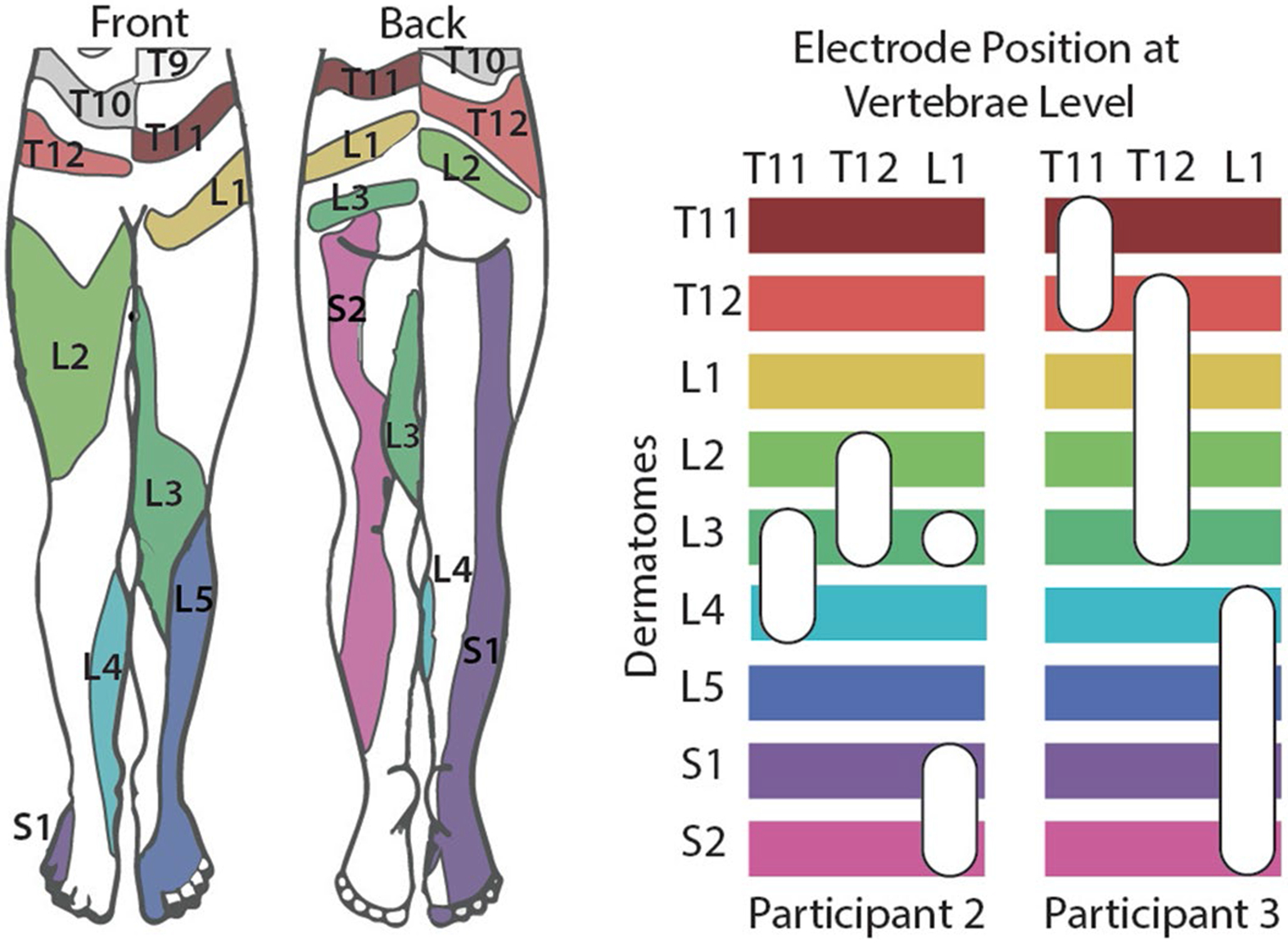
The left image shows the expected dermatomal innervation in the leg22. In the right, the horizontal bars indicate different dermatomes and the white ovals indicate the approximate electrode position that evoked sensations in that dermatome with respect to the vertebrae level. Participant 1 had substantial lead migration across weeks, making it challenging to precisely define the location of the electrodes with respect to vertebrae levels, so we have not included those results.
Extended Data Fig. 5 ∣. Percept quality of evoked sensations in the missing limb.
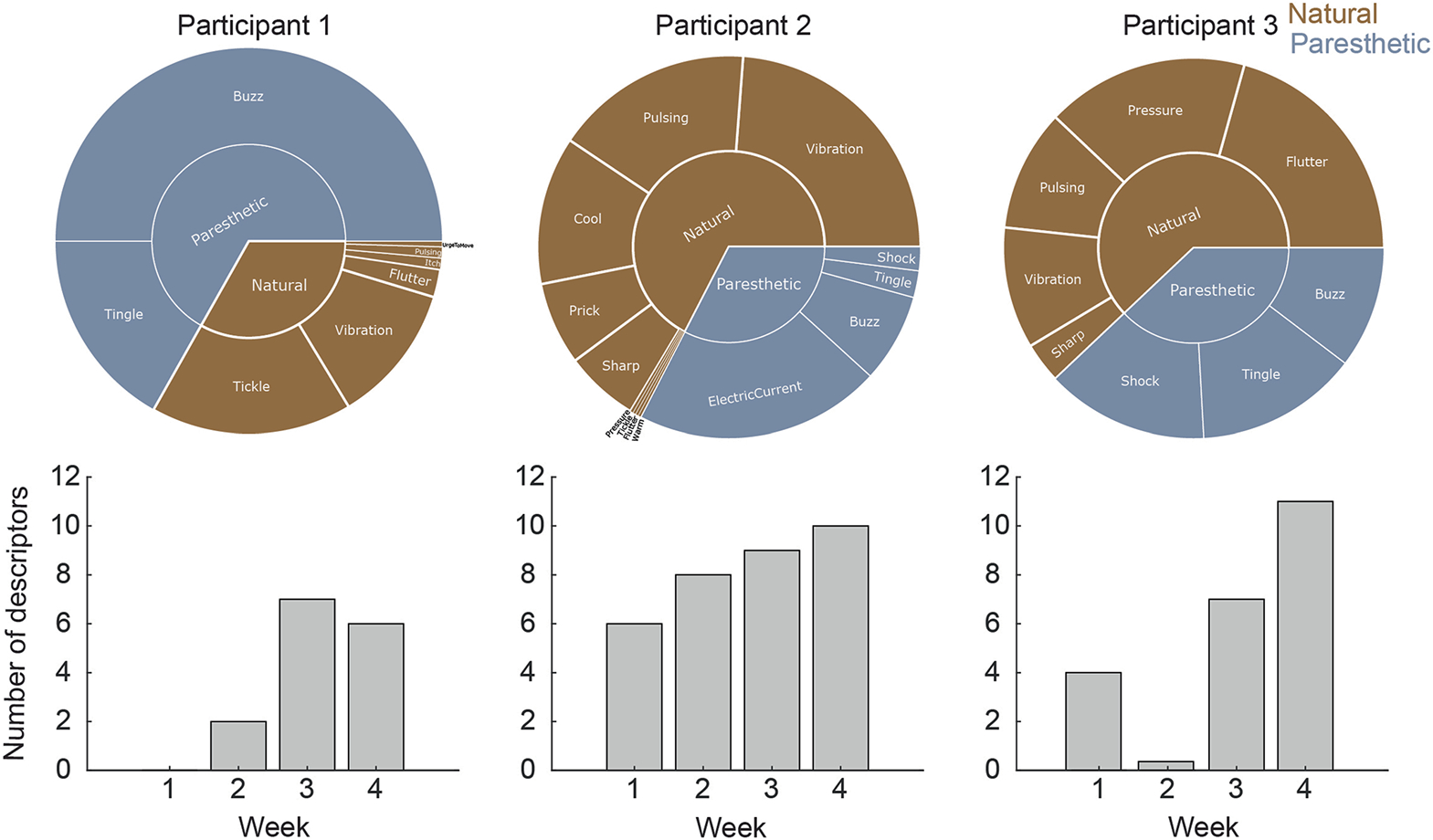
The participants were given a list of 13 natural descriptors and 5 paresthetic descriptors to describe the quality of the sensation. The top panel shows the frequency of each descriptor for the two evoked sensations for each participant shown in Fig. 2a. For all reported sensations, we stimulated via each electrode with a 1-sec long pulse train. The bottom panel shows the total number of descriptors used to describe the sensations each week.
Extended Data Fig. 6 ∣. Additional results from psychophysical discrimination assessment.

a, Variation of Weber fraction for different electrodes In Participant 1 and 2 as a function of the reference amplitude in the discrimination task. b, Variation of JND for the same electrodes in Participants 1 and 2 as a function of the reference amplitude. Participant 3 was discarded from these analyses due to insufficient data points.
Extended Data Fig. 7 ∣. Full results of Sensory Organization Test (SOT).

a, Participant 2 performed the SOT without stimulation (light blue) with sham stimulation (that is, stimulation in the residual limb only, gray) and with stimulation (stimulation in the prosthetic foot, dark blue). Sham stimulation substantially decreased performance for three of six conditions (with greater than minimum detectable change [MDC, 3.98]), suggesting that stimulation on the residual limb alone was not sufficient to improve performance. b, Participant 3 performed the SOT without stimulation (light magenta) and with stimulation (dark magenta). Both Participant 2 and Participant 3 exhibited improved performance on conditions with platform sway and eyes closed (+5.12 Participant 2, +9.60 Participant 3) and with visual surround sway (+4.04 Participant 2, +13.39 Participant 3). Both participants, however, exhibited decreased performance with stimulation during static standing with eyes closed (−6.25 Participant 2, −4.32 Participant 3). Additionally, Participant 3 had worse performance on static standing with eyes open with stimulation (−4.13). Change in median values reported. * represents a MDC, ** represents a clinically meaningful difference (>8.0).
Extended Data Fig. 8 ∣. McGill Pain Questionnaire results.
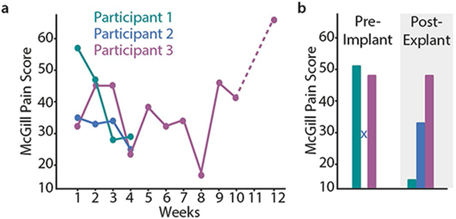
a, Weekly McGill Pain Questionnaire results. b, McGill Pain Questionnaire score before the implant and 1-month post-explant. The pre-implant score for Participant 2 was not recorded and we did not perform testing on week 11 for Participant 3 (indicated by the dashed line).
Supplementary Material
Acknowledgements
We dedicate this work to our dear friend and colleague Sliman J. Bensmaia, without whose support, insight and passion for science and mentorship this project would not have been possible. We wish to thank P. Sparto for his assistance with the blinded review of functional outcome measures. All authors acknowledge support for the research described in this study from the National Institutes of Health cooperative agreement UH3NS100541. B.A.P. acknowledges support for the research described in this study from National Institutes of Health training grant F30HD098794. B.B. acknowledges support for the research described in this study from Swiss National Science Foundation Doc.Mobility fellowship P1FRP3_188027.
Footnotes
Reporting summary
Further information on research design is available in the Nature Portfolio Reporting Summary linked to this article.
Competing interests
The authors declare no competing interests.
Extended data is available for this paper at https://doi.org/10.1038/s41551-023-01153-8.
Supplementary information The online version contains supplementary material available at https://doi.org/10.1038/s41551-023-01153-8.
Data availability
Source data for the figures in this study are available in the Data Archive for the Brain Initiative, with the identifier https://doi.org/10.18120/8qby-hk82 (ref. 59). The raw and analysed datasets generated during the study are available for research purposes from the corresponding author on reasonable request.
Code availability
The custom code used to generate figures for this manuscript is available at https://github.com/pitt-rnel/NatureBME2023_SCSLowerLimb.
References
- 1.Sexton AT & Fleming LL Lower extremity amputations. In Medical Management of the Surgical Patient: A Textbook of Perioperative Medicine (eds Walker HK, Lubin MF, Spell NO, Smith RB & Dodson TF) 741–743 (Cambridge University Press, 2006). [Google Scholar]
- 2.Viseux FJF The sensory role of the sole of the foot: review and update on clinical perspectives. Neurophysiol. Clin 50, 55–68 (2020). [DOI] [PubMed] [Google Scholar]
- 3.Petersen BA, Nanivadekar AC, Chandrasekaran S & Fisher LE Phantom limb pain: peripheral neuromodulatory and neuroprosthetic approaches to treatment. Muscle Nerve 59, 154–167 (2019). [DOI] [PubMed] [Google Scholar]
- 4.Kuffler DP Origins of phantom limb pain. Mol. Neurobiol 55, 60–69 (2018). [DOI] [PubMed] [Google Scholar]
- 5.Valle G. et al. Mechanisms of neuro-robotic prosthesis operation in leg amputees. Sci. Adv 7, eabd8354 (2021). [DOI] [PMC free article] [PubMed] [Google Scholar]
- 6.Petrini FM et al. Enhancing functional abilities and cognitive integration of the lower limb prosthesis. Sci. Transl. Med 11, eaav8939 (2019). [DOI] [PubMed] [Google Scholar]
- 7.Charkhkar H. et al. High-density peripheral nerve cuffs restore natural sensation to individuals with lower-limb amputations. J. Neural Eng 15, 056002 (2018). [DOI] [PubMed] [Google Scholar]
- 8.Petrini FM et al. Sensory feedback restoration in leg amputees improves walking speed, metabolic cost and phantom pain. Nat. Med 25, 1356–1363 (2019). [DOI] [PubMed] [Google Scholar]
- 9.Raspopovic S. et al. Restoring natural sensory feedback in real-time bidirectional hand prostheses. Sci. Transl. Med 6, 222ra19–222ra19 (2014). [DOI] [PubMed] [Google Scholar]
- 10.Dhillon GS & Horch KW Direct neural sensory feedback and control of a prosthetic arm. IEEE Trans. Neural Syst. Rehabil. Eng 13, 468–472 (2005). [DOI] [PubMed] [Google Scholar]
- 11.Charkhkar H, Christie BP & Triolo RJ Sensory neuroprosthesis improves postural stability during Sensory Organization Test in lower-limb amputees. Sci. Rep 10, 1–13 (2020). [DOI] [PMC free article] [PubMed] [Google Scholar]
- 12.Ortiz-Catalan M. et al. An osseointegrated human–machine gateway for long-term sensory feedback and motor control of artificial limbs. Sci. Transl. Med 6, 257re6–257re6 (2014). [DOI] [PubMed] [Google Scholar]
- 13.Horch K, Meek S, Taylor TG & Hutchinson DT Object discrimination with an artificial hand using electrical stimulation of peripheral tactile and proprioceptive pathways with intrafascicular electrodes. IEEE Trans. Neural Syst. Rehabil. Eng 19, 483–489 (2011). [DOI] [PubMed] [Google Scholar]
- 14.Tan DW et al. A neural interface provides long-term stable natural touch perception. Sci. Transl. Med 6, 257ra138–257ra138 (2014). [DOI] [PMC free article] [PubMed] [Google Scholar]
- 15.Davis TS et al. Restoring motor control and sensory feedback in people with upper extremity amputations using arrays of 96 microelectrodes implanted in the median and ulnar nerves. J. Neural Eng 13, 036001 (2016). [DOI] [PubMed] [Google Scholar]
- 16.Marasco PD, Schultz AE & Kuiken TA Sensory capacity of reinnervated skin after redirection of amputated upper limb nerves to the chest. Brain 132, 1441–1448 (2009). [DOI] [PMC free article] [PubMed] [Google Scholar]
- 17.Rossini PM et al. Double nerve intraneural interface implant on a human amputee for robotic hand control. Clin. Neurophysiol 121, 777–783 (2010). [DOI] [PubMed] [Google Scholar]
- 18.Dillingham TR, Pezzin LE & MacKenzie EJ Limb amputation and limb deficiency: epidemiology and recent trends in the United States. South. Med. J 95, 875–883 (2002). [DOI] [PubMed] [Google Scholar]
- 19.Kumar K & Rizvi S Historical and present state of neuromodulation in chronic pain. Curr. Pain Headache Rep 18, 387 (2014). [DOI] [PubMed] [Google Scholar]
- 20.Fanciullo GJ, Rose RJ, Lunt PG, Whalen PK & Ross E The state of implantable pain therapies in the United States: a nationwide survey of academic teaching programs. Anesth. Analg 88, 1311–1316 (1999). [DOI] [PubMed] [Google Scholar]
- 21.Chandrasekaran S. et al. Sensory restoration by epidural stimulation of the lateral spinal cord in upper-limb amputees. Elife 9, 1–26 (2020). [DOI] [PMC free article] [PubMed] [Google Scholar]
- 22.Lee MWL, McPhee RW & Stringer MD An evidence-based approach to human dermatomes. Clin. Anat 21, 363–373 (2008). [DOI] [PubMed] [Google Scholar]
- 23.Kim LH, McLeod RS & Kiss ZHT A new psychometric questionnaire for reporting of somatosensory percepts. J. Neural Eng 15, 13002 (2018). [DOI] [PubMed] [Google Scholar]
- 24.Graczyk EL et al. The neural basis of perceived intensity in natural and artificial touch (accepted). Sci. Transl Med 142, 1–11 (2016). [DOI] [PMC free article] [PubMed] [Google Scholar]
- 25.Page DM et al. Discriminability of multiple cutaneous and proprioceptive hand percepts evoked by intraneural stimulation with Utah slanted electrode arrays in human amputees. J. Neuroeng. Rehabil 18, 12 (2021). [DOI] [PMC free article] [PubMed] [Google Scholar]
- 26.Mastinu E. et al. Grip control and motor coordination with implanted and surface electrodes while grasping with an osseointegrated prosthetic hand. J. Neuroeng. Rehabil 16, 49 (2019). [DOI] [PMC free article] [PubMed] [Google Scholar]
- 27.Flesher SN et al. Intracortical microstimulation of human somatosensory cortex. Sci. Transl. Med 8, 361ra141–361ra141 (2016). [DOI] [PubMed] [Google Scholar]
- 28.Wrisley DM & Kumar NA Functional gait assessment: concurrent, discriminative, and predictive validity in community-dwelling older adults. Phys. Ther 90, 761–773 (2010). [DOI] [PubMed] [Google Scholar]
- 29.Koehler-McNicholas SR, Danzl L, Cataldo AY & Oddsson LIE Neuromodulation to improve gait and balance function using a sensory neuroprosthesis in people who report insensate feet—a randomized control cross-over study. PLoS ONE 14, e0216212 (2019). [DOI] [PMC free article] [PubMed] [Google Scholar]
- 30.Melzack R. The McGill Pain Questionnaire: major properties and scoring methods. Pain 1, 277–299 (1975). [DOI] [PubMed] [Google Scholar]
- 31.Cho TA Spinal cord functional anatomy. Contin. Lifelong Learn. Neurol 21, 13–35 (2015). [DOI] [PubMed] [Google Scholar]
- 32.Muret D & Makin TR The homeostatic homunculus: rethinking deprivation-triggered reorganisation. Curr. Opin. Neurobiol 67, 115–122 (2021). [DOI] [PubMed] [Google Scholar]
- 33.Selvarajah D. et al. Structural and functional abnormalities of the primary somatosensory cortex in diabetic peripheral neuropathy: a multimodal MRI study. Diabetes 68, 796–806 (2019). [DOI] [PubMed] [Google Scholar]
- 34.Ekman G. ös Weber’s law and related functions. J. Psychol 47, 343–352 (1959). [Google Scholar]
- 35.Kim S. et al. Behavioral assessment of sensitivity to intracortical microstimulation of primate somatosensory cortex. Proc. Natl Acad. Sci. USA 112, 15202–15207 (2015). [DOI] [PMC free article] [PubMed] [Google Scholar]
- 36.Petersen B, Sparto PJ & Fisher LE Clinical measures of balance and gait cannot differentiate somatosensory impairments in people with lower-limb amputation. Gait Posture 99, 104–110 (2023). [DOI] [PMC free article] [PubMed] [Google Scholar]
- 37.Jones MG et al. Neuromodulation using ultra low frequency current waveform reversibly blocks axonal conduction and chronic pain. Sci. Transl. Med 13, eabg9890 (2021). [DOI] [PubMed] [Google Scholar]
- 38.Lewandowski AS, Palermo TM, Kirchner HL & Drotar D Comparing diary and retrospective reports of pain and activity restriction in children and adolescents with chronic pain conditions. Clin. J. Pain 25, 299–306 (2009). [DOI] [PMC free article] [PubMed] [Google Scholar]
- 39.Saal HP & Bensmaia SJ Biomimetic approaches to bionic touch through a peripheral nerve interface. Neuropsychologia 79, 343–353 (2015). [DOI] [PubMed] [Google Scholar]
- 40.Okorokova EV, He Q & Bensmaia SJ Biomimetic encoding model for restoring touch in bionic hands through a nerve interface. J. Neural Eng 15, 66033 (2018). [DOI] [PMC free article] [PubMed] [Google Scholar]
- 41.Valle G. et al. Biomimetic intraneural sensory feedback enhances sensation naturalness, tactile sensitivity, and manual dexterity in a bidirectional prosthesis. Neuron 100, 37–45.e7 (2018). [DOI] [PubMed] [Google Scholar]
- 42.George JA et al. Biomimetic sensory feedback through peripheral nerve stimulation improves dexterous use of a bionic hand. Sci. Robot 4, 1–12 (2019). [DOI] [PubMed] [Google Scholar]
- 43.Sankar S. et al. Texture discrimination with a soft biomimetic finger using a flexible neuromorphic tactile sensor array that provides sensory feedback. Soft Robot. 8, 577–587 (2021). [DOI] [PubMed] [Google Scholar]
- 44.Stone AA & Broderick JE Real-time data collection for pain: appraisal and current status. Pain Med. 8, S85–S93 (2007). [DOI] [PubMed] [Google Scholar]
- 45.Gwaltney CJ, Shields AL & Shiffman S Equivalence of electronic and paper-and-pencil administration of patient-reported outcome measures: a meta-analytic review. Value Health 11, 322–333 (2008). [DOI] [PubMed] [Google Scholar]
- 46.Broderick JE et al. The accuracy of pain and fatigue items across different reporting periods. Pain 139, 146–157 (2008). [DOI] [PMC free article] [PubMed] [Google Scholar]
- 47.Garcia-Palacios A. et al. Ecological momentary assessment for chronic pain in fibromyalgia using a smartphone: a randomized crossover study. Eur. J. Pain 18, 862–872 (2014). [DOI] [PubMed] [Google Scholar]
- 48.Teirlinck CH, Sonneveld DS, Bierma-Zeinstra SMA & Luijsterburg PAJ Daily pain measurements and retrospective pain measurements in hip osteoarthritis patients with intermittent pain. Arthritis Care Res. 71, 768–776 (2019). [DOI] [PMC free article] [PubMed] [Google Scholar]
- 49.Toossi A. et al. Comparative neuroanatomy of the lumbosacral spinal cord of the rat, cat, pig, monkey, and human. Sci. Rep 11, 1955 (2021). [DOI] [PMC free article] [PubMed] [Google Scholar]
- 50.Nanivadekar A, Chandrasekaran S, Gaunt R & Fisher L RNEL PerceptMapper (2020). 10.5281/ZENODO.3939658 [DOI] [Google Scholar]
- 51.Prieto TE, Myklebust JB, Hoffmann RG, Lovett EG & Myklebust BM Measures of postural steadiness: differences between healthy young and elderly adults. IEEE Trans. Biomed. Eng 43, 956–966 (1996). [DOI] [PubMed] [Google Scholar]
- 52.Cripps A, Livingston SC & DeSantis BM The test–retest reliability and minimal detectable change of the Sensory Organization Test and Head-Shake Sensory Organization Test. J. Sports Med. Allied Health Sci 2, 2 (2016). [Google Scholar]
- 53.Wrisley DM, Marchetti GF, Kuharsky DK & Whitney SL Reliability, internal consistency, and validity of data obtained with the functional gait assessment. Phys. Ther 84, 906–918 (2004). [PubMed] [Google Scholar]
- 54.Beninato M, Fernandes A & Plummer LS Minimal clinically important difference of the functional gait assessment in older adults. Phys. Ther 94, 1594–1603 (2014). [DOI] [PubMed] [Google Scholar]
- 55.Pesarin F & Salmaso L The permutation testing approach: a review. Statistica 70, 481–509 (2010). [Google Scholar]
- 56.Powell MP et al. Epidural stimulation of the cervical spinal cord for post-stroke upper-limb paresis. Nat. Med 29, 689–699 (2023). [DOI] [PMC free article] [PubMed] [Google Scholar]
- 57.Sdrulla AD, Guan Y & Raja SN Spinal cord stimulation: clinical efficacy and potential mechanisms. Pain Pract. 18, 1048–1067 (2018). [DOI] [PMC free article] [PubMed] [Google Scholar]
- 58.Forouzanfar T, Weber WEJ, Kemler M & van Kleef M What is a meaningful pain reduction in patients with complex regional pain syndrome type 1? Clin. J. Pain 19, 281–285 (2003). [DOI] [PubMed] [Google Scholar]
- 59.Nanivadekar AC, et al. Dataset for ‘Restoration of sensory feedback from the foot and reduction of phantom limb pain via closed-loop spinal cord stimulation.’ Data Archive for the Brain Initiative (2023); 10.18120/8qby-hk82 [DOI] [Google Scholar]
Associated Data
This section collects any data citations, data availability statements, or supplementary materials included in this article.
Supplementary Materials
Data Availability Statement
Source data for the figures in this study are available in the Data Archive for the Brain Initiative, with the identifier https://doi.org/10.18120/8qby-hk82 (ref. 59). The raw and analysed datasets generated during the study are available for research purposes from the corresponding author on reasonable request.
The custom code used to generate figures for this manuscript is available at https://github.com/pitt-rnel/NatureBME2023_SCSLowerLimb.