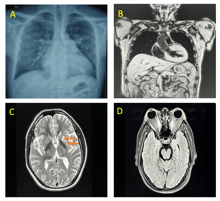
Figure 3. Investigations of the patient.
(A) Chest X-ray posteroanterior view revealing cardiomegaly. (B) Cardiac MRI study revealing asymmetrical hypertrophic cardiomyopathy. (C) MRI of the brain, axial cuts (arrows) showing chronic lacunar infarcts in left centrum semiovale and corona radiate. (D) MRI orbits showing enlargement of bilateral recti muscles with resultant bilateral proptosis.

