ABSTRACT
During the nineteenth century, New Zealand was promoted as a land of plenty, promising a ‘better life’, to encourage families to settle and develop the growing colony. This paper characterises the life-course of early settlers to New Zealand through historical epidemiological and osteological analyses of the St John’s burial ground in Milton, Otago. These people represent some of the first European colonists to Aotearoa, and their children. The analyses provided glimpses into the past of strenuous manual labour, repeated risk of injury, and oral and skeletal infections. Mortality of infants was very high in the skeletal sample and the death certificates outlined the varied risks of infection and accidents they faced. Osteobiographies of seven well-preserved adults demonstrated the detailed narratives that can be gleaned from careful consideration of individuals. The skeletal record indicates childhood stress affecting growth and risk of injury prior to migration. However, the historical record suggests that occupational risks of death to the working class were similar in the new colony as at home. The snapshot of this Victorian-era population provided by these data suggests that the colonial society transported their biosocial landscape upon immigration and little changed for these initial colonists.
KEYWORDS: Bioarchaeology, colonial, osteobiography, life-course, Otago
Introduction
Bioarchaeological enquiry into nineteenth century period cemeteries is well founded in the United Kingdom (UK). Much of this work has established the biosocial context of lived experiences during the Industrial Revolution in both urban and rural settings, and compared the health effects of increasing industrialisation between the two contexts (Gowland et al. 2018). Recurring themes are of increased hardship and detrimental health effects amongst the urban poor and a relatively ‘healthier’ lifestyle in rural settings (Roberts and Cox 2003). Families of higher social standing and greater wealth were afforded better access to State support, which resulted in better health and this may be reflected in the bioarchaeological record (Mays et al. 2009). Within this setting of burgeoning industrialisation and marked social hierarchy there was a push for Empire-building involving immigration to new colonies, including New Zealand (Ballantyne 2012). The sixteenth–nineteenth-century colonial drive by Europeans to new lands already settled by indigenous peoples had devastating consequences for the existing populations. Some effects were intentional, including marginalisation from ancestral lands and warfare, while others were unintentional such as the introduction of new infectious diseases (Pool 1991, 2015). While acknowledging the severe and largely irrevocable impacts of European colonisation on the tangata whenua of New Zealand, we also note that the biosocial experiences of colonisation for the European (and later Chinese) colonists remain largely unknown.
During the nineteenth century, there were many factors at play to encourage emigration from the Old World to the New. Push factors included population growth with resultant urban overcrowding, poor working conditions and hunger (seen most particularly in the Irish famines of the 1840s and 1879). Pull factors included the perceived benefits and opportunities of the New World colonies, considerably embellished by the booster literature of the period (Belich 2009). Between 1837 and 1852 some 200 publications were produced regarding New Zealand, many by the colonising organisations (Andrews 2009; Schrader 2016; Early New Zealand Books 2017). New Zealand was promoted as a land of plenty to encourage families to settle and develop the growing colony. The Otago settlement, in the south of New Zealand’s South Island, was a joint venture between the Lay Association of the Free Church of Scotland and the New Zealand Company, which acquired 144,600 acres of land in coastal Otago from Ngāi Tahu in 1844. The intention was to establish a Wakefield class settlement, where the community would be divided into a land-owning capitalist class, and a wage-earning working class (Hocken 1898; Olssen 1984; Schrader 2016). Land in the settlement was divided into urban, suburban and rural property, with the intention that agriculture would develop in the hinterland.
In south Otago, the small town of Milton was established in the 1850s as part of this agricultural hinterland and acted as a service town for the local farming community (Sumpter and Lewis 1949). Its communications roles were also important in Milton’s development: during the 1860s it was on one of the main routes to the Otago goldfields as thousands of people flocked to Central Otago from Dunedin (Sumpter and Lewis 1949; Olssen 1984). Milton was therefore linked to trade networks both along the coast and into the interior and was a local service centre, a role that it still fulfils today.
The St John’s Burial Ground was the first cemetery formally opened for Milton in 1860 and was last used in 1926. While this was primarily an Anglican burial ground, there is anecdotal evidence that burial of people from other denominations were welcome (Pers. Comm. V. Galletly), perhaps reflecting the necessity of inclusiveness in a fledgling colonial community. A bioarchaeological project of excavation and exhumation of 25 graves was conducted in 2016 at the request of an historical group formed of descendants (the TP60) and the Anglican Church (Buckley and Petchey 2018). This project had two aims. The first was to assist the Church and local community with identifying the extent of the burial ground and whether graves were located outside the current fenced area. The second, research-driven, aim was to build a biosocial profile of these early colonial settlers in their new physical and social environments. One of our questions was whether the promise of a new and better life was realised by the first-generation settlers and their children. Nineteenth century colonial cemeteries, such as the St John’s burial ground (SJM) are potentially an ideal source for assessing the biological effects of colonisation on the health of the colonisers.
The historical record, while biased to the voice of the controlling classes, provides a backdrop for placing these biosocial experiences in a firm contextual framework. Contextual information can be found in historic archival data, and summarised using epidemiological and demographic methods. These data sources include hospital administration records, doctor’s notes, records of mortality, burial records, and obituaries. While these types of historical records will rarely meet modern statistical standards, they provide information on individuals and the population they came from that is often not invisible in the skeletal record and considerably enrich the narrative regarding settler life and adaptation. Archival data can also aid in the identification of individuals and provide information of the health risks affecting the general population, potentially aiding in the differential diagnosis of observed pathology in human remains.
Records associated with the St. John’s burial ground show that although most of the people buried at St John’s were farmers or engaged in industry, some of the men were gold mining just before their death, or had settled at Milton after spending time in gold fields elsewhere (Findlay 2016). Mining was a dangerous occupation and it is to be expected that this would be reflected in bioarchaeological data in a frontier goldfields and early settler context, yet few bioarchaeological studies of these populations have been undertaken (White 2017). Our initial findings from the wider Otago bioarchaeology project attest to infectious disease (Snoddy et al. 2020), episodes of nutritional and metabolic stress (King et al. 2020), ill-health, and serious injury (Petchey et al. 2018) amongst European frontier men and women.
The aim of this paper is to characterise the lived experiences of early colonial settlers in Milton using osteological and historical epidemiological information. Through doing so we also begin to address the research question of whether life was ‘better’ in New Zealand than at home. The historical epidemiological information from death certificates is presented with a view to understanding the overall health risks to the population that may otherwise be invisible in the bioarchaeological record. We present the mortality profile of the skeletal sample, adult oral health data, non-specific stress, and adult skeletal pathology in the sample. From isotope analyses we know that all of the adults from SJM were first generation migrants to New Zealand (King et al. 2020), so their biological tissues hold the stories of home experiences as well as those in the colony.
Biosocial context of Milton
The settlement of Milton is located on the edge of the Tokomairiro Plain, 54 kilometres south of the main port city of Dunedin. This low-lying flood plain would have been damp and cold, especially during the winter months. While predominantly a farming community, Milton was also the location of a number of local industries including the brick and pipe works, flour mill, woollen mill and the short-lived flax mill, all of which are associated with increased risk of respiratory diseases. In addition to traffic to the inland goldfields there were also the small goldfields of Glenore and Canada Reef nearby. The inhabitants of the Milton area were therefore involved in a range of agricultural, industrial and service occupations, many of which involved a high level of risk. Perusal of the local newspaper, the Bruce Herald, chronicles the dangers faced by the settlers. Animals were part of everyday farming life, horses were probably the most common form of transport, and accidents were commonplace. For example, engineer Adam Johnston struck his head when he was thrown from a horse in January 1874, and died two days later, never having regained consciousness (Bruce Herald 27 January 1874). Other reported incidents include Thomas Turnnbull being gored by a cow (Bruce Herald 31 July 1867) and Alexander Mackay being kicked in the head by a colt (Bruce Herald 19 February 1868).
Another risk was drowning; many river and stream crossings had yet to be bridged and the risk was so high that drowning became known as the ‘New Zealand Death’. Around 100 deaths by drowning were reported nationwide a year in the 1890s (Bruce Herald 13 July 1865; Lake County Press 31 December 1885; Otago Witness 1 February 1894). Children were particularly at risk, not only from childhood diseases, but also from accidents; in 1868 a 20-month-old child drowned in a well and a 2-year-old child drowned in a creek (Bruce Herald 11 March 1868, 8 July 1868).
Materials and methods
Twenty-five graves were excavated from the SJM burial ground (Petchey et al. 2017). The preservation of the skeletons was highly variable, ranging from no bone in most of the subadult graves to excellent preservation in a small number of adults. Many of the subadults were represented by dentition only and some adult bone was extremely soft; in some cases, only the ‘shadow’ of bone remained. All of the people were buried in coffins, also of variable preservation, but most with coffin furniture (handles, decorative strips and metal coffin lid plates) preserved. Gold paint on the coffin lid plates was preserved well enough in four of the adult burials to positively identify the deceased (Petchey et al. 2017), and aspects of these individuals’ lives were documented in the TP60 historical accounts (Findlay 2016). While the identity of four adults is known, we do not name them in this paper. This decision is made out of respect for the deceased’s descendants and is not intended to diminish the dignity of the deceased. As such, each individual is referred by burial numbers as is standard archaeological practice.
Historical epidemiology
The data used for this analysis were derived from 77 death certificates of people buried at the ‘Tokomairiro’ or the ‘Anglican burial ground’. These were grouped and summarised using a number of different criteria, to create comparable data sets for observing trends and patterns of death in the Milton population. Counts of death were made on age brackets using the following criteria; the ages are grouped by 5 years from the age of 5–79. The age groups below 5 were grouped on single years and over the age of 80 as a single age group. Counts were also made on sex to calculate the male:female ratio, and month-of-death to indicate the most common season of death. The listed causes of death were reclassified using ICD-10 codes 2016 (WHO 2016) and were separated into sex and age groupings.
The primary methodological consideration when using these types of data is that the size of the population from which the cemetery sample is drawn is unknown and migration between Milton and other areas elsewhere in the Otago region may have been occurring. Therefore, there is no denominator to form mortality rates and as such an observed abnormality in the data cannot, with any certainty, be attributed to either a change in the mortality profile or a larger than average population from which the deaths were drawn. What can be observed is the size of death counts relative to other populations. There may be a number of reasons for differences in counts, only one of which may be a higher mortality rate. Finally, the data set of only 77 people is not representative of the entire living population and it is therefore unlikely that the deceased demographic profile is an unbiased sample of the historic living population of Milton.
Other, methodological considerations when handling historical data sources on disease are discussed in depth elsewhere (Alter and Carmichael 1999; Roberts and Manchester 2005; Roberts 2014). Briefly, many of the recorded deaths in the Milton population were made before full understanding of germ theory, so the people making the records had a different concept of health and disease than we do today and data were not collected with adherence to modern standards of record keeping.
Osteology methods
Age and sex estimations
Infants and children (subadults) are defined throughout as those under 15 years of age at death. Age at death of infants and children was estimated using dental formation stages (Moorrees et al. 1963a, 1963b). Dental methods for subadult age estimation are the most accurate and in most cases at SJM only dental remains survived. Where skeletal material was present, other methods based on skeletal development and estimated size of skeletal elements were used (Scheuer and Black 2000). A multifactorial approach was used in the estimation of the age at death of adults. Standard measures used were observations of late fusing epiphyses (sternal clavicle and sacrum) (Schaefer et al. 2009), pubic symphysis (Brooks and Suchey 1990) and auricular surface morphology (Lovejoy et al. 1985) and dental wear seriation (Smith 1984). Adults were classified in relative age groups of young, middle and old aged (Buikstra and Ubelaker 1994). The estimation of sex for adults was based on standard morphological observations of the pelvis (pubis and greater sciatic notch) (Phenice 1969; Milner 1992 and Walker in Buikstra and Ubelaker 1994) and cranium (Acsádi and Nemeskéri 1970). Preference was given to pelvis morphology as this is a more accurate reflection of sex. No attempt was made to sex infants and children due to the lack of sexual dimorphism in the pelvis and skull before puberty.
Stature
The eventual height (stature) attained by an adult is a reflection of a complex interplay between the genetic potential for height and the influence of the environment during growth (Eveleth and Tanner 1990). Therefore, adult stature can be used as a measure of population health. Long bone lengths were all measured in situ before being removed from the ground. If intact after lifting the bone was measured again in the laboratory and this was taken as the more accurate measurement. Stature was estimated from the lower limb bones (femur, fibula, or femur + fibula) where possible as these equations are accompanied by the smallest margin of error (Trotter and Gleser 1952, 1958). The tibia equations developed by Trotter and Gleser (1952, 1958) were not used as the accuracy of these have been disputed by subsequent researchers (Jantz et al. 1994).
The osteological health and disease parameters assessed in this study are outlined in Table SI1.
Results
Historical demographics and epidemiology
Year of burials
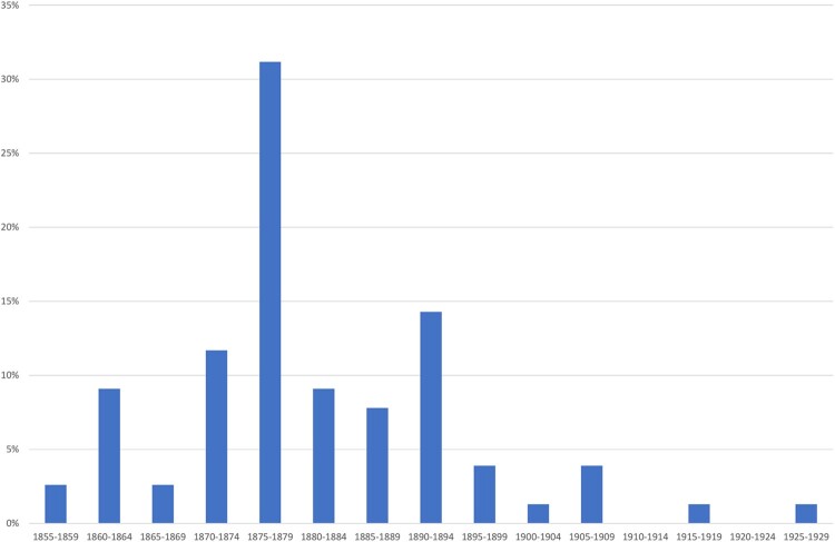
The year of burial at the Milton cemetery indicates that while the first burial occurred in 1857, it was during the 1870s that nearly half of the people were buried (Figure 1). Without a full population profile of the town it is difficult to confirm whether the population of Milton was more at risk in the 1870s, whether the population was simply larger, or alternatively, whether there were poorer burial records from the 1850s and 1860s.
Figure 1.
Year of death in five-year groupings (n = 77).
Demographics of the SJM burial ground death records
Censuses of the Otago population were taken in 1864 and 1867 over the whole district and these were published in the North Otago Times in 1868 (North Otago Times 1868) (Table 1). These data give a broad idea on the population that the SJM burial ground was drawn from but are not specific enough to calculate mortality rates. However, men between 21 and 39 years of age dominate the Otago European population at this time, which is likely a product of the gold rush and immigration. There was a growing number of young children, which is suggestive of the development of families in the region, and there were very few people over the age of 65 years.
Table 1. Census returns for the Otago Population 1864 and 1867 (North Otago Times 1868).
| Census of 1864 | Census of 1867 | |||||||
|---|---|---|---|---|---|---|---|---|
| Age | Male | Female | Male | Female | ||||
| Under 5 years | 3,516 | 7.2% | 3,359 | 6.9% | 4,563 | 9.4% | 4,367 | 9.0% |
| 5–9 years | 2,002 | 4.1% | 2,047 | 4.2% | 2,560 | 5.3% | 2,420 | 5.0% |
| 10–14 years | 1,311 | 2.7% | 1,167 | 2.4% | 1,634 | 3.4% | 1,807 | 3.7% |
| 15–20 years | 1,640 | 3.3% | 1,492 | 3.0% | 1,591 | 3.3% | 1,288 | 2.7% |
| 21–39 years | 20,375 | 41.6% | 6,780 | 13.8% | 14,833 | 30.7% | 6,851 | 14.2% |
| 40–54 years | 3,053 | 6.2% | 1,122 | 2.3% | 3,207 | 6.6% | 1,371 | 2.8% |
| 55–64 years | 469 | 1.0% | 240 | 0.5% | 539 | 1.1% | 328 | 0.7% |
| 65 and above | 100 | 0.2% | 67 | 0.1% | 186 | 0.4% | 124 | 0.3% |
| Unknown | 226 | 0.5% | 52 | 0.1% | 706 | 1.5% | 5 | 0.0% |
| Total Sex | 32,692 | 66.7% | 16,326 | 33.3% | 29,819 | 18,561 | ||
| Total | 49,018 | 48,380 | ||||||
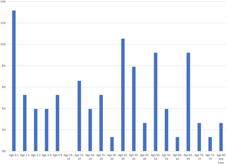
The expected mortality profile of a cemetery from an established population often has a U shape, in that age demographics at either extreme, very young and very old, are more commonly represented than other age groups as they are more susceptible to disease (Waldron 1994). At SJM, the 0–1.0 year age group is the most common age range of people buried in the cemetery (14%) and children aged less than 5 years account for 26% of the deceased. However, people in their late thirties (11%), early forties (8%) and early fifties (9%) are also present, with relatively few people aged 70 and over (Figure 2).
Figure 2.
Ages of death, St John’s burial ground (n = 76).
All of the adults in the Milton population were new migrants, as evidenced by the isotope data (King et al. 2020), which may explain a larger cohort of people in their thirties and forties in the community’s population rather than a greater risk of death in this age range. A larger number of young children (0–4 years), compared with children in the 5–14 age group suggests, a high infant mortality rate, a higher fertility rate, or both. The Otago censuses of 1864 and 1867 indicate a larger number of young children, and this trend likely continued into the mid-1870s, the period during which the majority of the burials occurred at SJM. The censuses do also suggest there were few older adults in Otago which is also reflected in the burial records.
Sex
There were slightly more males (53%) buried at SJM than females (47%). The 1867 census for the whole Otago region reported 29,819 (61.5%) males and 18, 561 (38.5%) females.
Month and season of death
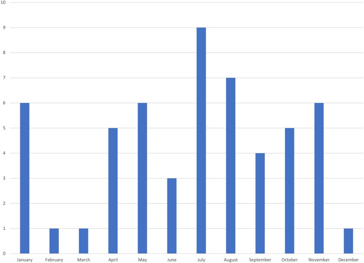
The month of death is recorded in 54 cases (Figure 3). The southern hemisphere winter months (June, July, August) recorded the highest number of deaths (35%) with nine (17%) deaths in July. There was only one death recorded in each of the warmer summer months of December, February and March, though six deaths occurred in January. Therefore, seasonal risk factors at Milton were potentially an important contributor to mortality in this area, as would be expected in this temperate environment.
Figure 3.
Month of death of burial at SJM.
Cause of death
There were 83 different causes of death noted on the death certificates; a number of people had multiple causes (Table S2). Of the listed causes of death, tuberculosis is the most common, accounting for 14% of deaths. Stroke and heart disease accounted for a further 13% of deaths and pneumonia, asthma and bronchitis killed 12% of the population. Together, pulmonary infections, including tuberculosis, pneumonia and bronchitis, with asthma killed over a quarter of the people in the record. Alcohol is only a listed cause of death in one case (Burial 4).
For the cohort over the age of 60, heart disease and stroke were the leading causes of death at Milton. Throughout the data set there is a large number of people who did not have a listed cause of death (R99 in Table S1).
Census of skeletal sample excavated
The sample of individuals excavated from the burial ground in 2016 range in age from preterm infants to old aged adults. The subadults of the sample constitute 62.9% (n = 17/27) of those excavated and over half of these died before one year of age. Three subadults (Burials 14, 15 and 17) were not able to have an age estimation assigned due to poor preservation but the small coffin sizes of these individuals suggest that they were of prenatal–perinatal age (Table 2). Two of the subadult graves represented the simultaneous burial of two individuals, each with their own coffin (Burials 3A and 3B and 20A and 20B). Another case of a possible double burial occurred with Burial 8, a 1–1.5-year-old young child, where an additional foetal cranial bone was identified in the laboratory. Ten adults were excavated, four males, five females and one (Burial 7) for which we could not assign sex. Most the adults were either middle aged or old at the time of death.
Table 2. Census of burials excavated at SJM.
| Age | Subadult | Male | Female | ?sex | Total (%) |
|---|---|---|---|---|---|
| Preterm (<37 fwks) | 1 | ||||
| Perinate (40 fwks-∼postpartum) | 2 | ||||
| 0–0.9 years (infant) | 3 | ||||
| Unknown infant (non-biological age estimation) | 3 | ||||
| 1.0–4.9 years (young child) | 7 | ||||
| 5.0–9.9 years (older child) | 1 | ||||
| 10–14.9 years (Adolescent) | 0 | ||||
| Subtotal subadults | 17 | 0 | 0 | 0 | 17 (62.9) |
| Young | 0 | 0 | 0 | 0 | |
| Mid | 2 | 2 | 0 | 4 | |
| Old | 2 | 1 | 1 | 4 | |
| Adult | 0 | 2 | 0 | 2 | |
| Subtotal adults | 4 | 5 | 1 | 10 (37.0) | |
| Total sample | 27 (100) |
fwks = foetal weeks.
Stature
Average stature in the SJM sample was 170.2 cm (5 ft 5″) for males and 161.83 cm (5 ft 3″) for females (Table 3). Several of the long bone measurements were taken in the field prior to exhumation due to the fragility of the remains, so the margin of error associated with the reported statures is likely slightly larger than listed.
Table 3. Stature estimates for SJM adults.
| Burial | Sex | Stature (cm) | SD (cm) |
|---|---|---|---|
| 4 | M | 173.35 | 3.62 |
| 11 | M | 169.83 | 3.94 |
| 13 | M | 173.64 | 3.94 |
| 21 | M | 163.90 | 3.87 |
| 6 | F | 157.84 | 3.72 |
| 7 | F | 163.77 | 3.72 |
| 10 | F | 157.96 | 3.72 |
| 22 | F | 158.35 | 3.57 |
| 23 | F | 160.93 | 3.72 |
| 29 | F | 174.17a | 3.57 |
See osteobiography below for B29.
M = Male; F = Female
Adult health and pathology
The skeletal preservation of the subadults was minimal, so any information on their health and diet can only be gleaned from the dentitions and ongoing isotope analyses presented in forthcoming publications. The preservation of seven of the adults was good enough to be able to compile descriptive osteobiographies of their life course. The other three adults were either not excavated or had extremely poor skeletal preservation. The profiles of all adults, including skeletal health and pathology observations are briefly presented in Table 4 and given in detail in SI1. Interpretations of the skeletal pathology observed in the seven well-preserved adults are discussed together in an attempt to place these individuals within the context of the wider community and begin to assess ‘population’ health of these early settlers.
Table 4. Descriptive summaries of all SJM adults, including age and sex, pathology and skeletal preservation.
| Burial number | Age | Sex | ID known? (yes, suspected, unknown) | Death certificate? | Material present | Skeletal pathology | comments |
|---|---|---|---|---|---|---|---|
| 4 | 41 years | Male | yes | Yes. Cause of death alcoholism and chloroform abuse. | Poorly preserved bones most surface unobservable. Some teeth | Unobservable | Positively identified in situ. Local doctor. |
| 6 | 36 years | Female | yes | Yes. Cause of death complications of childbirth | Good preservation of complete skeleton. Some damage to cranium. | Well-developed entheses of upper limb. Maxillary sinusitis | Positively identified in situ. Wife of local doctor. |
| 7 | Old | Male? | No | – | Complete very poorly preserved post cranial skeleton. Edentulous maxilla fragment. | Unobservable in postcranial bones. Moderate hyperostosis frontalis interna on left. | Very elaborate coffin plates covering most of the body. Only the cranium and a long bone fragment were sampled the rest remained unexcavated due to poor preservation |
| 9 | 47 years | Male | Yes | Yes. Cause of death tuberculosis. ‘Pthisis pulmondes’ |
No skeletal or dental remains preserved | – | Positively identified in situ. Drill instructor. |
| 10 | Old | Female | No | – | Very good preservation of limbs and cranial material. Thorax damaged by large coffin plate. | Thin diaphyseal cortices and generally light bone. Retention of metopic suture. | |
| 11 | Mid | Male | Suspected | Possible. Cause of death injuries due to mining accident | Excellent preservation of skeletal and dental. Hair preserved. | Multiple perimortem and antemortem trauma. | Possible identification from multiple sources. Miner. |
| 12 | Adulta | Male | Yes | Not excavated | – | Grave and identity confirmed from discovery of semi buried headstone. Not excavated. | |
| 13 | Mid? | Male | No | – | Good preservation in situ. Cranium damaged. Did not survive well after drying. | Multiple antemortem fractures and enthesopathies. Generally very robust and marked entheses. Most bone surfaces eroded. | |
| 21 | 42 years | Male | Yes | Yes. Cause of death Tuberculosis. ‘Pneumonic pthisis haemorrhage’ | Very well preserved skeletal and dental material though fragmented and disarticulated. Hair and brain tissue preserved. | Skeletal tuberculosis lesions of hips and cranium (Snoddy et al. 2020). | Positively identified in situ. Labourer. Invalid for 12 months before death. |
| 22 | Adult | Female? | No | Very poorly preserved skeletal material. Dentition present. | Unobservable | Not fully exhumed due to poor preservation. Bone samples taken. | |
| 23 | Adult | Female? | No | Very poorly preserved skeletal and dental. Hair and complete calcified brain present. | Unobservable | Not fully exhumed due to poor preservation. Cranium and dentition lifted. | |
| 29 | Mid | Female | No | Well preserved skeletal and dental. Damage to facial bones. | Some form of skeletal dysplasia (bowing of limbs and deformity of ribs). Possible perimortem sharp force injury to sternum. |
aOnly the name of this individual was visible on the headstone. His age was not visible and no further information about his life was present in the historic record.
Oral health
Summary statistics for the census of the dentition are presented in Table 5. There were four middle-aged adult males (B4, B11, B13, B21) and two middle aged adult females (B6 and B29) and an individual of unknown age and sex (B23) with preserved dentition. Many of the teeth were brittle with cracked or flaking enamel and blue/black staining, indicating poor preservation in the burial environment.
Table 5. Census of the teeth and alveoli present for St John’s Milton adults.
| Sex | Age | N1 Ind | Total alveoli and teeth | Alveoli/N1 | N2 Ind | Total teeth | Teeth/N2 | Teeth/alveoli % |
|---|---|---|---|---|---|---|---|---|
| Male | Middle | 4 | 90 | 22.5 | 4 | 61 | 15.3 | 67.8 |
| Female | Middle | 2 | 41 | 20.5 | 2 | 23 | 11.5 | 56.1 |
| Old | 1 | 12 | 12 | 0 | 0 | |||
| Subtotal | 3 | 53 | 17.7 | 2 | 23 | 11.5 | 43.4 | |
| Unk. | Unk. | 1 | 14 | 14 | 1 | 10 | 10 | 71.4 |
| Total | 8 | 157 | 19.6 | 7 | 94 | 13.4 | 59.9 |
Unk. Unknown age or sex.
N1 number of total individuals with observable alveoli and teeth.
N2 number of total individuals with observable teeth.
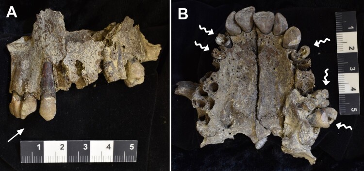
Tooth wear increases with age, but as all the individuals with at least one quarter of the dentition present were from the middle-aged adult age range, tooth wear can be compared at an intra-population level. Generally, the anterior teeth displayed heavier wear (‘moderate’ grades 4 and 5) compared with the premolars and molars (more ‘mild’ wear grades 2 and 3) (Table S3). Pipe facets were observed in the dentition of all four males and one middle-aged adult female (B6) (Figure 4; Table S4). In most cases, tobacco staining could not be assessed because of the blue/black staining presumably from the burial environment. Only the teeth of B23 displayed brown staining that may be associated with tobacco use. Antemortem chipping was only observed on one mandibular molar of B6 (possibly associated with a faulty amalgam filling) (Table S4).
Figure 4.
Burial 11. Buccal view of left maxilla (A) and inferior view of maxilla (B). Straight arrow indicates pipe facets between the lateral incisor and canine. Wavy arrows indicate massive carries destroying most of the tooth crowns in multiple teeth. Crowding of the anterior maxillary teeth is also apparent (B).
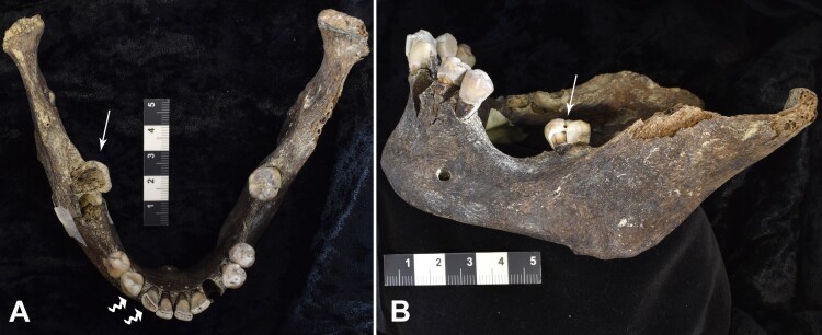
The overall rates of calculus were low, although over half (57.1%) were affected by mild calculus on at least a few teeth (Table S5). However, the lack of calculus may have been a result of the loss of the calculus deposits in the burial environment or detachment during excavation and cleaning. There were high rates of caries for both the males and females but for the males, most of the carious lesions occurred in the posterior teeth and for the females, the anterior teeth (Figure 5; Tables S7 and S8). This trend may be biased because of the relatively few posterior teeth present in the females due to antemortem tooth loss (AMTL) and poor preservation. All individuals were affected by AMTL (Table S6). Of the 37 instances of AMTL, 35 affected the molars (Figure 4). Nearly two-thirds of the SJM individuals (62.4%) had alveolar lesions (Table S5), likely caused by exposure of the tooth pulp by caries, though the overall rates were potentially reduced because many of the teeth affected by caries were either lost or removed during life. All the SJM individuals were affected by periodontal disease, especially in the posterior quadrants of the dental arch.
Figure 5.
Burial 21. Superior view of mandible (A) and buccal view of left mandible (B). Straight arrows indicate massive caries on the right third molar and pit and fissure caries on left third molar. Wavy arrows identify pipe wear facets between the right lateral incisor and canine. Antemortem tooth loss of all other molars and the right premolar is apparent by the gaps and bone remodelling closing the tooth sockets and substantial loss of alveolar bone.
All but one SJM adult had linear enamel hypoplasia (LEH) (85.7%). Most of the LEH were located on the canines and incisors (26/27 of observed LEH). The anterior teeth of males were more affected compared to females, but this may be partly a reflection of the smaller sample size for female teeth (Table S9).
Discussion
What was life (and death) like in colonial Milton?
The historical epidemiology and osteology analyses both reflect a maturing population with a high infant mortality, which is in keeping with the historical record. The relatively poor representation of very old adults in death certificates is not surprising given the early colonial context where these immigrants were more likely working aged adults and their families. There are few deaths related to alcohol and accidents, a pattern which is more common in gold mining contexts (Roberts 2006). This environment was not, however, without some specific risks. The colder months of the year brought higher death counts, and respiratory disease, particularly pulmonary tuberculosis, was a major cause of death. The high mortality rates from pulmonary complaints may be related to occupational health, an environmental risk factor, an epidemic or an unidentified pathogen that was not recognised at the time. Milton was reported as having ‘always a damp vapour rising, highly prejudicial to the health of the inmates, and especially the children’ (MacBean Stewart 1875). This description may relate to the theory that disease was spread via miasma or bad air, a perception that persisted in European society until being disproven in the second half of the nineteenth century by germ theory. Pneumonia, and other respiratory complaints, were commonly listed causes of death for both sexes, however the higher number of cases in men, may suggest an occupational health risk.
Illnesses related to industrialisation were rife in Victorian England (Wohl 1983). Mills producing textiles were particularly dangerous, causing chronic and often fatal respiratory illness, and this has been demonstrated in the bioarchaeological record (Gowland 2018). We know from the historical record that the township of Milton was home to various industries which were detrimental to respiratory health (flour milling, textiles, and pottery production) (Elwood 1965; Thomas and Stewart 1987; Mohammadien et al. 2013). In some cases, respiratory conditions may have been contracted back ‘home’ in England but exacerbated by the colonial climate and unstable food resources in the early stages of the settlement.
The causes for respiratory illness leading to death are probably a multi-factorial combination of a high prevalence of tuberculosis, the local environment causing the ‘bad air’ (i.e. the low lying flood plain with limited drainage created damp conditions), occupational risk and possibly vitamin D deficiency, which is endemic in the modern Otago population (Wheeler et al. 2018). The ubiquitous evidence of tobacco use (pipe facets) in all four males and one of two females would also have contributed to respiratory disease in the population, and would not have been recognised as a cause of illness at this time.
Interestingly, skeletal tuberculosis was present in only one of the individuals excavated from the burial ground (B21). Similarly, maxillary sinusitis, a skeletal marker of respiratory irritation (Roberts and Manchester 2005) was recorded in a single adult female (B6). The poor skeletal preservation of the burials would partly explain the lack of skeletal evidence for respiratory disease in the sample. In these cases, the historical archives can aid in interpretation of mortality trends. However, the use of the historical archives to interpret mortality trends is not equal for all diseases. For example, the variety of terminology used for diagnosis of pulmonary and extra-pulmonary tuberculosis in the nineteenth century, makes it particularly challenging when studying that disease. This challenge is compounded for small data sets such as those assessed here. These limitations can be mitigated by studying larger data sets of disease reports, which can then be correlated with cemetery records, ensuring the diagnosis usage fits the known epidemiological characteristics of the modern interpretation of the disease. An examples of how this can be applied includes Roberts (2014). While the archival record uses diagnoses that may not always be familiar to the modern reader, and need their own interpretation, they do report on soft tissue and systemic complaints present in a population at the time a cemetery was active; thus providing an extra level of insight on population health for the bioarchaeologist. If individuals cannot be positively identified, however, much of the information regarding the individual experience is accessible through osteological analysis alone as biological tissues lock in the individual lifecourse. If positive identification of individuals is possible, death certificates, hospital administration records and other personal data will also contribute to understanding the individual lived experience, highlighting the importance of the identification of individuals in historical studies.
Erosive joint lesions were observed in two of the individuals (B13 and B29). A differential diagnosis of these lesions would include gout and rheumatoid arthritis (Buckley 2007). In historical Europe gout was associated with dietary excess, but is rarely recorded in British populations contemporary with SJM (Roberts and Cox 2003; Brickley and Buteux 2006).
Activity-related bone changes
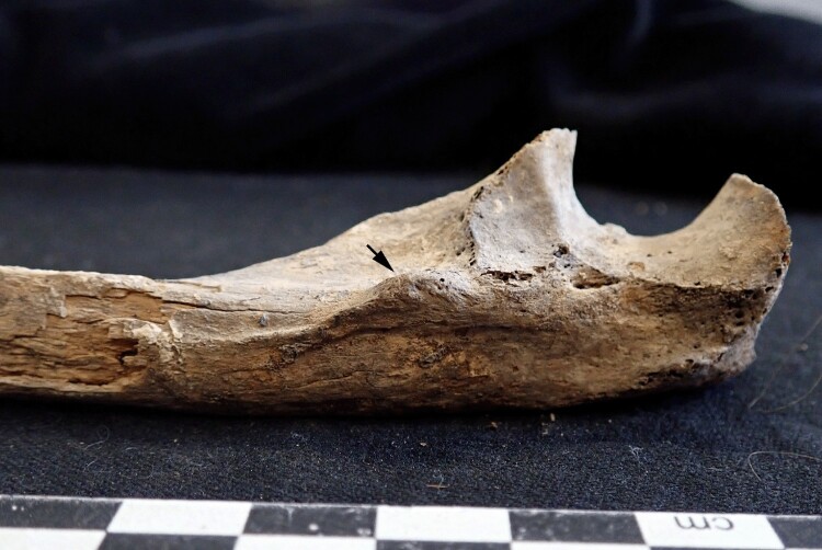
Muscle attachment sites were not systematically recorded in this study, however, all individuals, including the females, displayed high robusticity of entheses (Figure 6). Similarly, while few individuals had observable joint surfaces and those that could be assessed were free of the bone changes associated with osteoarthritis (e.g. eburnation, see TS1). While the causes of osteoarthritis are multi-factorial, including advancing age (Arden and Nevitt 2006; Domett et al. 2017), even Burial 10 (an old female) had no evidence of subchondral bone change. The lack of osteoarthritis in these people is difficult to explain, as high frequencies of the disease are present in contemporary British samples (Brickley and Buteux 2006). This does also highlight the difficulties of using arthritis as a marker of activity due to its multifactorial aetiology. However, while entheseal development is not directly correlated to specific activities (Foster et al. 2014), the evidence of highly developed muscle attachment sites in most of the observable individuals suggests prolonged and strenuous general activity.
Figure 6.
The proximal right ulna of Burial 6. The black arrow indicates the distinctly enlarged bony ridge where the supinator and pronator muscles attached. These muscles are responsible for the rotatory movements of the forearm. All of the muscle attachment sites associated with actions of the forearm, wrist, and hands were enlarged in this individual, suggesting strenuous use.
Evidence of childhood and work-related injury
Traumatic injuries were observed in three of the seven (42.8%) well preserved adults. While the sample is small, this is a very high frequency of trauma and warrants further discussion. Most of these bone fractures occurred well before death so the very low frequency of death by accidents (other than drowning) in the death certificates is perhaps not surprising.
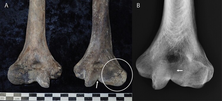
Burial 11 had multiple perimortem and antemortem injuries. The healed fracture of his left humerus (Figure 7) is most likely from a childhood injury (Wadsworth 1964). These types of fractures are the result of extreme varus (inward) indirect force applied to the elbow from a fall (Wadsworth 1964; Hamza et al. 2019). A common complication of this injury is a valgus (outward) deformity of the elbow which effects the carrying angle, restricts extension of the elbow, and can lead to ulnar neuritis (numbness of the medial side of the forearm and hand) (Skak et al. 2001). There is a slight fishtail deformity of the lateral condyle of the humerus in B11 indicating abnormal development of the growth centre during healing and the bone being shorter on this side supports this finding. However, the deformity is not severe and suggests an effective treatment of this common fracture during his childhood at ‘home’ in England. The clinical implications of this injury suggest that B11 was partially disabled in this arm. He likely had limited extension (straightening) ability in this elbow and may have had difficulty carrying heavy weights with this arm. He may have also experienced some loss of sensation in this hand making precision movements of the hand difficult. A similar case was reported in an adult male from St Martin’s, Birmingham, (Brickley and Buteux 2006), but was not illustrated. The flattening of the tip of B11’s left thumb may be work-related (Brickley and Buteux 2006) and risk of injury to this hand may have been increased due to the deformity of the elbow and any loss of sensation.
Figure 7.
Distal humerii of the Burial 11 (A). A depressed linear defect is observable on in apex of the trochlear space (White arrow) and extends to the posterior trochlear surface. The capitulum of the left humerus (white circle) is distorted laterally and inferiorly. The capitululum is considerably enlarged compared the right hand side (pictured left) which is the normal rounded shape for articulation with the radial head. The distortion of the capitulum is clinically described as a ‘fishtail deformity’. B, Radiograph of left distal humerus. The white arrow shows the remains of the fracture line extending from the linear defect in the trochlear apex. This injury likely occurred during childhood (<14–19 years).
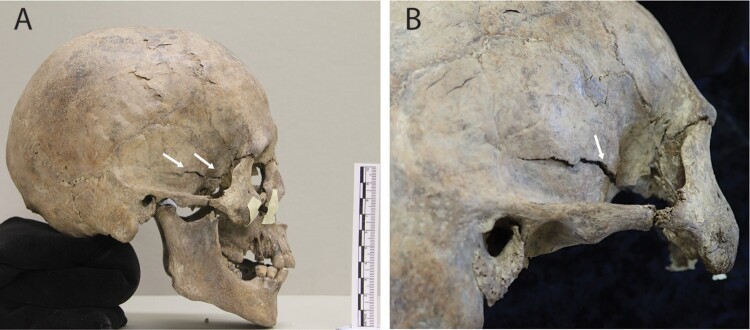
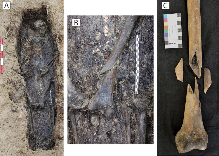
The perimortem injury to the right temporal region of B11’s head is a linear longitudinal basal skull fracture (Figure 8). These fractures are invariably caused by a blow to the front or side of the head and are usually fatal if untreated as they are associated with brain-stem damage (Kerman et al. 2002; Ta’ala et al. 2006; Wedel and Galloway 2014). The perimortem fracture to the left thigh bone was comminuted (the shaft is broken into more than two fragments) (Figure 9), which is usually associated with a direct force to the bone, such as crushing (Lovell 1997; Wedel and Galloway 2014). The femoral shaft is the most highly mineralised and strongest area of the skeleton because of locomotor requirements (Lovell 2008). Therefore, it takes extreme force to fracture this bone and breaks are usually associated with severe blood loss from damage to associated vasculature (Clarke et al. 1955). Today, fractures of the femoral shaft are commonly associated with motor vehicle accidents, car-pedestrian collisions and plane crashes (Salminen et al. 2000; Wedel and Galloway 2014).
Figure 8.
Burial 11. Perimortem (around death) longitudinal fracture of the squamosal portion of the right temporal and greater wing of the sphenoid bone (A; white arrows). This fracture showed minimal displacement, the edges were sharp and stained a similar colour to the rest of the bone (B; white arrow). This injury was likely fatal.
Figure 9.
A, Burial 11 in situ in his coffin. His left femur has rotated outwards and the distal part of the shaft is fractured perimortem. B, A closer view of the perimortem fracture shows the oblique perimortem fracture line on the posterior aspect of the bone. The oblique posterior fracture pattern and staining of the fractured bone edges is highly suggestive of perimortem rather than postmortem trauma to the bone C. Anterior view of the femur with the shattered bone pieces (comminuted). The fracture line extends upwards through the shaft. This type of bone fracture is caused by direct blunt force trauma, commonly crushing accidents.
Due to the perimortem injuries suffered by Burial 11, alongside his age and sex, his identity is suspected to be that of a gold miner described in the records, who died at the small mining settlement of Canada Reef in a mining accident. He was 37 years old when he died and was born in Oxford, England in c1840 (Findlay 2016). The strontium isotope work conducted on this individual is consistent with an origin in southern England (King et al. 2020). A newspaper account of the inquest into his death describes the accident: One to two tons of rock and earth fell on him while he was working underground in the mine. A schoolmaster who first attended the injured miner stated ‘I noticed a wound on his temple three quarters of an inch from the left eye. Above his left ear the skull seemed to be fractured’ (Tuapeka Times 1877). Despite the fact the injury to the deceased is described on the left rather than right hand side, the location of the wound is the same, and the observed fracture is consistent with this type of trauma (Kranioti 2015). No mention was made of other injuries to his body at the inquest, perhaps not surprising given the wound to his head was fatal. Finally, this deceased miner is the only individual in the historical records found where death occurred from such extreme trauma, further supporting his identification.
Rib fractures are most commonly associated with blunt force trauma to the chest and in modern times are often caused by motor vehicle accidents (Wedel and Galloway 2014). A nineteenth century equivalent could be injury associated with horse drawn vehicles and horseback riding (Brickley and Buteux 2006). There are numerous newspaper accounts in this period of either from being thrown from a horse, kicked by a horse, or people being killed and injured in cart accidents (either being run over by them or thrown from them). Other causes of trauma to the chest are work related, assault, and interpersonal violence, especially domestic violence (Wedel and Galloway 2014). The clinical implications of rib fractures are usually overlooked in bioarchaeology as mundane, however, they signify an inability to work for some weeks at the very least, and in more extreme circumstances, life-threatening injury to organs or vessels (Brickley 2006).
Burial 13 had three well healed antemortem fractures to the ribs. These injuries represent at least one period of significant discomfort and disability during this man’s life. The two rib injuries of Burial 11 occurred in separate incidents, one close to the time of death and the other was well-healed. The transverse alignment of these fractures antemortem fractures all indicate direct force causing the injury (Lovell 2000). The perimortem sharp force injury to a lower right rib fragment in Burial 11 may have contributed to this man’s demise by damaging internal organs, and adds to the perimortem femoral and cranial fractures as evidence for his death by crushing in a rock fall.
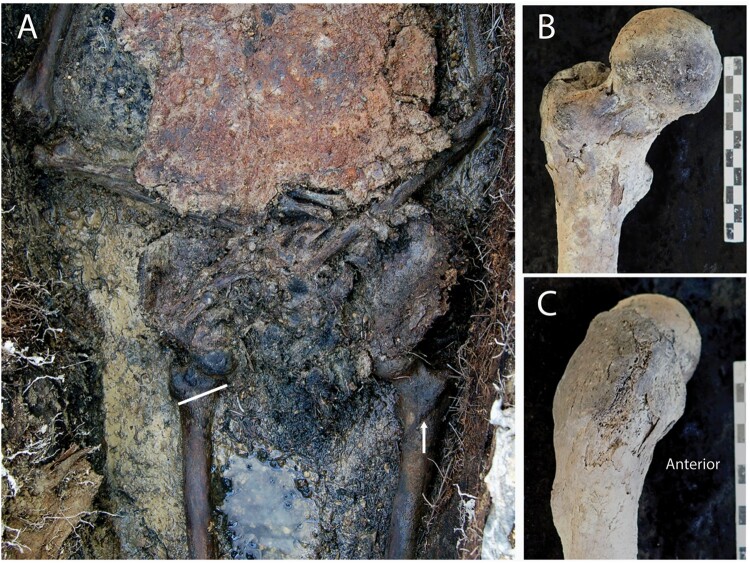
The structural changes to the right femur (and possibly the right tibia) observed in Burial 13 (Figure 10) may be due to a plastic deformation fracture during childhood (Glencross and Stuart-Macadam 2000). This type of fracture results in a permanent bending deformity of the affected bone without the typical callus formation (Lavery et al. 2007). However, fractures of any type to the proximal femur and affecting the hip joint are very rare in children (Brousil and Hunter 2013), and other causes such as some form of dysplasia should also be considered.
Figure 10.
The right femur of Burial 13. A, In situ image of the pelvis and hip area showing the transverse (abnormal) orientation of the right intertrochanteric line (attachment for the iliofemoral ligament of the hip joint) compared with the oblique (normal) orientation on the left side. B and C, The proximal third of the right femur is significantly bowed anteriorly. No fracture callus was appreciated in radiography of the bone.
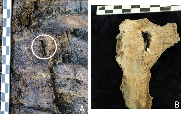
The overall pattern of trauma in Burial 11 (four fractures) and Burial 13 (three, possibly five fractures) attests to repeated exposure to risks of injury throughout life. This story of injury recidivism through the life-course is not an uncommon one for this time period in England (Brickley and Buteux 2006), and in other Otago sites (Petchey et al. 2018). Whether the childhood trauma was related to manual labour while still in Britain is unknown, but child labour was common amongst the poor and working class (Gowland et al. 2018). The possible perimortem sharp force injury (Figure 11) to Burial 29’s chest would be unlikely to have caused her death, but any other injuries to soft tissues are not visible in the skeletal record. This defect was visible during the excavation process and the margins of the break are uniformly coloured with the overlying cortex, suggesting that this occurred before the remains were interred. However, without further imaging of this trauma, it is not possible to firmly differentiate it from post mortem damage and therefore remains speculative. Her story is the subject of ongoing analysis and will be reported in full in a forthcoming paper.
Figure 11.
Burial 29 sternum. A, A triangular perimortem sharp force injury (2.0 cm in length) to the anterior sternum photographed in situ. B, The same lesion in clean and dry bone. There is a fragment of cortical bone pushed into the cancellous bone (arrow) supporting the perimortem timing of the injury.
Evidence of community care
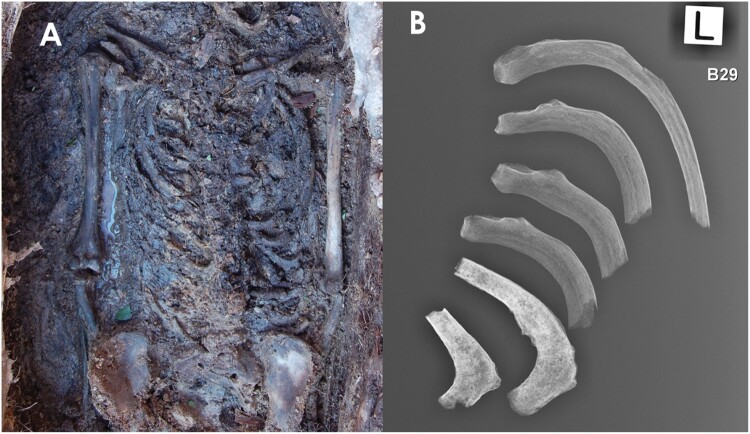
The evidence for chronic disease and/or potentially disabling trauma in the SJM population allows us to also consider evidence of care (both medical and community) for those afflicted with debilitating conditions in this community (Tilley 2015). In the case of Burial 21, as already mentioned, we know from historical accounts that he and his family were supported by an example of an incipient social welfare system (Snoddy et al. 2020). Burial 29 was afflicted with some form of skeletal dysplasia, one that she likely suffered from since birth (Figure 12). Her dietary life history and relatively elaborate coffin furniture suggest that she may have been born into a family with greater wealth than others in the community (King et al. 2020), perhaps buffering her from the need to perform duties required of those of the lower classes for survival. The antemortem childhood trauma in Burials 11 and 13 also indicate that they experienced periods of disability requiring medical intervention but also a prolonged period of familial and community support back ‘home’ in England. Furthermore, their healed rib fractures would have limited their ability to work for several weeks at least, a theme touched on in other cases in Otago (Petchey et al. 2018). While evidence of care for the disabled has been found in skeletons thousands of years old (e.g. Tilley and Oxenham 2011) these cases from SJM also offer tantalising glimpses into the social history of our colonial past.
Figure 12.
Burial 29 pathological ribs. A, The thorax of Burial 29 in situ. B, Radiograph of left ribs. The lower ribs show the calcification of the costal cartilage.
Dental evidence for stress and disease
The very high frequency of LEH (85.7%) in the sample is indicative of physiological stress during tooth development. There are few comparative data from post-medieval period England, however the lower status ‘earth cut graves’ from St Martins, Birmingham had a similar high prevalence (73.5%) compared with higher status ‘vault burials’ (47%) (Brickley and Buteux 2006). All of the adults with appropriate dentition (n = 7) from SJM have been isotopically identified as non-local, and thus are likely first generation colonists to New Zealand (King et al. 2020). As these episodes of stress occurred during childhood, they represent periods of stress at ‘home’ rather than in the colony and suggest that affected individuals were of a working-class background.
Isotopic analysis of the SJM burials suggests that their diet was broadly similar between individuals, based on the consumption of terrestrial crops (e.g. wheat, barley, staple vegetables), with variable amounts of domestic animal meat (pork, beef, mutton) – similar to the diet of contemporary Britain. There is some suggestion from both isotopic analysis and historical sources (Otago Witness 1868, 1869) that the availability of farmed meat was limited in the earliest days of the settlement. However, the availability of wild wetland resources nearby means that colonists seem to have supplemented their farmed diet to ameliorate nutritional stress in the colony (King et al. in press).
The oral health of a population can inform on diet, extra-masticatory use of teeth (e.g. pipe smoking), and general health (Hillson 2008). Poor oral health (particularly gum disease) can have severe systemic health consequences (Beck et al. 2000). The oral health of the adults from SJM is relatively typical of the time period, and fits well with the description of British populations by Roberts and Cox (2003, p. 324):‘At the outbreak of the First World War many of the working classes still had appalling dental health reflecting a soft cariogenic diet, a lack of oral hygiene and inadequate dental treatment’.
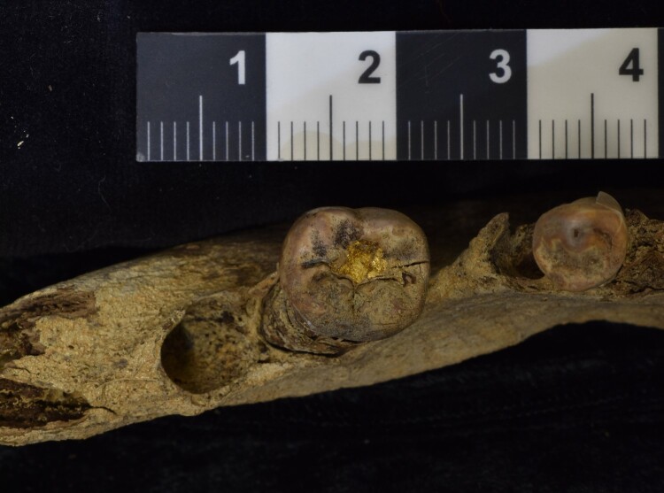
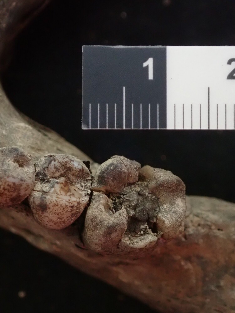
The high rates of AMTL, especially in the posterior teeth, suggest that many molars were lost as a result of tooth decay and periodontal disease. Two individuals had evidence of dental intervention (the doctor and his wife), although whether this was back home (Bavaria for the doctor) or in New Zealand is not known. The use of gold for filling cavities, as seen in Burial 4 (Figure 13), predates the use of amalgam (the type of filling seen in his wife Burial 6) (Figure 14), which was not developed until the early nineteenth century (Roberts and Cox 2003). In post-medieval England, dental care was available mostly to the middle and upper classes (Roberts and Cox 2003), and the higher status of this couple in the community would be reflected in this dental evidence regardless of their positive identification during excavation.
Figure 13.
Burial 4. Gold filling present on occlusal surface of left second molar.
Figure 14.
Amalgam in the occlusal surface of the left first molar in Burial 6. The crown of the tooth has fractured antemortem.
All the men had evidence for pipe facets and one of the two women (B6) also displayed pipe facets on her teeth. Smoking is a major risk factor associated with chronic destructive periodontal disease (Bergström 2004), though it is also associated with a number of genetic and environmental factors. However, with the ubiquitous tobacco use at SJM, it is likely that both tobacco smoking and tartar build-up from diet and poor oral hygiene influenced disease progression.
Conclusion
This paper aimed to characterise the life-course of early settlers to New Zealand through historical epidemiological and osteological analyses. These analyses have provided glimpses into the past of strenuous manual labour, repeated risk of injury, and oral and skeletal infections. Mortality of infants was very high in the skeletal sample and the death certificates outline the varied risks of infection and accidents they faced.
The osteobiographies of the seven adults also demonstrate the detailed narratives that can be gleaned from careful consideration of individuals. These individual stories are often lost in population-based analyses of larger samples. The question of whether the people’s health was ‘better’ in the colony than back at home is complicated. Certainly the skeletal record indicates childhood stress affecting growth and risk of injury prior to migration. However, the historical record suggests that occupational risks of death to the working class were similar in the new colony as at home. The snapshot of this Victorian-era population provided by these data suggests that the colonial society transported their biosocial landscape on immigration and little changed for these initial colonists.
Supplementary Material
Acknowledgements
Funding for the excavation was provided by a Grant-in-Aid from the Department of Anatomy, University of Otago. The ongoing analyses were funded by a Marsden Fund Grant awarded to HB and PP (18-UOO-028) and a University of Otago Research Grant. We are particularly grateful to Wayne Stevenson for the donation of his time and use of his digger, and Grant Love (the farmer) for the use of his haybarn, water supply and access to his land. This project is a collaboration between many individuals and groups. The TP60 group who instigated the work at St. Johns Cemetery and provided a vast amount of background research are Robert Findlay, Kath Croy, Isobel Michelle, Mary-Anne Miller and Rev. Vivienne Galletly. Other locals provided a great deal of support, including Dudley Finch and Geoff Finch. Bishop Kelvin Wright has supported the project wholeheartedly, and provided his permission for the excavation to be undertaken. Rachel Wesley of Te Rūnaka o Ōtākou provided Māori tikanga guidance, and participated in the excavation. Richard Walter and Phil Latham of the Otago Archaeology & Anthropology programme provided much of the excavation and field equipment. Lee Maher of Pacific Radiology for her assistance with radiography.
Funding Statement
Funding for the excavation was provided by a Grant-in-Aid from the Department of Anatomy, University of Otago. The ongoing analyses were funded by a Marsden Fund Grant awarded to HB and PP [18-UOO-028] and a University of Otago Research Grant.
Disclosure statement
No potential conflict of interest was reported by the author(s).
References
- Acsádi G, Nemeskéri J.. 1970. History of human life and mortality. Budapest: Akadémiai Kiadó. [Google Scholar]
- Alter G, Carmichael A.. 1999. Classifying the dead: toward a history of the registration of causes of death. Journal of the History of Medicine. 54:114–132. [DOI] [PubMed] [Google Scholar]
- Andrews J. 2009. No other home than this: a history of European New Zealanders. Nelson: Craig Potton Publishing. [Google Scholar]
- Arden N, Nevitt M.. 2006. Osteoarthritis: epidemiology. Best Practice and Research Clinical Rheumatology. 20(1). DOI: 10.1016/j.berh.2005.09.007. [DOI] [PubMed] [Google Scholar]
- Ballantyne T. 2012. Webs of empire: locating New Zealand’s colonial past. Wellington: Bridget Williams Books. [Google Scholar]
- Beck J, Slade G, Offenbacher S.. 2000. Oral disease, cardiovascular disease and systemic inflammation. Periodontology. 23(1):110–120. [DOI] [PubMed] [Google Scholar]
- Belich J. 2009. Replenishing the earth. The settler revolution and the rise of the Anglo world, 1783–1939. Oxford: Oxford University Press. [Google Scholar]
- Bergström J. 2004. Tobacco smoking and chronic destructive periodontal disease. Odontology. 92(1):1–8. [DOI] [PubMed] [Google Scholar]
- Brickley M. 2006. Rib fractures in the archaeological record: a useful source of sociocultural information? International Journal of Osteoarchaeology. 16:61–75. [Google Scholar]
- Brickley M, Buteux S.. 2006. St Martin's uncovered: investigations in the churchyard of St Martins-in-the-Bull Ring, Birmingham, 2001. Oxford: Oxbow Books. [Google Scholar]
- Brooks S, Suchey J.. 1990. Skeletal age determination based on the os pubis: a comparison of the Ascádi-Nemeskéri and Suchey-Brooks methods. Human Evolution. 5:227–238. [Google Scholar]
- Brousil J, Hunter J.. 2013. Femoral fractures in children. Current Opinion in Pediatrics. 25(1):52–57. [DOI] [PubMed] [Google Scholar]
- Bruce Herald . 1865. Jul 13.
- Bruce Herald . 1867. Jul 31.
- Bruce Herald . 1868. Feb 19.
- Bruce Herald . 1868. Jul 8.
- Bruce Herald . 1868. Mar 11.
- Bruce Herald . 1874. Jan 27.
- Buckley H. 2007. Possible gouty arthritis in Lapita-associated skeletons from Teouma, Efate Island, Central Vanuatu. Current Anthropology. 48(5):741–749. [Google Scholar]
- Buckley H, Petchey P.. 2018. Human skeletal remains and bioarchaeology in New Zealand. In: O'Donnabhain B., Lozada M. C., editors. Archaeological human remains: legacies of imperialism, communism and colonialism. Cham (Switzerland: ): Springer; p. 93–110. [Google Scholar]
- Buikstra J, Ubelaker D, editors. 1994. Standards for data collection from human skeletal remains. Fayetteville: Arkansas Archaeological Survey. 206 p. [Google Scholar]
- Clarke R, Topley E, Fear C.. 1955. Assessment of blood-loss in civilian trauma. The Lancet. 268(6865):629–638. [DOI] [PubMed] [Google Scholar]
- Domett K, Evans C, Chang N, Tayles N, Newton J.. 2017. Interpreting osteoarthritis in bioarchaeology: highlighting the importance of a clinical approach through case studies from prehistoric Thailand. Journal of Archaeological Science: Reports. 11:762–773. [Google Scholar]
- Early New Zealand Books . 2017. Early New Zealand Books. New Zealand: University of Auckland. http://www.enzb.auckland.ac.nz. [Google Scholar]
- Elwood P. 1965. Respiratory symptoms in men who had previously worked in a flax mill in Northern Ireland. British Journal of Industrial Medicine. 22:38–42. [DOI] [PMC free article] [PubMed] [Google Scholar]
- Eveleth PB, Tanner JM.. 1990. Worldwide variation in human growth. 2nd ed. Cambridge: Cambridge University Press. [Google Scholar]
- Findlay R. 2016. A history of the St. John's burial ground, Back Road, Tokomairiro, Otago. Auckland (New Zealand: ): Robert M. Findlay. [Google Scholar]
- Foster A, Buckley H, Tayles N.. 2014. Using enthesis robusticity to infer activity in the past: a review. Journal and Archaeological Method and Theory. 21:511–533. [Google Scholar]
- Glencross B, Stuart-Macadam P.. 2000. Childhood trauma in the archaeological record. International Journal of Osteoarchaeology. 10:198–209. [Google Scholar]
- Gowland R. 2018. ‘A mass of crooked alphabets’: the construction and othering of working class bodies in industrial England. In: Stone PK, editor. Bioarchaeological analyses and bodies, bioarchaeology: New ways of knowing anatomical and archaeological skeletal collections. Cham (Switzerland): Springer International Publishing; p. 147–160. [Google Scholar]
- Gowland R, Caffell A, Newman S, Levene A, Holst M.. 2018. Broken childhoods: rural and urban non-adult health during the industrial revolution in Northern England (eighteenth–nineteenth centuries). Bioarchaeology International. 2(1):44–62. [Google Scholar]
- Hamza M, Hussein J, Mahdi M.. 2019. Outcome of operative treatment of displaced lateral condyle fractures of the humerus in children in relation to time of presentation. International Journal of Developmental Research. 9(7):28975–28988. [Google Scholar]
- Hillson S. 2008. Dental pathology. In: Katzenberg MA, Saunders S, editors. Biological anthropology of the human skeleton. New York (NY: ): Wiley-Liss; p. 295–334. [Google Scholar]
- Hocken T. 1898. Contributions to the early history of New Zealand. (Settlement of Otago). London: Sampson, Low, Marston and Company. [Google Scholar]
- Jantz R, Hunt D, Meadows L.. 1994. Maximum length of the tibia – how did trotter measure it. American Journal of Physical Anthropology. 93(4):525–528. [DOI] [PubMed] [Google Scholar]
- Kerman M, Cirak B, Dagtekin A.. 2002. Management of skull base fractures. Neurosurgery Quarterly. 12(1):23–41. [Google Scholar]
- King C, Buckley H, Petchey P, Kinaston R, Millard A, Zech J, Roberts P, Matisoo-Smith E, Nowell G, Gröcke D.. 2020. A multi-isotope, multi-tissue study of colonial origins and diet in New Zealand. American Journal of Physical Anthropology. 172:605–620. [DOI] [PubMed] [Google Scholar]
- King C, Petchey P, Kinaston R, Gröcke D, Millard A, Wanhalla A, Brooking T, Matisoo-Smith E, Buckley H.. in press. A land of plenty? Colonial diet in rural New Zealand. Historical Archaeology. 55:1. [Google Scholar]
- Kranioti E. 2015. Forensic investigation of cranial injuries due to blunt force trauma: current best practice. Research and Reports in Forensic Medical Science. 5:25–37. [Google Scholar]
- Lake County Press . 1885. Dec 31.
- Lavery M, Huang M, Annunziato A.. 2007. Plastic deformity in a 9-year-old boy. Clinical Orthopaedics and Related Research. 462:238–241. [DOI] [PubMed] [Google Scholar]
- Lovejoy C, Meindl R, Pryzbeck T, Mensforth R.. 1985. Chronological metamorphosis of the auricular surface of the ilium: a new method for the determination of adult skeletal age at death. American Journal of Physical Anthropology. 68:15–28. [DOI] [PubMed] [Google Scholar]
- Lovell N. 1997. Trauma analysis in paleopathology. Yearbook of Physical Anthropology. 40:139–170. [Google Scholar]
- Lovell N. 2008. Analysis and interpretation of skeletal trauma. In: Katzenberg MA, Saunders SR, editors. Biological anthropology of the human skeleton. 2nd ed. Hoboken (NJ: ): Wiley-Liss; p. 341–386. [Google Scholar]
- Lovell N. 2000. Paleopathological description and diagnosis. In: Saunders S, editor. Biological anthropology of the human skeleton. New York: Wiley-Liss; p. 217–248. [Google Scholar]
- MacBean Stewart F. 1875. ‘Medical Officer of Health, Milton, to His Worship the Mayor’. Reports from the Boards of Health in the Various Provinces, H-22, AJHR.
- Mays S, Brickley M, Ives R.. 2009. The effects of socioeconomic status on endochondral and appositional bone growth, and acquisition of cortical bone in children from 19th century Birmingham, England. American Journal of Physical Anthropology. 140(3):410–416. [DOI] [PubMed] [Google Scholar]
- Milner G. 1992. Determination of skeletal age and sex: a manual prepared for the Dickson Mounds Reburial Team. Lewiston (IL: ): Dickson Mounds Museum. [Google Scholar]
- Mohammadien H, Hussein M, El-Sokkary R.. 2013. Effects of exposure to flour dust on respiratory symptoms and pulmonary function of mill workers. Egyptian Journal of Chest Diseases and Tuberculosis. 62:745–753. [Google Scholar]
- Moorrees C, Fanning E, Hunt E.. 1963a. Formation and resorption of three deciduous teeth in children. American Journal of Physical Anthropology. 21:205–213. [DOI] [PubMed] [Google Scholar]
- Moorrees C, Fanning E, Hunt E.. 1963b. Age variation of formation stages for ten permanent teeth. Journal of Dental Research. 42(6):1490–1502. [DOI] [PubMed] [Google Scholar]
- North Otago Times. 1868. Social and domestic. Volume X, Issue 290, 3 March 1868. https://paperspast.natlib.govt.nz/newspapers/NOT18680303.2.45?items_per_page=50&page=20194&query=auckland+star
- Olssen E. 1984. A history of Otago. Dunedin: John McIndoe.
- Otago Witness . 1868. Mar 21. Dear meat.
- Otago Witness . 1869. Jan 16. The price of meat.
- Otago Witness . 1894. Feb 1.
- Petchey P, Buckley H, Kinaston R, Smith B.. 2017. A nineteenth century settlers’ graveyard: preliminary report on the excavation of St. John’s cemetery, Back Road, Milton, Otago. Archaeology in New Zealand. 60(1):19–30. [Google Scholar]
- Petchey P, Buckley H, Scott R.. 2018. Life, death and care on the Otago goldfields: A preliminary glimpse. Journal of Pacific Archaeology. 9(2):44–58. [Google Scholar]
- Phenice T. 1969. A newly developed visual method of sexing in the os pubis. American Journal of Physical Anthropology. 30:297–301. [DOI] [PubMed] [Google Scholar]
- Pool I. 1991. Te Iwi Māori: a New Zealand population past, present and projected. Auckland: Auckland University Press. [Google Scholar]
- Pool I. 2015. Colonization and development in New Zealand between 1769 and 1900: the seeds of Rangiatea. Charbit Y, Pool I, editors. Switzerland: Springer. [Google Scholar]
- Roberts P. 2006. Gold fever. Disease and its cultural relationship: a case study on the development of the Colony of Victoria, 1950 to 1900 [unpublished thesis]. The Australian National University.
- Roberts P. 2014. Diagnosis as an artefact: a case study to determine the meaning of ‘ague’ and ‘remittent fever’ in nineteenth century Victoria. The Artefact. 37:3–17. [Google Scholar]
- Roberts C, Cox M.. 2003. Health and disease in Britain from prehistory to the present day. Thrupp: Sutton Publishing Limited. [Google Scholar]
- Roberts C, Manchester K.. 2005. The archaeology of disease. Ithaca (NY: ): Cornell University Press. [Google Scholar]
- Salminen S, Pihlajamaki H, Avikainen V, Bostman O.. 2000. Population based epidemiologic and morphologic study of femoral shaft fractures. Clinical Orthopaedics and Related Research. 372(372):241–249. [DOI] [PubMed] [Google Scholar]
- Schaefer M, Black S, Scheuer L.. 2009. Juvenile osteology: a laboratory and field manual. London: Academic Press. [Google Scholar]
- Scheuer L, Black S.. 2000. Developmental juvenile osteology. London: Academic Press. [Google Scholar]
- Schrader B. 2016. The big smoke. New Zealand cities 1840–1920. Wellington: Bridget Williams Books. [Google Scholar]
- Skak S, Olsen S, Smaabrekke A.. 2001. Deformity after fracture of the lateral humeral condyle in children. Journal of Paediatric Orthopaedics Part B. 10:142–152. [PubMed] [Google Scholar]
- Smith B. 1984. Patterns of molar wear in hunter-gatherers and agriculturalists. American Journal of Physical Anthropology. 63:39–56. [DOI] [PubMed] [Google Scholar]
- Snoddy A, Buckley H, King C, Kinaston R, Nowell G, Gröcke D, Duncan W, Petchey P.. 2020. ‘Captain of all these men of death’: an integrated case study of tuberculosis in nineteenth-century Otago. New Zealand. Bioarchaeology International. 3(4):217–237. [Google Scholar]
- Sumpter D, Lewis J.. 1949. Faith and toil. The story of Tokomairiro. Christchurch: Otago Centennial Historical Publications. [Google Scholar]
- Ta’ala S, Berg G, Haden K.. 2006. Blunt force cranial trauma in the Cambodian killing fields. Journal of Forensic Sciences. 51(5):996–1001. [DOI] [PubMed] [Google Scholar]
- Thomas T, Stewart P.. 1987. Mortality from lung cancer and respiratory disease among pottery workers exposed to silica and talc. American Journal of Epidemiology. 125(1):35–43. [DOI] [PubMed] [Google Scholar]
- Tilley L. 2015. Theory and practice in the bioarchaeology of care. Cham (Switzerland): Springer International. [Google Scholar]
- Tilley L, Oxenham M.. 2011. Survival against the odds: modelling the social implications of care provision to serious disabled individuals. International Journal of Paleopathology. 1:35–42. [DOI] [PubMed] [Google Scholar]
- Trotter M, Gleser G.. 1952. Estimation of stature from long bones of American Whites and Negroes. American Journal of Physical Anthroplogy. 10(4):463–514. [DOI] [PubMed] [Google Scholar]
- Trotter M, Gleser G.. 1958. A re-evaluation of estimation of stature based on measurements of stature taken during life and of long bones after death. American Journal of Physical Anthropology. 16(1):79–123. [DOI] [PubMed] [Google Scholar]
- Tuapeka Times . 1877. Nov 14. Inquest.
- Wadsworth T. 1964. Premature epiphyseal fusion after injury of the capitulum. The Journal of Bone and Joint Surgery. 46B(1):46–49. [PubMed] [Google Scholar]
- Waldron T. 1994. Counting the dead: the epidemiology of skeletal populations. Surrey: Wiley. [Google Scholar]
- Wedel V, Galloway A.. 2014. Broken bones: anthroplogical analysis of blunt force trauma. Proquest Ebook Central, Charles C. Thomas Publisher. [Google Scholar]
- Wheeler B, Taylor B, de Lange M, Harper M, Jones S, Mekhail A, Houghton L.. 2018. A longitudinal study of 25-hydroxy vitamin D and parathyroid hormone status throughout pregnancy and exclusive lactation in New Zealand mothers and their infants at 45° S. Nutrients. 10:86–12. [DOI] [PMC free article] [PubMed] [Google Scholar]
- White P. 2017. The archaeology of American mining. Gainsville (FL: ): University Press of Florida. [Google Scholar]
- WHO . 2016. ICD-10 version 2016. Available from International Statistical Classification of Diseases and Related Health Problems 10th Revision. [PubMed]
- Wohl A. 1983. Endangered lives; public health in Victorian England. Cambridge (MA: ): Harvard University Press. [Google Scholar]
Associated Data
This section collects any data citations, data availability statements, or supplementary materials included in this article.


