

Abstract
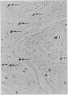
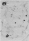
Mast cell counts were carried out on sections of human carotid bodies from 39 subjects showing one of four stages of histological change associated with aging, and in five subjects showing different forms of histopathology in the carotid body associated with disease. There was no relation between mast cell density and age or the histological changes associated with aging of glomic tissue. The normal range of mast cell density calculated in terms of the 80% confidence limits was 18.5 to 67.5/mm2. In three middle aged subjects with carotid bodies of normal histological appearance there was an abnormally high density of 83 to 96/mm2. In two elderly subjects showing age changes of fibrosis and accumulation of lymphocytes there was an abnormally low density of 12/mm2 or less. Mast cell density was not related to different types of carotid body hyperplasia. The mast cells were essentially stromal in location, usually closely applied to the walls of small glomic blood vessels, and were rarely found in intimate association with glomic chief cells. This suggests that mast cells are not directly concerned with the functions of glomic cells but does not preclude the possibility that they may have some effect on regulating glomic blood vessels and thus participate in the distribution of blood supply within the carotid body.
Full text
PDF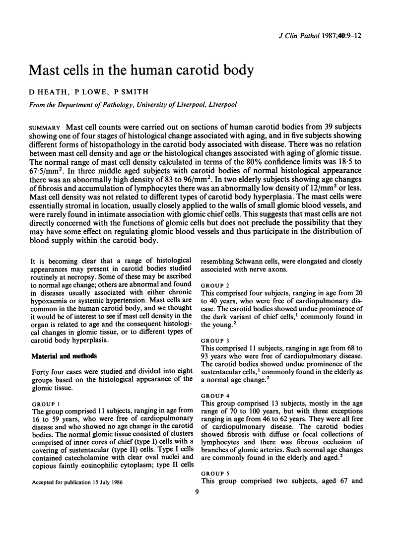
Images in this article
Selected References
These references are in PubMed. This may not be the complete list of references from this article.
- Arias-Stella J., Bustos F. Chronic hypoxia and chemodectomas in bovines at high altitudes. Arch Pathol Lab Med. 1976 Dec;100(12):636–639. [PubMed] [Google Scholar]
- Grimley P. M., Glenner G. G. Histology and ultrastructure of carotid body paragangliomas. Comparison with the normal gland. Cancer. 1967 Sep;20(9):1473–1488. doi: 10.1002/1097-0142(196709)20:9<1473::aid-cncr2820200914>3.0.co;2-i. [DOI] [PubMed] [Google Scholar]
- HEATH I. D. Staining of sulphated mucopolysaccharides. Nature. 1961 Sep 30;191:1370–1371. doi: 10.1038/1911370a0. [DOI] [PubMed] [Google Scholar]
- Heath D., Smith P., Hurst G. The carotid bodies in coarctation of the aorta. Br J Dis Chest. 1986 Apr;80(2):122–130. doi: 10.1016/0007-0971(86)90032-x. [DOI] [PubMed] [Google Scholar]
- Heath D., Smith P., Jago R. Dark cell proliferation in carotid body hyperplasia. J Pathol. 1984 Jan;142(1):39–49. doi: 10.1002/path.1711420109. [DOI] [PubMed] [Google Scholar]
- Heath D., Smith P., Jago R. Hyperplasia of the carotid body. J Pathol. 1982 Oct;138(2):115–127. doi: 10.1002/path.1711380203. [DOI] [PubMed] [Google Scholar]
- Heath D., Trueman T., Sukonthamarn P. Pulmonary mast cells in mitral stenosis. Cardiovasc Res. 1969 Oct;3(4):467–471. doi: 10.1093/cvr/3.4.467. [DOI] [PubMed] [Google Scholar]
- Hurst G., Heath D., Smith P. Histological changes associated with ageing of the human carotid body. J Pathol. 1985 Nov;147(3):181–187. doi: 10.1002/path.1711470306. [DOI] [PubMed] [Google Scholar]
- PRYSE-DAVIES J., DAWSON I. M. SOME MORPHOLOGIC, HISTOCHEMICAL, AND CHEMICAL OBSERVATIONS ON CHEMODECTOMAS AND THE NORMAL CAROTID BODY, INCLUDING A STUDY OF THE CHROMAFFIN REACTION AND POSSIBLE GANGLION CELL ELEMENTS. Cancer. 1964 Feb;17:185–202. doi: 10.1002/1097-0142(196402)17:2<185::aid-cncr2820170208>3.0.co;2-1. [DOI] [PubMed] [Google Scholar]
- Smith P., Hurst G., Heath D., Drewe R. The carotid bodies in a case of ventricular septal defect. Histopathology. 1986 Aug;10(8):831–840. doi: 10.1111/j.1365-2559.1986.tb02581.x. [DOI] [PubMed] [Google Scholar]
- Smith P., Jago R., Heath D. Anatomical variation and quantitative histology of the normal and enlarged carotid body. J Pathol. 1982 Aug;137(4):287–304. doi: 10.1002/path.1711370404. [DOI] [PubMed] [Google Scholar]