



Abstract
Purpose of review:
Differences of sex development (DSD) are a group of chromosomal, gonadal, and anatomic conditions that are not often diagnosed during pregnancy. Families and clinicians need diagnostic guidance that supports all aspects of the care from the prenatal to postnatal period.
Recent findings:
Noninvasive prenatal screening (NIPS) is obtained by sampling cell-free fetal DNA in the mother’s bloodstream in the first trimester. While its primary purpose is to screen for genetic aneuploidies, it is also used to determine the sex of the fetus. When screening ultrasound shows genital anatomy that is discordant with the sex determination by NIPS, a DSD workup is warranted. The use of this relatively new screening tool may result in a higher number of prenatal referrals than in the past.
Summary:
This review summarizes suggested prenatal counseling, neonatal management, and postnatal workup of the most common DSD diagnoses. All of these diagnoses are rare, but the common features that families face are addressed with particular emphasis on psychosocial support and a measured shared decision-making approach.
Keywords: Difference of sex development, genital difference, noninvasive prenatal screening, noninvasive prenatal testing, shared decision making
1. Introduction
Differences of sexual development (DSD) are a diverse group of conditions affecting the upper and lower reproductive tracts. The etiology of these conditions can be genetic, biochemical, developmental or a combination of these causes. The most common DSD seen by pediatricians is congenital adrenal hyperplasia, for which newborn screening is universally done. Other diagnoses in newborns would be hypospadias and other anatomical abnormalities of XY infants. Adolescents can also present for the first time with a DSD if they undergo a workup for primary amenorrhea. Common diagnoses in this situation include androgen insensitivity and vaginal agenesis. The pediatrician may be the first to diagnose a DSD and knowledge of the conditions is helpful to ensure referral to a center with expertise and multidisciplinary care.
Prior to the introduction of noninvasive prenatal screening (NIPS), anatomical abnormalities of the internal and external reproductive organs were not routinely or accurately diagnosed on prenatal screening ultrasound. An estimated 25–50% of pregnant women in the U.S. undergo NIPS, usually at 10 to 12 weeks of gestation, at which time fetal sex chromosomes are documented. If a discrepancy is then identified on screening ultrasound, the family may be referred for further testing, imaging, and counseling.(1, 2)
Diagnoses resulting in a discrepancy between NIPS and prenatal imaging include XY sex chromosomes with female-appearing or atypical genitalia, XX sex chromosomes with male-appearing or atypical genitalia, and XY/XO sex chromosomes with mixed gonadal features. Because of the more recent introduction and use of NIPS, there is a current gap in the literature addressing how to offer a consistent evaluation of discordant prenatal findings. Additionally, there is a lack of consensus on how much of the diagnostic workup can be or should be done prenatally. Although these diagnoses are rare, delivery teams may encounter them and should know how to sensitively address the topic of genital or reproductive tract differences with families.
We will review how atypical genitalia may be prenatally identified by ultrasound or NIPS and how to approach these discrepancies during the prenatal period. We provide a guide in the form of a checklist for both clinicians and parents regarding expectations of postnatal workup. This division of care into prenatal, neonatal, and postnatal phases can be explained in a manner so parents understand the steps of their child’s journey and that their child will become the key decision maker when possible. (Figure 1)
Figure 1.

Stepwise approach to counseling and evaluation of prenatal and postnatal findings of DSD
2. Overview of Sex Development
DSD are a group of chromosomal, gonadal, and anatomic anomalies that can affect the internal and external reproductive tract.(3) Many conditions represent an interruption in normal development where some organs are poorly or incompletely formed. Families are not expected to understand early fetal development, but an appropriate explanation is as follows:
Gonadal development begins with structures that are identical in XX and XY fetuses and differentiation into testes or ovaries starts in the 5th and 6th week of development.
If a fetus has XY sex chromosomes, the testes typically form and the presence of testosterone and anti-Mullerian hormone will work to further develop male features and prevent female structures from forming.
If a fetus has XX sex chromosomes, ovaries typically form and the Mullerian structures, including fallopian tubes and the uterus, should develop.
Inherited genetic alterations may affect the process of gonadal development.
Additionally, there are multiple known genes that can affect fetal reproductive health. (Figure 2) The close relationship between kidney and gonadal development underlies the potential for kidney and ureteral anomalies, so the genitourinary system may be evaluated before and after birth. Online resources for families are available demonstrating gonadal and reproductive development.(4, 5)
Figure 2.

Genes and the pathways involved in embryonic development.
3. Prenatal Identification of Possible Genital Difference
Retrospective review of postnatally confirmed cases of DSD reveals that prenatal detection is not common, although the detection rate may be increasing due to discrepancy between the results of NIPS and imaging findings. Other reasons for prenatal referral for consultation with pediatric specialists, aside from discordant results, are abnormal ultrasound findings when obtained for reasons unrelated to the reproductive tract and in the absence of NIPS and genetic abnormalities. The detection rate of a DSD on prenatal ultrasound alone is 15–24%.(6)
NIPS has been found to be accurate in the determination of fetal chromosomal sex and if discrepancies are noted, they remain rare.(7, 8) Less than 2% of live births have any genital or reproductive difference, and less than 1% of infants need a medical or surgical reconstructive intervention.(9) For further investigation of sex chromosomes, karyotyping can be performed via chorionic villus sampling or amniocentesis, but it is not often done if the fetus is otherwise developing normally. In the case of congenital adrenal hyperplasia (CAH), an autosomal recessive DSD, parents can be tested to determine if they carry the CYP21A2 gene, and this can be detected in cell-free fetal DNA in the first trimester(6, 10). Most genetic testing is reserved for the postnatal period, when the biochemical and anatomical findings can inform the workup. Advanced fetal imaging that may be performed includes magnetic resonance imaging (MRI), three-dimensional ultrasound, and dynamic imaging to assess bladder and urethral function.
In general, no changes need to be made to the delivery plan, except for a baby suspected to have CAH. For these infants, possible electrolyte abnormalities due to enzyme deficiencies involved in steroidogenesis could lead to cardiovascular collapse. For this reason, these babies should be monitored in the neonatal intensive care unit during neonatal workup. All delivery teams should receive education and training in how to lessen stress for families expecting a baby with a reproductive difference (or atypicality). Emphasis should be placed on congratulating them on their newborn without undue attention to issues regarding gender of rearing and without repeated genital examinations.
3.1. Consults / Interdisciplinary Team
When introducing a guide for parents, consultation with the pediatric subspecialists who will care for them during the perinatal and postnatal period should happen as early as possible. This starts with a care coordinator who can assemble all diagnostic specialists, such as radiologists, neonatologists, and geneticists, and consultations can then extend to the rest of the clinical team including psychologists, endocrinologists, urologists, gynecologists, and palliative care specialists. The DSD team works closely with the obstetrician and maternal fetal medicine team to form a birth plan and a strategy for postnatal diagnostics.
3.2. Algorithm for Parental Education and Counseling
Expectant parents want to know how their child’s condition will affect the child’s long-term health. Decision-making guides are helpful because they start with a review of the family’s knowledge of the condition, while considering the specific cultural values and family dynamics. Caregivers often do not know what to ask, especially when a condition is rare or unfamiliar, so a list of questions is helpful. The natural history of the suspected difference is also helpful to anticipate the effects on the patient throughout a lifetime. In the case of a DSD, families have questions about puberty and future fertility, and these can be addressed in these initial visits. The pace at which to receive the information may depend on the family.
Parents should be made aware that the postnatal evaluation will include biochemical and imaging studies and that the results can take weeks to obtain and interpret. An explanation of the mini puberty of infancy, a period from two to six months of age where gonadal function and hormone production can be measured, will help reset the timeline for investigation and help set expectations. Emphasis on the overall health of the baby and encouraging parents to bond with their newborn in a positive way is important as information is gathered. While families understandably desire an accurate diagnosis and treatment plan within the first few days of their baby’s life, a measured approach that takes time is more realistic. These points should be emphasized throughout the process, and treatment to address grief and stress should be offered.
Meeting with a mental health provider during the process can help caregivers adjust to the diagnosis, understand the importance of sharing information both with the child or their own peer groups, and understand long-term implications for the child with regards to mental, physical, and sexual health. It is imperative the patient knows and understands what is going on with their own body or diagnosis, and mental health providers can help facilitate those conversations between clinicians and parents or parents and patients. Discussions regarding appropriate gender of rearing or potential sex assignment should be conducted with the clinical team to determine an optimal way forward for the child with an understanding that fluidity may be warranted.
If enough reliable information is available prenatally, the differential diagnosis can be narrowed, and the postnatal workup can be customized. It should be noted that a significant false positive rate exists with both NIPS and imaging, and in some cases, no notable genital difference is identified at birth. In the absence of other abnormal findings, up to 30% of genital anomalies identified on ultrasound alone are not confirmed at birth(11). This is because imaging of the small structures of the pelvis and perineum can be inaccurate.
4. Neonatal and Postnatal Evaluation of Genital Difference
The first step after the baby is born is to determine whether neonatal monitoring is needed.(12) This should be reserved for concern for CAH and for infants that have other significant anomalies, such as congenital heart disease, neurologic deficits, or gastrointestinal issues. In the absence of those concerns, infants can stay with the parents in a couplet care model.
Initial investigative and diagnostic studies are genetic testing and imaging, which are based on physical examination. Attention is paid to presence of syndromic features, abdominal distention, which would be a sign of obstruction, and ability for the infant to pass urine and meconium. Once these major findings are ruled out, a focused exam of the genitalia should document presence of a clitoris, phallus, or clitorophallic structure. The appearance of the labia or scrotum and whether those structures appear well-formed and separated, versus less separated or fused, will help determine next steps. Most infants with DSD, except patients with a persistent cloaca, will have a separate anus. The location of the opening for urine will help determine whether a urogenital sinus is present versus two separate openings for the urethra and vagina within the introitus.
In evaluating the groins and scrotum or labia, the areas should be palpated to evaluate for the presence of gonads. If there are two descended gonads, they are likely testicles. If there is asymmetry with one palpable gonad on one side or if neither gonad is palpable, further investigation is needed to determine the composition of the gonadal structures. The more dysgenetic-appearing the gonads are, the more concern there is for malignant transformation early in life, while fully formed, normal-appearing gonads are less likely to have malignant change. Additional imaging, if not already performed, and diagnostic laparoscopy may be indicated during the first year of life to help assess malignancy risk (13).
Imaging modalities for the newborn evaluation of a DSD may include plain radiographic films of the abdomen to identify distended structures and ultrasound of the pelvis, bladder, and kidneys to document presence of a uterus or gonads bilaterally and to identify potential abnormalities of the size and location of the kidneys.
After sex chromosome analysis is obtained and confirmed, further genetic workup may include whole exome sequencing or a more targeted evaluation. DSD genetic panels are available that address the most common mutations, which can be ordered after discussion with the family. Sophisticated testing is not available at all centers and may require external testing. Out-of-pocket costs and the availability of a qualified genetic specialist to review the significance of the results should be considered prior to ordering genetic testing.
A summary of the postnatal anatomical findings, biochemical workup, genetic evaluation and expected imaging results are presented in Figure 3 for common DSD conditions.
Figure 3.
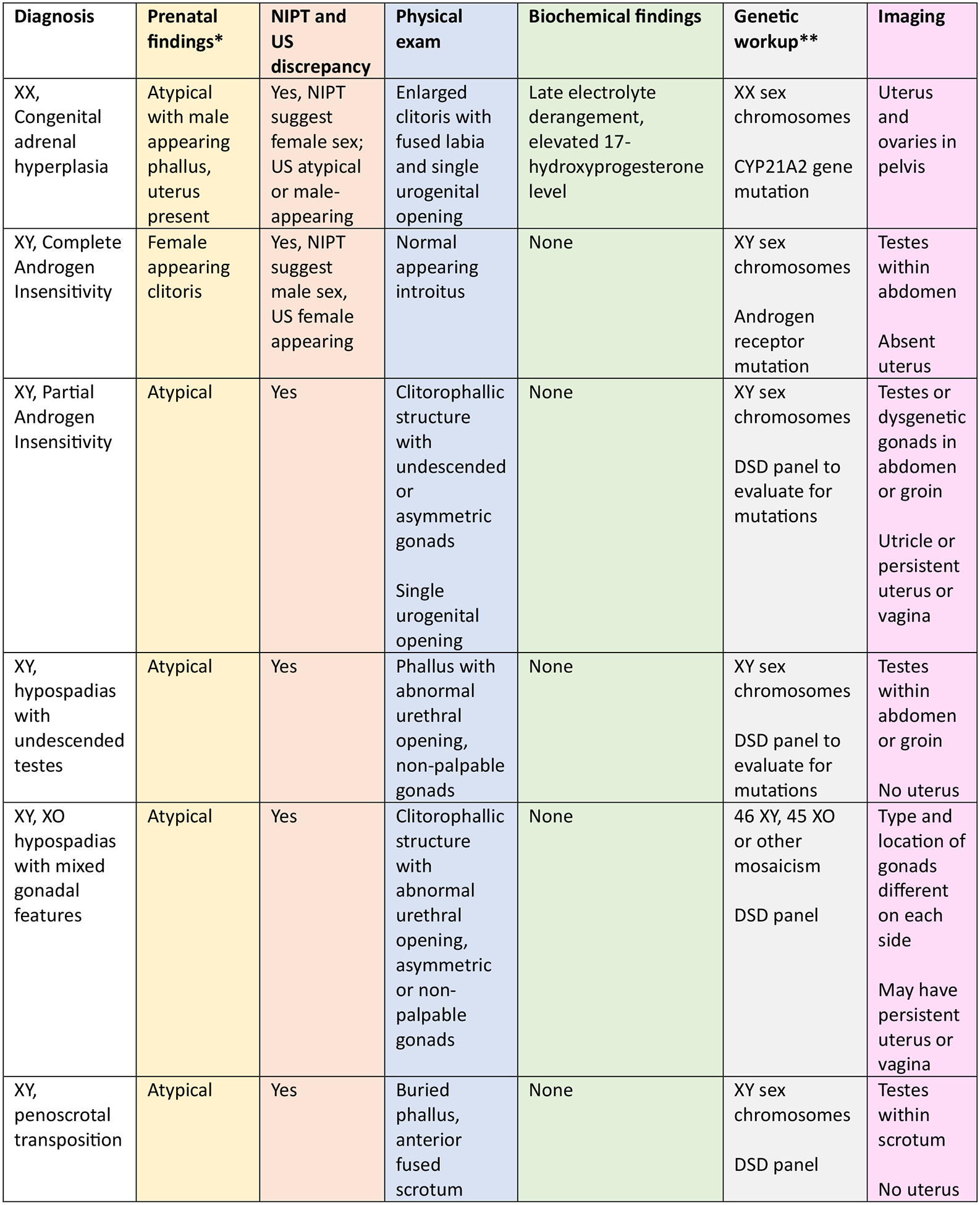
Prenatal and postnatal findings for the most common DSD diagnoses
*Findings are for typical/common appearance of these conditions. Variations from mild to severe exist for all structures.
**Karotype for sex chromosomes may take 2 to 3 days. DSD panel can be sent as an outpatient and results are not as rapid.
An important aspect to the long-term care of the family and patient is peer support. Lee et al (2016) discusses the value of peer support, which not only provides mental health support in allowing a person to not feel alone in their journey but can also be a source of information sharing that is imperative in the care of individuals with DSD. Allowing a family to meet another family or adult patient with a similar or same diagnosis can positively impact long-term outcomes and potentially relieve stress of the uncertainty or unknown (ref).
5. Medical and Surgical Interventions
There is a wide variety of possible postnatal findings and specific needs should be tailored to each diagnosis. For example, patients with CAH may have biochemical abnormalities in steroid synthesis and will require replacement therapy, which is the primary focus for these patients’ care. Discussion of surgical reconstruction is delayed until the diagnosis is confirmed. The approach to surgical reconstruction is beyond the scope of this review, but adherence to a shared decision-making model allows the family to consider a full set of options, including no reconstruction in the absence of urinary obstruction(14–17). A risk assessment of the gonads and their malignant potential will depend on the level of differentiation and the location within the pelvis, groin, or scrotum(18). Difficult decisions regarding gonadectomy should be made in a measured way with consideration for fertility potential and fertility preservation (19).
When possible, the patient, at all stages of development, should be part of the discussion regarding genital reconstruction and management of the gonads. This requires ongoing education and psychosocial care, including sexual health counseling. Assent for procedures should be obtained when possible from adolescents (20).
6. Anticipating needs throughout the lifespan
Discussing the long-term health needs of a newborn with a genital difference helps give context to a family dealing with the trauma of a new diagnosis and can help them see how many parts of their child’s life will not be affected by the condition. Like any child, achieving developmental, educational, and social milestones are important and those can be times when issues related to a medical condition may surface. Fortunately, these transitional times can be anticipated. Developmental periods like infancy, toddler years, potty-training, entry into preschool or elementary school, participation in sports, entry into middle school and high school, puberty and dating, and future career goals all serve to allow individual optimization, goal setting, and assessment for personal success. Creation of a care team that walks with the individual with a DSD and their family along this journey allows for the best outcome.
7. Conclusion
Upon referral to a fetal and neonatal care team, families facing a prenatal diagnosis of a genital difference should have the diagnosis and imaging confirmed as efficiently as possible. The family should be offered the chance to meet with any team members that the baby might need postnatally. The information can be overwhelming and multiple visits may be required to explain the care plan. The effect of stress on the pregnancy and the family should be assessed and psychosocial support should be offered as a standard of care. Peer support should be offered when the diagnosis is confirmed as meeting other parents and patients can be comforting and provide unique perspectives(21). A clinical roadmap that includes the prenatal, postnatal, and long-term follow up plans should be followed.
Key Points:
Discrepancies between sex chromosome findings on noninvasive prenatal screening (NIPS) and unexpected or atypical genitalia on second trimester ultrasound have become more common.
Families should be referred to a multidisciplinary DSD team for discussion of a measured approach to prenatal testing and postnatal workup for babies with atypical genitalia or reproductive tract findings.
Emphasis should be placed on psychosocial support for the family and the affected child with the goal of preserving autonomy and monitoring physical and sexual health.
Peer support is a key adjunct in caring for families and individuals with DSD and should be introduced early in the decision-making process.
Acknowledgements.
We would like to acknowledge our team members who contributed to the development of the Prenatal Guide for Families, including Reeti Chawla, Rachel Lombardo, Margaret Pearson, Gwen Grimsby, Janett Coronado, and Amy Williamson.
Financial support and sponsorship.
This research is supported by grants from the Eunice Kennedy Shriver National Institute for Child Health and Human Development R01 HD086583 and HD093450.
Footnotes
Conflicts of Interest: EM Weidler is the Executive Director of Accord Alliance.
References
- 1.**.Jayashankar SS, Nasaruddin ML, Hassan MF, et al. Non-Invasive Prenatal Testing (NIPT): Reliability, Challenges, and Future Directions. Diagnostics (Basel). 2023;13(15):2570. [DOI] [PMC free article] [PubMed] [Google Scholar]; This article aims to review the reliability of NIPT while discussing the current challenges prevalent among different communities worldwide while using this relatively new tool.
- 2.LeFevre NM, Sundermeyer RL. Fetal Aneuploidy: Screening and Diagnostic Testing. Am Fam Physician. 2020;101(8):481–8. [PubMed] [Google Scholar]
- 3.Lee PA, Houk CP, Ahmed SF, Hughes IA. Consensus statement on management of intersex disorders. International Consensus Conference on Intersex. Pediatrics. 2006;118(2):e488–500. [DOI] [PubMed] [Google Scholar]
- 4.*.Allen NG, Krishna KB, Lee PA. Differences of Sex Development: What Neonatologists Need to Know. Clin Perinatol. 2022;49(1):207–18. [DOI] [PubMed] [Google Scholar]; The neonatologist is often the first health care provider to interact with parents of newborns with DSD and must be familiar with the approach to patients with DSD. The initial workup in the neonatal intensive care unit is reveiwed.
- 5.University Health Network and the Hospital for Sick Children. How Sex Development Works. 2015. [Available from: https://pie.med.utoronto.ca/htbw/module.html?module=sex-development.
- 6.Adam MP, Fechner PY, Ramsdell LA, et al. Ambiguous genitalia: what prenatal genetic testing is practical? Am J Med Genet, Part A. 2012;158a(6):1337–43. [DOI] [PubMed] [Google Scholar]
- 7.Wright CF, Wei Y, Higgins JPT, Sagoo GS. Non-invasive prenatal diagnostic test accuracy for fetal sex using cell-free DNA a review and meta-analysis. BMC Res Notes. 2012;5(1):476. [DOI] [PMC free article] [PubMed] [Google Scholar]
- 8.Dhamankar R, DiNonno W, Martin KA, Demko ZP, et al. Fetal Sex Results of Noninvasive Prenatal Testing and Differences With Ultrasonography. Obstet Gynecol. 2020;135(5):1198–206. [DOI] [PMC free article] [PubMed] [Google Scholar]
- 9.Blackless M, Charuvastra A, Derryck A, et al. How sexually dimorphic are we? Review and synthesis. Am J Hum Biol. 2000;12(2):151–66. [DOI] [PubMed] [Google Scholar]
- 10.New MI, Tong YK, Yuen T, et al. Noninvasive prenatal diagnosis of congenital adrenal hyperplasia using cell-free fetal DNA in maternal plasma. J Clin Endocrinol Metab. 2014;99(6):E1022–30. [DOI] [PMC free article] [PubMed] [Google Scholar]
- 11.*.van Bever Y, Groenenberg IAL, Knapen M,, et al. Prenatal ultrasound finding of atypical genitalia: Counseling, genetic testing and outcomes. Prenat Diagn. 2023;43(2):162–82. [DOI] [PubMed] [Google Scholar]; The authors looked retrospectively at the genetic testing done after abnormal US findings of atypical genitalia with and without other anomalies. Atypical genitalia were postnatally confirmed in 91% (group 1) and 64% (group 2), indicating a high rate of false positive US diagnosis of ambiguous genitalia.
- 12.**.Fluck C, Guran T. Ambiguous Genitalia in the Newborn. In: Feingold KR, Anawalt B, Blackman MR, editors. EndoText. South Dartmouth, MA: MDText.com, Inc.; 2023. [Google Scholar]; An extremely detailed review of the known diagnoses resulting in upper and lower reproductive tract disorders that includes the recognition that having a child with ambiguous genitalia can be very stressful for parents. The importance of early education, access to expert care through DSD network teams, psychological support, shared decision-making, and promoting acceptance and inclusivity is emphasized.
- 13.Cools M, Pleskacova J, Stoop H, et al. Gonadal pathology and tumor risk in relation to clinical characteristics in patients with 45,X/46,XY mosaicism. J Clin Endocrinol Metab. 2011;96(7):E1171–80. [DOI] [PubMed] [Google Scholar]
- 14.Chawla R, Rutter M, Green J, Weidler EM. Care of the adolescent patient with congenital adrenal hyperplasia: Special considerations, shared decision making, and transition. Semin Pediatr Surg. 2019;28(5):150845. [DOI] [PMC free article] [PubMed] [Google Scholar]
- 15.Weidler EM, Baratz A, Muscarella M, et al. A shared decision-making tool for individuals living with complete androgen insensitivity syndrome. Semin Pediatr Surg. 2019;28(5):150844. [DOI] [PMC free article] [PubMed] [Google Scholar]
- 16.Chawla R, Weidler EM, Hernandez J, et al. Utilization of a shared decision-making tool in a female infant with congenital adrenal hyperplasia and genital ambiguity. J Pediatr Endocrinol Metab. 2019;32(6):643–6. [DOI] [PubMed] [Google Scholar]
- 17.Sandberg DE, Gardner M, Kopec K, et al. Development of a decision support tool in pediatric Differences/Disorders of Sex Development. Semin Pediatr Surg. 2019;28(5):150838. [DOI] [PMC free article] [PubMed] [Google Scholar]
- 18.Weidler EM, Pearson M, van Leeuwen K, Garvey E. Clinical management in mixed gonadal dysgenesis with chromosomal mosaicism: Considerations in newborns and adolescents. Semin Pediatr Surg. 2019;28(5):150841. [DOI] [PMC free article] [PubMed] [Google Scholar]
- 19.Finlayson C, Fritsch MK, Johnson EK, et al. Presence of Germ Cells in Disorders of Sex Development: Implications for Fertility Potential and Preservation. J Urol. 2017;197(3):937–43. [DOI] [PMC free article] [PubMed] [Google Scholar]
- 20.*.Lai K, Rubalcava NS, Weidler EM, van Leeuwen K. Paediatric surgeons’ current knowledge and practices of obtaining assent from adolescents for elective reconstructive procedures. J Med Ethics. 2023;49(9):602–6. [DOI] [PubMed] [Google Scholar]; This study evaluated providers’ knowledge of assent and found that there are opportunities to teach reconstructive surgeons about the American Academy of Pediatrics guidelines on obtaining assent in adolescents.
- 21.Joo JH, Bone L, Forte J, et al. The benefits and challenges of established peer support programmes for patients, informal caregivers, and healthcare providers. Fam Pract. 2022; 39(5):903–912. [DOI] [PMC free article] [PubMed] [Google Scholar]