Summary
A complex liver lesion presents a significant challenge in terms of diagnosis and management. This case is an illustrative example, highlighting the steps involved in managing such complex scenarios. This patient, in her early 20s, presented with a fever associated with worsening abdominal pain, as well as a background history of chronic abdominal pain, anorexia, vomiting, constipation and weight loss. The radiology revealed an irregular complex cyst in the liver with biliary and vascular invasion, raising concerns about hepatocellular carcinoma. The diagnosis was changed to alveolar echinococcosis after the infectious diseases consultant gave helpful advice, and echinococcosis antibodies were found. We subsequently started the patient on albendazole therapy. Following prudent advice from hepatobiliary surgeons and given the complexity of the hepatic lesion, a liver transplant was considered the best management option due to the extensive involvement of the biliary and venous systems. The combined approach of albendazole and a liver transplant marked a transformative phase for this patient, putting an end to her prolonged suffering.
Keywords: Infections, Portal hypertension, Hepatitis and other GI infections, Pain
Background
Complex cystic lesions of the liver represent a heterogeneous group of disorders that differ in aetiology, prevalence and clinical manifestations. Usually, solid components in such lesions appear in the context of malignancy often accompanied by necrotic areas. Diagnosing intricate, complex cystic lesions in the liver can be a significant challenge for healthcare professionals. The literature reports several causes of such lesions, including hepatocellular carcinoma (HCC), haemangiomas, cystadenomas and infective causes such as hydatid cysts.1 2 In the majority of cases, these lesions are typically diagnosed incidentally. However, a minority of such lesions can cause serious symptoms and can be associated with complications leading to high morbidity and mortality.1,4
This case illustrates a prime example of a complex hepatic cyst in a patient, presenting numerous diagnostic dilemmas. The case will demonstrate the steps taken to reach the final diagnosis and the effective management of the patient by a multidisciplinary team (MDT). Due to overlapping features, it is very difficult to differentiate hepatic alveolar echinococcosis (AE) from the atypical presentation of HCC clinically and radiologically. Opting for a fine needle aspiration or liver biopsy can further complicate AE. This case will also highlight indications of liver transplantation in hepatic AE cases.
Case presentation
A female patient in her late 20s was admitted due to worsening right upper quadrant (RUQ) pain, vomiting and fever.
The patient had enjoyed excellent health until 4 years ago when she began to experience occasional abdominal pain in the RUQ associated with loss of appetite, feelings of nausea, vomiting and constipation. Acetaminophen somewhat alleviated the pain, which was typically a dull ache, occasionally cramps and without radiation. She denies any diarrhoea, jaundice, retrosternal burning, weight loss, lumps or bumps, cough, shortness of breath, chest pain, urinary symptoms or skin rash. She had recently relocated here.
There was no other significant medical or surgical history. She mentioned no notable family medical history, and there were no instances of liver, breast or bowel cancer in her family. However, she did acknowledge substantial exposure to farm animals, especially cows, sheep and dogs, and she had direct contact with them for feeding purposes and handling their litter. She denied smoking, alcohol or illicit drug use.
On examination, she was febrile with a temperature of 38.2°C, the blood pressure was 125/82 mm Hg, the pulse was 102 beats per minute and the respiratory rate was 16 breaths per minute. In-room air, oxygen saturation was 98%. In the right hypochondria, the abdomen was soft and distended, with associated mild tenderness. There was a mild enlargement of the liver, which was firm in consistency. The bowel sounds were normal, and there was no guarding or rebound tenderness at the costovertebral angle.
Investigations
Initial blood investigations showed mild normocytic anaemia, mild thrombocytopaenia and raised liver function tests (LFTs). While awaiting imaging results and due to the raised LFTs, we planned a non-invasive liver screen, which included an infectious hepatitis profile and an autoimmune screening, both of which returned negative. Blood culture and stool for ova and cyst examinations were unremarkable. Key investigations relevant to the case are mentioned in table 1.
Table 1. List of key Investigations and Follow up investigation (4 months post Surgery).
| List of key investigation | Follow-up investigations (4 months postsurgery) | |
| Bilirubin (total) | 25 µmol/L (3–21) | 8 µmol/L (3–21) |
| Alkaline phosphatase | 837 U/L (35–104) | 128 U/L (35–104) |
| Gamma-glutamyl transferase | 613 U/L (6–42) | 117 U/L (6–42) |
| Alanine transaminase | 299 U/L (10–35) | 19 U/L (10–35) |
| C reactive protein | 3 mg/L (0–5) | 2 mg/L (0–5) |
| White cell count | 4.12×10⁹/L (3.9–11.1) | 8.82×10⁹/L (3.9–11.1) |
| Haemoglobin | 96 g/L (12–16 g/L) | 112 g/L (12–16 g/L) |
| Platelets | 109×109/L, (>150×109/L) | 468×109/L (>150×109/L) |
| MCV | 85 FL (80–100 FL) | 93 FL (80–100 FL) |
| Neutrophils | 2.42×10⁹/L (1.7–6.1) | 5.08×10⁹/L (1.7–6.1) |
| Lymphocytes | 1.09×10⁹/L (1.0–3.2) | 2.55×10⁹/L (1.0–3.2) |
| Eosinophils | 0.24×10⁹/L (0.03–0.46) | 0.41×10⁹/L (0.03–0.46) |
| Albumin | 30 g/L (35–50) | 37 g/L (35–50) |
| Echinococcus (haemagglutination) | 640 (normal <160) | Not performed |
| Echinococcus serology EIA | 24 (positive: >11) | Not performed |
EIAenzyme immunoassayMCVmean corpuscular volume
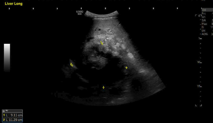
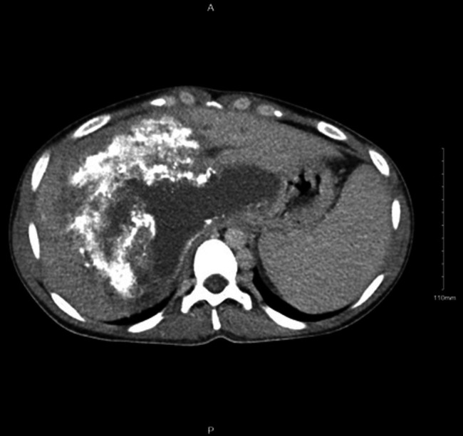
Transabdominal ultrasonography of the liver (figure 1) (MOU2) showed a 9×11×14 cm complex cystic structure in segment 7 with an irregular outline. There was splenomegaly with splenic varices. Based on ultrasound findings, initial screening with tumour markers, including alfa fetoprotein, Ca 19-9 and Ca 15-3, was planned, but it came back negative. Further imaging with CT of the thorax, abdomen and pelvis was arranged with contrast that showed (figure 2) (MOU3) a complex calcified mixed solid cystic hepatic mass of 14×16×12 cm, resulting in a significant extrinsic mass effect characterised by intrahepatic biliary ductal dilatation, vascular invasion of the hepatic veins, portal vein confluence and inferior vena cava (IVC) with resultant portal hypertension and IVC obstruction. No lymphadenopathy was seen.
Figure 1. Ultrasound liver showing a cystic structure in segment seven in right lobe with dimensions of 9×11×14 cm.

Figure 2. CT scan of liver showing complex calcified mixed solid cystic hepatic mass 14×16×12 cm resulting in significant extrinsic mass effect.

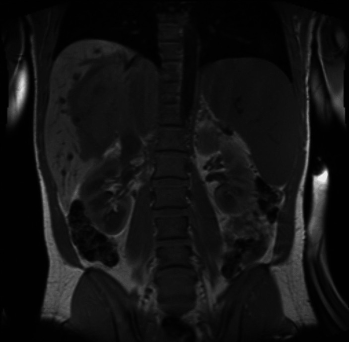
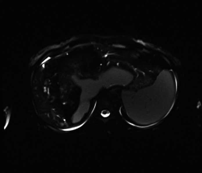
The case was discussed with consultant gastroenterologists and hepatobiliary surgeons; they advised MRI liver, MR cholangiopancreatography (MRCP) and MDT discussion. MRI and MRCP of the liver showed (figures3 4) (MOU4) a large cystic, solid, partly calcified mass in the liver (18×16 cm in transverse dimension) having a malignant appearance involving multiple segments of both lobes of the liver and causing bilobar intrahepatic biliary obstruction as well as occlusion of the suprahepatic IVC and portal vein at the porta hepatis. Although there was no definite invasion of the IVC or porta hepatis, there was evidence of portal hypertension with splenomegaly and prominent retroperitoneal and upper abdominal varices.
Figure 3. Coronal MRI scan of liver showing large cystic and solid partly calcified mass in the liver (18×16 cm in transverse dimension that involves segments 1, 2, 4A, 4B, 5, 6, 7 and 8), having malignant appearance, involving multiple segments of the right and left lobes of the liver.

Figure 4. Axial MRI scan of liver showing large cystic and solid partly calcified mass in the liver.

The infectious diseases consultant recommended further investigations with echinococcal serology, as the patient was brought up in an endemic area with extensive exposure to farm animals and as imaging showed a complex hepatic cyst with calcification and an area of necrosis that can also be seen in hepatic AE. Screening tests using EIA for IgG reported a value of 24 (with a cut-off of 11) while the echinococcal indirect haemagglutination test showed a result of 640 (with a cut-off of 160). The sensitivity of these tests is 80%–90% and 85%–90%, respectively, with a specificity of 98%–100% for diagnosing hepatic echinococcosis.5 6 Currently, the most accurate serological diagnosis is achieved by combining these tests.7 It made the case more complex, raising thoughts about AE.
Differential diagnosis
Hepatocellular Carcinoma is a highly aggressive tumour that typically develops in individuals with chronic liver disease. In the early stages, it can be asymptomatic, but over time, can present with reduced appetite, weight loss and a dull ache in the RUQ. In atypical cases, imaging scans detect an irregular cystic mass with the involvement of venous or biliary systems, as seen in this case.4 However, in our case, there was no history of chronic liver disease preceding the development of liver cancer and positive echinococcal serology changed the diagnosis.
Hepatocellular adenoma was another differential, as these are commonly found in women of childbearing age using oestrogen-containing pills. They are often asymptomatic but can present with RUQ pain.8 On imaging, these lesions can be heterogeneous, with calcifications resulting from internal bleeding, necrosis or fibrosis,9 giving a picture of a complex cyst as in this case. MRI can aid in distinguishing HCA from other liver lesions due to its high fat or glycogen content.10 11
Pyogenic liver abscess was another differential, as the patient presented with typical features of fever, RUQ pain and tenderness, raised LFT and raised inflammatory markers.12 On imaging, abscesses can appear more complex, with localised subcollections or an irregular border. Peripheral rim enhancement and surrounding oedema relatively specific to liver abscesses not seen in our case.13
Acute hepatitis is based on symptoms of fever, RUQ pain and raised LFTs, one can think of acute hepatitis as a cause either due to infective aetiology or autoimmune aetiology. We did an infective and autoimmune workup that came back negative.
Acutecholecystitis can also present with similar symptoms of pain in RUQ, fever, nausea and vomiting, but ultrasound examination showed the presence of a complex cystic lesion with no gallbladder wall oedema or gallstones.
Acute cholangitis can also have a similar presentation, but on LFTs, there should be an obstructive picture, which was against our case, and ultrasound findings were consistent with a complex hepatic cyst.
Alveolar Ecginococcusliver cysts are caused by Echinococcus, also called hydatid cysts. They are often asymptomatic. Symptoms, when present, usually arise from the enlarging cyst’s mass effect or complications such as infection, rupture into the peritoneum, or bile duct obstruction or venous invasion, giving a picture of HCC. Positive serology can aid in diagnosis as seen in our case.
Treatment
Following an MDT discussion involving hepatobiliary surgeons, the gastroenterology team and an infectious disease consultant, the diagnosis of AE was established. Infectious diseases recommend treatment with the antiparasitic agent albendazole, given at 400 mg two times per day (10 mg/kg in two divided doses). The plan was to continue until surgery and then postsurgery.
Meanwhile, it was determined that a liver transplant (LT) coupled with antiparasitic therapy would be the most suitable treatment option due to the extent of the lesion and the involvement of the biliary and portal systems. The plan was to continue albendazole, with a follow-up appointment in the clinic to prepare for surgery. The patient was also initiated on carvedilol for portal hypertension. While waiting for surgery, the patient was screened with an MRI brain and CT thorax to exclude disseminated hydatid disease.
Subsequently, the patient underwent LT surgery within a few months. Her liver was sent to histopathology for further examination, which showed an intact cyst with a maximum dimension of 170 mm. Sectioning revealed calcification and necrosis. Microscopically dense fibrous tissue, extensive calcification, abundant multinucleated giant cells and non-viable parasites were seen. The following histopathology case was discussed in MDT, and it was decided to continue to treat as AE.
Outcome and follow-up
Following an LT, regular follow-up was arranged with the infectious disease department and hepatobiliary surgeons. Albendazole was planned to continue for at least 2 years. On follow-up, she showed drastic signs of recovery, including a resolution of portal hypertension signs. Her blood also showed improvement, with LFTs settling down and improvements in haemoglobin and platelet count. Blood results at 4 months follow-up postsurgery are given in table 1.
Discussion
According to our experience, when we encounter CT findings of a complex irregular calcified hepatic lesions with features such as an extrinsic mass effect, our initial suspicion usually leans towards HCC. Similarly, due to irregular margins and invasive features, it is very difficult to distinguish radiologically AE from atypical HCC. However, key distinguishing features of AE on imaging can be the presence of calcification, hypoattenuating cystic components, and the absence of contrast enhancement or very mild perilesional enhancement.14 15 This case was found unusual for two reasons. First, instead of malignancy, this case turned out to be a complex hydatid cyst. Second, very few patients with hydatid cysts undergo LT due to extensive liver involvement.
Echinococcosis is a zoonotic illness resulting from an infection with the Echinococcus tapeworm. There are four species known to cause infections in humans, with Echinococcus granulosus and Echinococcus multilocularis being the most prevalent. They give rise to cystic echinococcosis and AE, respectively.11 It is worth noting that this disease continues to be endemic in selected regions around the world.
The initial stage of primary infection with echinococcosis is typically asymptomatic for years.14 16 The liver is affected in around two-thirds of patients. These cysts tend to increase in size over the years.17 18 Complications such as, portal hypertension was also seen in a few other case reports with gastrointestinal bleeding, and cholestasis due to a hydatid cyst.19 20
In endemic areas, screening with imaging and serology to diagnose echinococcosis is found to be very useful.21 However, in non-endemic regions, its significance is uncertain.22 Since the introduction of treatment with albendazole, the prognosis has improved significantly if diagnosed early.23
The treatment approach to (AE) typically involves a combination of albendazole and surgery.13 Due to the early asymptomatic nature of the disease, it is usually diagnosed in later stages when complications start occurring.24,28 In the case of surgery for AE, it is crucial to remove infected tissues as completely as possible. This often necessitates the complete excision of parasitic tissue, and, in some instances, radical resection of host tissue may also be required. Surgery usually involves standard radical resection with reconstructive and plastic interventions on the great vessels and biliary ducts.29 However, radical resection is not feasible in cases with a large volume of damaged parenchyma, invasion of the afferent and/or efferent hepatic vessels, the retrohepatic segment of the IVC, and the right atrium,24 25 30 31 severe liver failure, or recurrent life-threatening cholangitis. In such cases, LT is the only option left.32,35
There are no cumulative data showing the percentage of patients requiring LT for AE. However, a case report by Aliakbarian et al, analysing 27 case studies from 1994 to 2017, identified 150 cases of AE where LT was required as a curative treatment.36
The advantage of giving albendazole as a routine preoperative treatment is uncertain.37 Antiparasitic therapy (albendazole 400 mg two times per day) should be continued for at least 2 years postsurgery, and monitoring should be continued for at least 10 years, as unrecognised or invisible parasites can regrow after years, especially in post-LT immunosuppressed patients.38 This postoperative administration of albendazole is done to lower the risk of recurrence, even in cases where it appears that the infection has been cured.39,41 Chances of recurrence are even higher with immunosuppression; in the literature, a study reported recurrence in two out of nine LT patients over a 7-year period.42 Another study showed recurrence in 5 out of 23 LT patients over a 21-year follow-up period.33
If echinococcosis is left untreated, the prognosis is severe. More than 90% of patients are at risk of dying within 10 years of the onset of clinical symptoms.43 This highlights the critical importance of early diagnosis and timely treatment in improving the chances of survival for individuals affected by this disease.
Learning points.
Alveolar echinococcosis(AE) can manifest as complex hepatic lesions that resemble hepatocellular carcinoma, posing a diagnostic challenge for medical teams. However, a thorough clinical history and a careful diagnostic approach can result in an accurate diagnosis.
Following diagnosis, the management plan includes detailed points involving the use of antiparasitic agents along with surgical intervention.
A liver transplant can be a curative option for hepatic AE and can improve prognosis. However, the risk of recurrence, especially due to immunosuppression, necessitates long-term regular follow-up for these patients.
Footnotes
Funding: The authors have not declared a specific grant for this research from any funding agency in the public, commercial or not-for-profit sectors.
Case reports provide a valuable learning resource for the scientific community and can indicate areas of interest for future research. They should not be used in isolation to guide treatment choices or public health policy.
Provenance and peer review: Not commissioned; externally peer reviewed.
Patient consent for publication: Consent obtained directly from patient(s).
Contributor Information
Alvina Zanib, Email: alvinazanib@yahoo.com.
Ahmed Abdelaziz Ahmed, Email: DR_AHMEDTMTAM@HOTMAIL.COM.
Antonella Salvia Milos, Email: a.salvia.milos@gmail.com.
Syed Shabbar Musavi, Email: syed.musavi@hse.ie.
References
- 1.Salemis NS, Georgoulis E, Gourgiotis S, et al. Spontaneous rupture of a giant non parasitic hepatic cyst presenting as an acute surgical abdomen. Ann Hepatol. 2007;6:190–3. [PubMed] [Google Scholar]
- 2.Tong KS, Hassan R, Gan J, et al. Simple hepatic cyst rupture exacerbated by anticoagulation. BMJ Case Rep. 2019;12:12.:e230243. doi: 10.1136/bcr-2019-230243. [DOI] [PMC free article] [PubMed] [Google Scholar]
- 3.Miyamoto M, Oka M, Izumiya T, et al. Nonparasitic solitary giant hepatic cyst causing obstructive jaundice was successfully treated with monoethanolamine oleate. Int Med. 2006;45:621–5. doi: 10.2169/internalmedicine.45.1408. [DOI] [PubMed] [Google Scholar]
- 4.Enke T, Manatsathit W, Merani S, et al. Ciliated Hepatic Foregut Cyst: a Report of a Case Incidentally Discovered during Transplant Evaluation. Case Rep Gastrointest Med. 2019;2019 doi: 10.1155/2019/7828427. [DOI] [PMC free article] [PubMed] [Google Scholar]
- 5.Zhang W, Wen H, Li J, et al. Immunology and immunodiagnosis of cystic echinococcosis: an update. Clin Dev Immunol. 2012;2012:101895. doi: 10.1155/2012/101895. [DOI] [PMC free article] [PubMed] [Google Scholar]
- 6.Biava MF, Dao A, Fortier B. Laboratory diagnosis of cystic hydatic disease. World J Surg. 2001;25:10–4. doi: 10.1007/s002680020002. [DOI] [PubMed] [Google Scholar]
- 7.Ortona E, Riganò R, Margutti P, et al. Native and recombinant antigens in the immunodiagnosis of human cystic echinococcosis. Parasite Immunol. 2000;22:553–9. doi: 10.1046/j.1365-3024.2000.00336.x. [DOI] [PubMed] [Google Scholar]
- 8.Ben Ari Z, Cohen-Ezra O, Weidenfeld J, et al. Ciliated hepatic foregut cyst with high intra-cystic carbohydrate antigen 19-9 level. World J Gastroenterol. 2014;20:16355–8. doi: 10.3748/wjg.v20.i43.16355. [DOI] [PMC free article] [PubMed] [Google Scholar]
- 9.Wilson JM, Groeschl R, George B, et al. Ciliated hepatic cyst leading to squamous cell carcinoma of the liver - A case report and review of the literature. Int J Surg Case Rep. 2013;4:972–5. doi: 10.1016/j.ijscr.2013.07.030. [DOI] [PMC free article] [PubMed] [Google Scholar]
- 10.Ziogas IA, van der Windt DJ, Wilson GC, et al. Surgical Management of Ciliated Hepatic Foregut Cyst. Hepatology. 2020;71:386–8. doi: 10.1002/hep.30877. [DOI] [PubMed] [Google Scholar]
- 11.Wen H, Vuitton L, Tuxun T, et al. Echinococcosis: advances in the 21st Century. Clin Microbiol Rev. 2019;32:32. doi: 10.1128/CMR.00075-18. [DOI] [PMC free article] [PubMed] [Google Scholar]
- 12.Ito A, Schantz PM, Wilson JF. Em18, a new serodiagnostic marker for differentiation of active and inactive cases of alveolar hydatid disease. Am J Trop Med Hyg. 1995;52:41–4. doi: 10.4269/ajtmh.1995.52.41. [DOI] [PubMed] [Google Scholar]
- 13.Vuitton DA, Azizi A, Richou C, et al. Current interventional strategy for the treatment of hepatic alveolar echinococcosis. Expert Rev Anti Infect Ther. 2016;14:1179–94. doi: 10.1080/14787210.2016.1240030. [DOI] [PubMed] [Google Scholar]
- 14.Graeter T, Kratzer W, Oeztuerk S, et al. Proposal of a computed tomography classification for hepatic alveolar echinococcosis. World J Gastroenterol. 2016;22:3621–31. doi: 10.3748/wjg.v22.i13.3621. [DOI] [PMC free article] [PubMed] [Google Scholar]
- 15.Bulakci M, Yilmaz E, Cengel F, et al. Disseminated alveolar hydatid disease resembling a metastatic malignancy: a diagnostic challenge-a report of two cases. Case Rep Radiol. 2014;2014:638375. doi: 10.1155/2014/638375. [DOI] [PMC free article] [PubMed] [Google Scholar]
- 16.Ammann RW, Eckert J. Cestodes Echinococcus. Gastroenterol Clin North Am. 1996;25:655–89. doi: 10.1016/s0889-8553(05)70268-5. [DOI] [PubMed] [Google Scholar]
- 17.Frider B, Larrieu E, Odriozola M. Long-term outcome of asymptomatic liver hydatidosis. J Hepatol. 1999;30:228–31. doi: 10.1016/s0168-8278(99)80066-x. [DOI] [PubMed] [Google Scholar]
- 18.Moro PL, Gilman RH, Verastegui M, et al. Human hydatidosis in the central Andes of Peru: evolution of the disease over 3 years. Clin Infect Dis. 1999;29:807–12. doi: 10.1086/520440. [DOI] [PubMed] [Google Scholar]
- 19.Pielok L, Karczewski M, Cierach W, et al. Portal hypertension as a result of the incomplete surgically treated advanced alveolar echinococcosis: a case description. BMC Gastroenterol. 2007;50:57–9. doi: 10.14712/18059694.2017.60. [DOI] [PMC free article] [PubMed] [Google Scholar]
- 20.Portal hypertension and cholestasis due to Echinococcus alveolaris: a case report: Yuksel Seckin; Yasir Furkan Cagin; Oguzhan Yildirim; Yilmaz Bilgic; Yahya Atayan; Mehmet Erdogan. Nasuhi Engin Aydin Int Med J. 2016 [Google Scholar]
- 21.Brunetti E, Kern P, Vuitton DA, et al. Expert consensus for the diagnosis and treatment of cystic and alveolar echinococcosis in humans. Acta Trop. 2010;114:1–16. doi: 10.1016/j.actatropica.2009.11.001. [DOI] [PubMed] [Google Scholar]
- 22.Wójcik-Fatla A, Sroka J, Zając V, et al. Study on Toxoplasma Gondii, Leptospira Spp., Coxiella Burnetii, and Echinococcus Granulosus Infection in Veterinarians from Poland. J Vet Res. 2018;62:477–83. doi: 10.2478/jvetres-2018-0069. [DOI] [PMC free article] [PubMed] [Google Scholar]
- 23.Torgerson PR, Schweiger A, Deplazes P, et al. Alveolar echinococcosis: from a deadly disease to a well-controlled infection. Relative survival and economic analysis in Switzerland over the last 35 years. J Hepatol. 2008;49:72–7. doi: 10.1016/j.jhep.2008.03.023. [DOI] [PubMed] [Google Scholar]
- 24.Skipenko OG, Shatverian GA, Bagmet NN, et al. Alveococcosis of liver: retrospective analysis of treatment of 51 patients. Khir Zh im NI Pirogova. 2012;12:4–13. [PubMed] [Google Scholar]
- 25.Porshennikov IA, Bykov AYu, Pavlik VN, et al. Liver transplantation and liver resection with vascular reconstruction for advanced alveococcosis. Ann hir gepatol. 2016;21:11–24. doi: 10.16931/1995-5464.2016211-24. [DOI] [Google Scholar]
- 26.Buttenschoen K, Gruener B, Carli Buttenschoen D, et al. Palliative operation for the treatment of alveolar echinococcosis. Langenbecks Arch Surg. 2009;394:199–204. doi: 10.1007/s00423-008-0367-6. [DOI] [PubMed] [Google Scholar]
- 27.Kawamura N, Kamiyama T, Sato N, et al. Long-term results of hepatectomy for patients with alveolar echinococcosis: a single-center experience. J Am Coll Surg. 2011;212:804–12. doi: 10.1016/j.jamcollsurg.2011.02.007. [DOI] [PubMed] [Google Scholar]
- 28.Moore J, Gupta V, Ahmed MY, et al. Hydatid cyst disease: optimal management of complex liver involvement. S Med J. 2011;104:222–4. doi: 10.1097/SMJ.0b013e318205e686. [DOI] [PubMed] [Google Scholar]
- 29.Zagaynov VE, Kiselev NM, Gorokhov GG, et al. Modern methods of surgical treatment of diffuse liver alveococcosis. Ann hir gepatol. 2016;21:44–52. doi: 10.16931/1995-5464.2016144-52. [DOI] [Google Scholar]
- 30.Ayifuhan A, Tuerganaili A, Jun C, et al. Surgical treatment for hepatic alveolar echinococcosis: report of 50 cases. Hepatogastroenterol. 2012;59:790–3. doi: 10.5754/hge10545. [DOI] [PubMed] [Google Scholar]
- 31.Buttenschoen K, Carli Buttenschoen D, Gruener B, et al. Long-term experience on surgical treatment of alveolar echinococcosis. Langenbecks Arch Surg. 2009;394:689–98. doi: 10.1007/s00423-008-0392-5. [DOI] [PubMed] [Google Scholar]
- 32.Koch S, Bresson-Hadni S, Miguet J-P, et al. Experience of liver transplantation for incurable alveolar echinococcosis: a 45-case European collaborative report. Transplantation. 2003;75:856–63. doi: 10.1097/01.TP.0000054230.63568.79. [DOI] [PubMed] [Google Scholar]
- 33.Bresson-Hadni S, Blagosklonov O, Knapp J, et al. Should possible recurrence of disease contraindicate liver transplantation in patients with end-stage alveolar echinococcosis? A 20-year follow-up study. Liver Transpl. 2011;17:855–65. doi: 10.1002/lt.22299. [DOI] [PubMed] [Google Scholar]
- 34.Ozdemir F, Ince V, Barut B, et al. Living donor liver transplantation for Echinococcus Alveolaris: single‐center experience. Liver Transpl. 2015;21:1091–5. doi: 10.1002/lt.24170. [DOI] [PubMed] [Google Scholar]
- 35.Yetişir F, Dogan SM, Mamedov R, et al. Replacement of Vena Cava up to the Right Atrium during Living Donor Liver Transplantation for Echinococcus alveolaris. Case Rep Transplant. 2014;2014:801657. doi: 10.1155/2014/801657. [DOI] [PMC free article] [PubMed] [Google Scholar]
- 36.Aliakbarian M, Tohidinezhad F, Eslami S, et al. Liver transplantation for hepatic alveolar echinococcosis: literature review and three new cases. Infect Dis (Lond) 2018;50:452–9. doi: 10.1080/23744235.2018.1428823. [DOI] [PubMed] [Google Scholar]
- 37.Guidelines for treatment of cystic and alveolar echinococcosis in humans. WHO Informal Working Group on Echinococcosis. Bull World Health Organ. 1996;74:231. [PMC free article] [PubMed] [Google Scholar]
- 38.Reuter S, Jensen B, Buttenschoen K, et al. Benzimidazoles in the treatment of alveolar echinococcosis: a comparative study and review of the literature. J Antimicrob Chemother. 2000;46:451–6. doi: 10.1093/jac/46.3.451. [DOI] [PubMed] [Google Scholar]
- 39.Brunetti E, White AC. Cestode infestations: hydatid disease and cysticercosis. Infect Dis Clin North Am. 2012;26:421–35. doi: 10.1016/j.idc.2012.02.001. [DOI] [PubMed] [Google Scholar]
- 40.Stettler M, Fink R, Walker M, et al. In vitro parasiticidal effect of Nitazoxanide against Echinococcus multilocularis metacestodes. Antimicrob Agents Chemother. 2003;47:467–74. doi: 10.1128/AAC.47.2.467-474.2003. [DOI] [PMC free article] [PubMed] [Google Scholar]
- 41.Reuter S, Buck A, Grebe O, et al. Salvage treatment with amphotericin B in progressive human alveolar echinococcosis. Antimicrob Agents Chemother. 2003;47:3586–91. doi: 10.1128/AAC.47.11.3586-3591.2003. [DOI] [PMC free article] [PubMed] [Google Scholar]
- 42.Sulima M, Wołyniec W, Oładakowska-Jedynak U, et al. Liver Transplantation for Incurable Alveolar Echinococcosis: an Analysis of Patients Hospitalized in Department of Tropical and Parasitic Diseases in Gdynia. Transplant Proc. 2016;48:1708–12. doi: 10.1016/j.transproceed.2016.01.087. [DOI] [PubMed] [Google Scholar]
- 43.Ozdemir F, Ince V, Barut B, et al. Living donor liver transplantation for Echinococcus Alveolaris: single-center experience. Liver Transpl. 2015;21:1091–5. doi: 10.1002/lt.24170. [DOI] [PubMed] [Google Scholar]


