Abstract
A 13-year-old boy visited our clinic due to a malunion following a phalangeal bicondylar T-shaped fracture in the proximal interphalangeal (PIP) joint of his small finger. Imaging studies showed over 2 mm of fracture displacement and ulnar deviation of the radial condyle. The patient underwent surgical correction four weeks after the initial injury. The malunited fragments were reduced to their near-anatomical positions, and an extra-articular osteotomy was performed to realign the angular deformity. Solid bone union was successfully achieved eight weeks after the corrective surgery. This intra- and extra-articular double-level osteotomy is a good option for pediatric phalangeal bicondylar T-shaped malunions.
Keywords: extra-articular osteotomy, intra-articular osteotomy, intra- and extra-articular double-level osteotomy, phalangeal bicondylar t-shaped malunion, phalangeal bicondylar t-shaped fracture
Introduction
Treatment of malunion after complex articular fractures of the phalanx is challenging, since the malunion is often associated with avascular osteonecrosis and/or proximal interphalangeal (PIP) joint contracture [1]. For extra-articular malunion, extra-articular osteotomy is usually performed to correct the bone alignment and rotation [2]. On the other hand, intra-articular osteotomy is utilized for intra-articular malunion with an incongruent articular surface [3].
However, complex malunion might lead to a double-level deformity, including both intra-articular joint incongruity and extra-articular malalignment. Hence, a single approach might not be sufficient to restore normal finger function in such cases. In this report, we describe a pediatric case of phalangeal malunion after a bicondylar T-shaped fracture that was successfully treated using intra- and extra-articular double-level osteotomy, and discuss the effectiveness of this technique for complex phalangeal malunion.
Case presentation
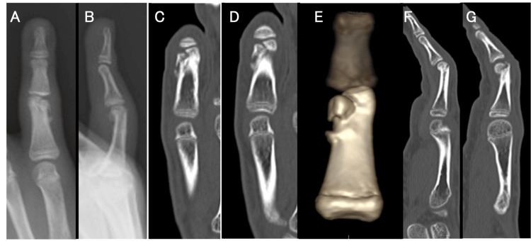
A 13-year-old boy stubbed his right little finger into a tree while riding a bicycle. He visited a nearby orthopedic clinic and was diagnosed with a proximal phalangeal fracture in the small finger. Initially, since the fracture was not displaced, he underwent conservative treatment with immobilization and splinting. The patient visited the same clinic 3 weeks later because of sustained pain in the affected finger. Although there was no rotational or sagittal angular deformity, >2 mm of fracture displacement and ulnar deviation of the radial condyle, which had not appeared on the initial radiographs, were noted on the images (Figure 1).
Figure 1. Preoperative radiographs and CT images.
Radiographs (A, B) and CT scan images (C, D, E, F, G) showed that the ulnar condyle was proximally shortened, the articular step-off was 2 mm, and the radial head demonstrated 10° of ulnar deviation.
According to the findings on the CT scan, a displaced fracture with callus formation was observed, indicating malunion after a phalangeal bicondylar T-shaped fracture. The range of motion of his PIP joint was limited to 70° in flexion and -20° in extension, with pain. Since the articular displacement and joint malalignment were considered unacceptable, surgical correction was planned for 4 weeks after the initial injury.
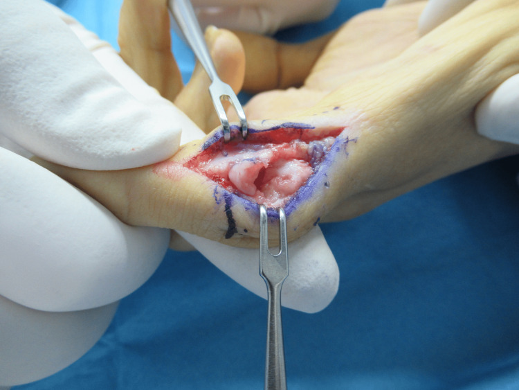
During the operation, the proximal phalanx was exposed through a mid-lateral incision on the ulnar side. First, intra-articular osteotomy was performed using a chisel along the fracture line under direct visualization of the joint and with fluoroscopic guidance. The malunited fragment was reduced to the anatomical position and temporarily fixed with Kirschner wires (K-wires) under fluoroscopic guidance. Then, an ulnar-side open-wedge extra-articular osteotomy was performed using a chisel with fluoroscopy (Figure 2).
Figure 2. Intraoperative images.
Intraoperative images showing the shortened ulnar condyle stretched distally to improve the congruity of the joint's articular surface. Next, an ulnar-side open-wedge extra-articular osteotomy was performed to correct the ulnar deviation.
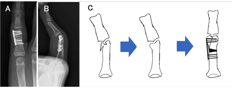
A small fragment locking plate was applied to the ulnar side of the phalanx and fixed with screws and additional K-wires. Cancellous bone from the distal radius was grafted at the open-wedge osteotomy site (Figure 3).
Figure 3. Intraoperative images.
A small fragment locking plate was applied to the ulnar side of the phalanx and fixed with screws and additional Kirschner wires. (C) Graphical illustration of this procedure.
Image credits: Hirotaka Akezuma.
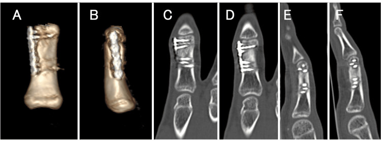
Functional rehabilitation was initiated immediately after the surgery. Buddy taping was applied, and mild range of motion exercises were initiated on the day after the surgery. Buddy taping was continued for 8 weeks postoperatively. With this treatment, solid bone union was successfully obtained at 8 weeks after the corrective surgery (Figure 4).
Figure 4. CT images at the three-month follow-up.
(A, B, C, D, E, F) CT images at the three-month follow-up showed the ideal position of the proximal phalanx.
The plate was removed 11 months postoperatively. At one year after the injury, the patient no longer experienced any pain and the range of motion of his PIP joint was 90° in flexion and -10° in extension (Figure 5).
Figure 5. Photographs at the nine-month follow-up and radiographs at the 12-month follow-up.
(A, B) Photographs showing good active range of motion of the finger at the nine-month follow-up. (C, D) Radiographs following implant removal 12 months after osteotomy, showing bone union at the proximal phalangeal fragment with good bony alignment.
Discussion
In this report, we described a pediatric case of phalangeal malunion after a bicondylar T-shaped fracture that was successfully treated with intra- and extra-articular double-level osteotomy. Phalangeal bicondylar T-shaped fracture is a relatively rare condition, for which various fixation methods have been utilized for acute fractures. Previous reports described fixations performed using external fixators, screws, and plates [4-6]. In terms of approach, a previous report demonstrated that the fracture was exposed via a dorsal incision and reduced under direct vision between the central slip and lateral band, and then fixed with interfragmentary screws [6]. In another report, surgery was performed using a bilateral approach [5]. Unlike acute fractures, there are few specific reports of treatment for malunion after bicondylar T-shaped fracture. For simple malunions, extra-articular osteotomy for phalangeal extra-articular fracture malunion has been utilized to restore alignment of the phalanx [1, 7]. Then, the osteotomized fragments were fixed with plates and intraosseous wires. There have also been multiple reports of intra-articular osteotomy for intra-articular phalangeal hemicondylar fracture malunion [1, 3, 7-9]. Creating small bone fragments might lead to avascular necrosis (AVN) of the fragments. Also, small phalangeal bone fragments are often difficult to reduce correctly and secure during surgery [10-12]. To prevent AVN, a previous report described that condylar advancement osteotomy was useful for preventing AVN of small bone fragments [9]. However, this technique can be applied to hemicondylar fracture malunion but cannot be used for bicondylar fracture malunion.
In the present case, we used combined double-level osteotomy: intra-articular osteotomy at the fracture site and extra-articular osteotomy at the bone shaft slightly proximal to the fracture line in order to simultaneously restore both articular congruity and malalignment. In previous reports on distal radius malunion, several cases underwent intra- and extra-articular double-level corrective osteotomy and reported good results [13]. Alternatively, 3D intra-articular osteotomy was also performed for the treatment of malunion after distal radius fractures [14, 15]. However, for phalangeal malunion, a 3D intra-articular osteotomy at the fracture site appears challenging because cutting the small fracture fragments accurately is likely difficult, and the osteotomy might endanger the blood supply to the bone fragments. In contrast, during our double-level osteotomy, the operating surgeon can focus on each procedural step sequentially, intra-articular osteotomy and fixation, as well as extra-articular alignment correction, and it could be a more feasible option than 3D osteotomy.
Conclusions
We report a case of successfully treating a pediatric phalangeal bicondylar T-shaped malunion using a combined intra- and extra-articular double-level osteotomy. This technique proved effective in addressing both intra-articular incongruity and extra-articular malalignment, thereby restoring proper finger functionality. The intra-articular osteotomy at the fracture site, combined with an extra-articular osteotomy at the bone shaft, allowed for precise realignment. Additionally, the use of a small fragment locking plate and Kirschner wires ensured stable fixation. While this approach may offer a viable solution for similar complex pediatric phalangeal malunions, further studies are needed to confirm its applicability.
Disclosures
Human subjects: Consent was obtained or waived by all participants in this study.
Conflicts of interest: In compliance with the ICMJE uniform disclosure form, all authors declare the following:
Payment/services info: All authors have declared that no financial support was received from any organization for the submitted work.
Financial relationships: All authors have declared that they have no financial relationships at present or within the previous three years with any organizations that might have an interest in the submitted work.
Other relationships: All authors have declared that there are no other relationships or activities that could appear to have influenced the submitted work.
Author Contributions
Concept and design: Hirotaka Akezuma, Kazutoshi Kubo, Yoshifumi Kudo, Keikichi Kawasaki
Drafting of the manuscript: Hirotaka Akezuma, Ichiro Okano, Kazutoshi Kubo, Keikichi Kawasaki
Critical review of the manuscript for important intellectual content: Hirotaka Akezuma, Yoshifumi Kudo, Keikichi Kawasaki
Acquisition, analysis, or interpretation of data: Ichiro Okano
Supervision: Ichiro Okano, Keikichi Kawasaki
References
- 1.Corrective osteotomy for post-traumatic malunion of the phalanges in the hand. Büchler U, Gupta A, Ruf S. J Hand Surg Br. 1996;21:33–42. doi: 10.1016/s0266-7681(96)80009-x. [DOI] [PubMed] [Google Scholar]
- 2.In situ osteotomy for extra-articular malunion of the proximal phalanx. Trumble T, Gilbert M. J Hand Surg Am. 1998;23:821–826. doi: 10.1016/S0363-5023(98)80156-1. [DOI] [PubMed] [Google Scholar]
- 3.Intra-articular osteotomy for malunited proximal interphalangeal joint fracture. Yang C, Wei J, Xu J, Xu L, Lu J. Ann Plast Surg. 2019;83:34–39. doi: 10.1097/SAP.0000000000001858. [DOI] [PubMed] [Google Scholar]
- 4.The use of the s-quattro dynamic external fixator for the treatment of intra-articular phalangeal fractures: a review of the literature. Bhamra JS, Khan WS, Pastides P. Open Orthop J. 2012;6:54–59. doi: 10.2174/1874325001206010054. [DOI] [PMC free article] [PubMed] [Google Scholar]
- 5.Condylar fractures of the proximal and middle phalanges. Shewring DJ, Miller AC, Ghandour A. J Hand Surg Eur Vol. 2015;40:51–58. doi: 10.1177/1753193413508514. [DOI] [PubMed] [Google Scholar]
- 6.Articularly placed interfragmentary screw fixation of difficult condylar fractures of the hand. Tan JS, Foo AT, Chew WC, Teoh LC. J Hand Surg Am. 2011;36:604–609. doi: 10.1016/j.jhsa.2010.12.004. [DOI] [PubMed] [Google Scholar]
- 7.Corrective osteotomy of malunited fractures of phalanges and metacarpals. Gollamudi S, Jones WA. J Hand Surg Br. 2000;25:439–441. doi: 10.1054/jhsb.2000.0418. [DOI] [PubMed] [Google Scholar]
- 8.Extra-articular osteotomy for malunited unicondylar fractures of the proximal phalanx. Harness NG, Chen A, Jupiter JB. J Hand Surg Am. 2005;30:566–572. doi: 10.1016/j.jhsa.2004.12.003. [DOI] [PubMed] [Google Scholar]
- 9.Condylar advancement osteotomy for correcting condylar malunion of the finger. Teoh LC, Yong FC, Chong KC. J Hand Surg Br. 2002;27:31–35. doi: 10.1054/jhsb.2001.0694. [DOI] [PubMed] [Google Scholar]
- 10.Phalangeal neck fractures in children: classification and outcome in 66 cases. Al-Qattan MM. J Hand Surg Br. 2001;26:112–121. doi: 10.1054/jhsb.2000.0506. [DOI] [PubMed] [Google Scholar]
- 11.Phalangeal neck fractures in adults. Al-Qattan MM. J Hand Surg Br. 2006;31:484–488. doi: 10.1016/j.jhsb.2006.06.005. [DOI] [PubMed] [Google Scholar]
- 12.Treatment outcomes of unstable proximal phalangeal head fractures of the finger in children. Bergeron L, Gagnon I, l'Ecuyer C, Caouette-Laberge L. Ann Plast Surg. 2005;54:28–32. doi: 10.1097/01.sap.0000141945.03201.c9. [DOI] [PubMed] [Google Scholar]
- 13.Corrective osteotomy for combined intra- and extra-articular distal radius malunion. Buijze GA, Prommersberger KJ, González Del Pino J, Fernandez DL, Jupiter JB. J Hand Surg Am. 2012;37:2041–2049. doi: 10.1016/j.jhsa.2012.07.013. [DOI] [PubMed] [Google Scholar]
- 14.Best approaches in distal radius fracture malunions. Evans BT, Jupiter JB. Curr Rev Musculoskelet Med. 2019;12:198–203. doi: 10.1007/s12178-019-09540-y. [DOI] [PMC free article] [PubMed] [Google Scholar]
- 15.[Correction of intraarticular malunion of the distal radius based on a computer-assisted virtual planning] Pillukat T, Osorio M, Prommersberger KJ. Handchir Mikrochir Plast Chir. 2018;50:310–318. doi: 10.1055/a-0751-2959. [DOI] [PubMed] [Google Scholar]


