Abstract
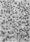
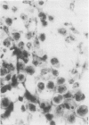
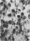
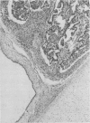
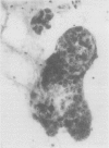
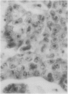
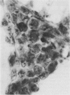
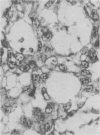
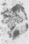
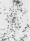
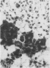
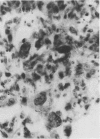
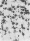
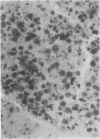
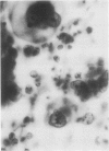
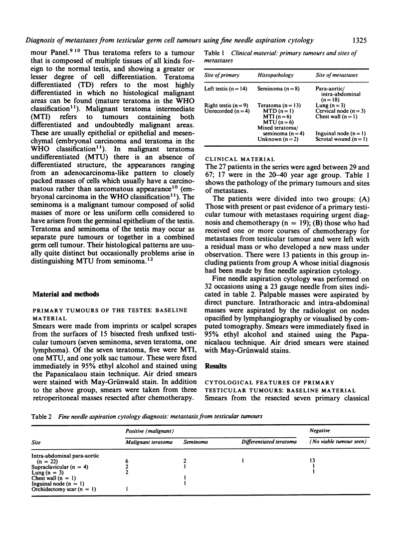
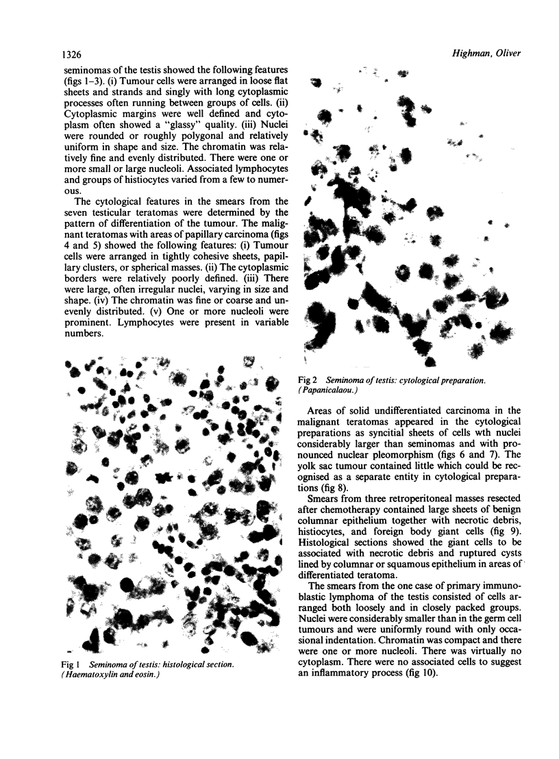
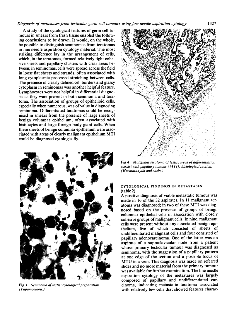
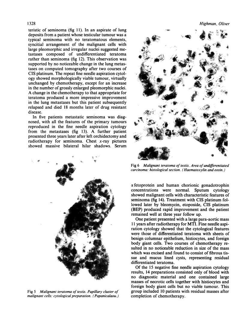
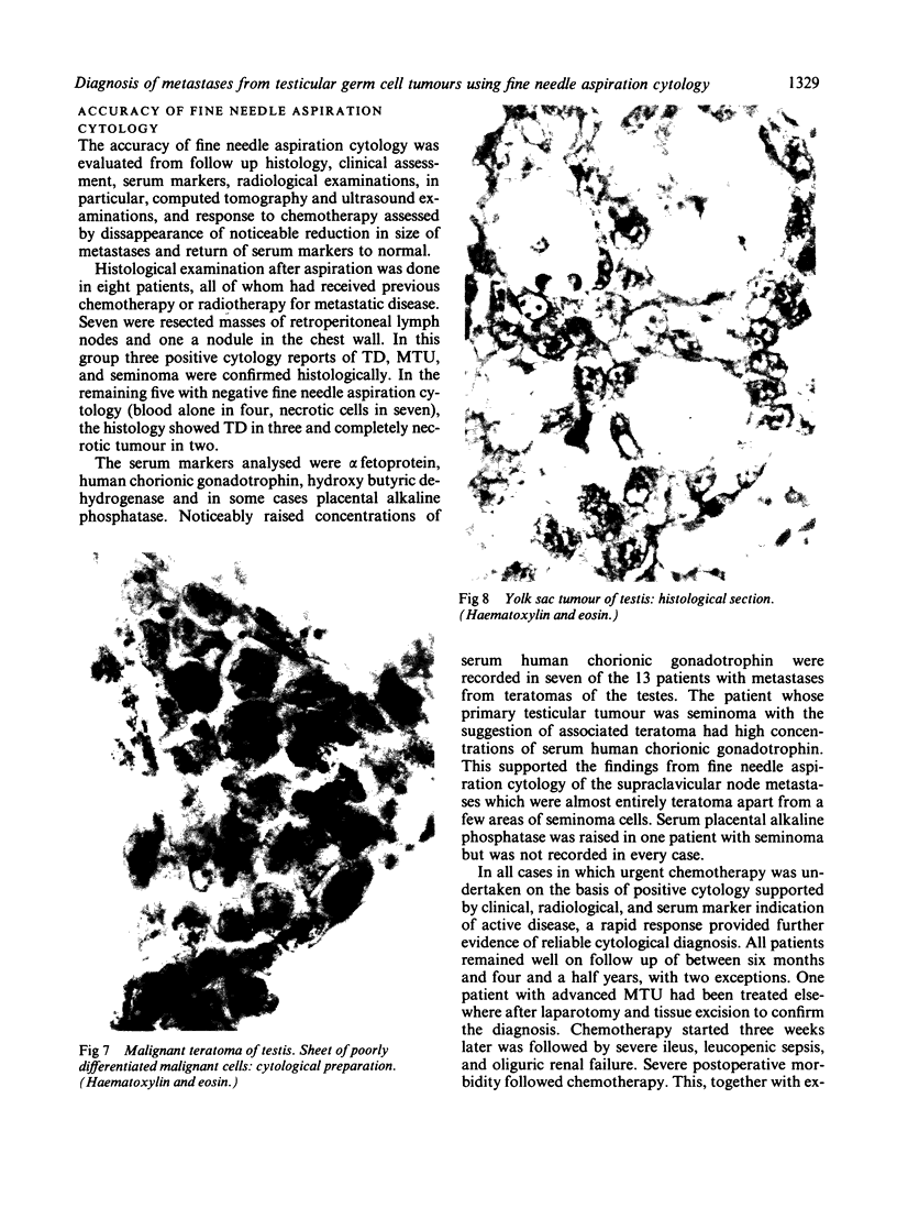
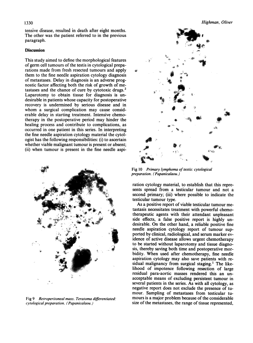
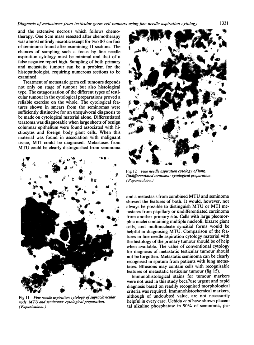
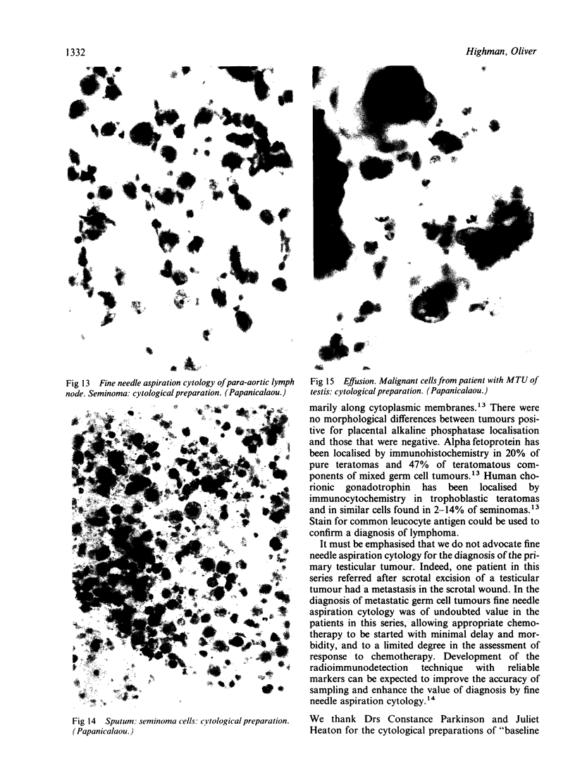
The cytological features of testicular germ cell tumours were established in smears from 15 freshly resected tumours. These features were applied to the fine needle aspiration cytology diagnosis of metastases in 27 patients referred for chemotherapy. There were 16 positive reports in 32 aspirates of which 13 were taken before chemotherapy and three in patients with residual or new masses after chemotherapy. Teratomas and typical seminomas showed certain characteristic morphological features in cytological preparations which when present in fine needle aspiration cytology material enabled tumour types to be diagnosed. Spermatocytic and anaplastic seminoma were not represented in this series. It is unlikely that these could be distinguished from malignant teratoma undifferentiated (MTU) in the fine needle aspiration cytology material. Metastases from carcinomatous areas in MTU and malignant teratoma intermediate (MTI) may not be distinguishable in fine needle aspiration cytology material from metastatic adenocarcinoma or undifferentiated carcinoma from a different primary site. Positive cytological findings are of value to the oncologist in the management of patients with metastases from testicular germ cell tumours; negative cytology does not exclude the presence of viable tumour. The sampling of small foci of viable tumour in large necrotic masses persisting after chemotherapy is a problem for radiologists, cytologists, and histopathologists. This paper does not advocate the use of fine needle aspiration cytology for the diagnosis of primary testicular tumour.
Full text
PDF
Images in this article
Selected References
These references are in PubMed. This may not be the complete list of references from this article.
- Alderdice J. M., Johnston S. R. Embryonal carcinoma of testis simulating seminoma. J Clin Pathol. 1987 Apr;40(4):434–437. doi: 10.1136/jcp.40.4.434. [DOI] [PMC free article] [PubMed] [Google Scholar]
- Droese M., Rahlf G. Punktionszytologie maligner Hodentumoren? Urologe A. 1979 Nov;18(6):338–342. [PubMed] [Google Scholar]
- Ekelund L., Göthlin J. Fine needle biopsy of metastases at retrograde pyelography, directed by fluoroscopy. Report of a case with malignant teratoma of the testis. Scand J Urol Nephrol. 1976;10(3):261–262. [PubMed] [Google Scholar]
- Oliver R. T., Highman W. J., Kellett M. J., Curling O. M., Dacie J. E. The value of fine needle aspiration cytology in the management of metastatic germ cell tumours. Br J Urol. 1985 Apr;57(2):200–203. doi: 10.1111/j.1464-410x.1985.tb06424.x. [DOI] [PubMed] [Google Scholar]
- Wajsman Z., Gamarra M., Park J. J., Beckley S. A., Pontes J. E., Murphy G. P. Fine-needle aspiration of metastatic lesions and regional lymph nodes in genitourinary cancer. Urology. 1982 Apr;19(4):356–360. doi: 10.1016/0090-4295(82)90188-1. [DOI] [PubMed] [Google Scholar]
- Wajsman Z., Gamarra M., Park J. J., Beckley S., Pontes J. E. Transabdominal fine needle aspiration of retroperitoneal lymph nodes in staging of genitourinary tract cancer (correlation with lymphography and lymph node dissection findings). J Urol. 1982 Dec;128(6):1238–1240. doi: 10.1016/s0022-5347(17)53442-4. [DOI] [PubMed] [Google Scholar]
- Zornoza J., Handel P., Lukeman J. M., Jing B. S., Wallace S. Percutaneous transperitoneal biopsy in urologic malignancies. Urology. 1977 Apr;9(4):395–398. [PubMed] [Google Scholar]


