Abstract
Background
Surgical extraction of impacted mandibular third molars is the most commonly performed procedure in oral surgery; its associated complications include sensory nerve damage, swelling, and trismus. This study aimed to evaluate the effects of hyaluronic acid (HA) on healing of the socket following extraction of the lower impacted third molar tooth in 40 dental patients.
Material/Methods
This prospective, double-blind, randomized, controlled study was carried out on 40 adult healthy patients indicated for surgical removal of bilateral impacted mandibular third molars with equal surgical difficulty (moderate surgical difficulty according to the Koerner index. Patients with right mandibular third molars were included into the study (HA) group and those with left mandibular third molars were included into the control group. Surgical removal of impacted teeth was performed at different times for each patient for proper measurement of postoperative clinical variables, including pain, swelling, and mouth opening.
Results
Postoperative pain evaluation results using the visual analog scale (VAS) showed reduced pain levels at all observation periods. Postoperative swelling peaked in intensity within 12–48 hours, resolving between the 5th and 7th days, and there was no significant difference in pre- and postoperative measurements of interincisal opening between both groups (P>0.05).
Conclusions
We found that intra-socket application of hyaluronic acid after surgical extraction of impacted mandibular third molars promoted normal wound healing, and there was a clinical benefit of reduced postoperative pain and swelling.
Keywords: Pain, Edema, Hyaluronic Acid, Tooth, Impacted, Surgery, Oral, Tooth Extraction
Introduction
Surgical extraction of mandibular impacted third molars is one of the most commonly performed procedures in oral surgery, and it is frequently associated with postoperative complications such as delayed healing, swelling, pain, dry socket, inferior alveolar nerve dysesthesia, and damage to the second molar [1–3].
Hyaluronic acid (HA) is the main non-fibrillar constituent of connective tissues, commonly distributed in the extracellular matrix found in various body tissues, including connective tissue, epithelium, and nerve tissues. It is involved in numerous critical biological processes, such as signaling, regulation of cellular adhesion, proliferation, and differentiation. In addition, HA can be used safely in medicine because it is nonimmunogenic and nontoxic [4–6].
Numerous studies have demonstrated that HA is an effective means of speeding up the healing process of wounds by encouraging the formation of granulation tissue, reducing harmful inflammation during the healing phase, and facilitating angiogenesis and re-epithelialization. It also facilitates mesenchymal cell chemotaxis, proliferation, and gradual differentiation. Consequently, it is essential for tissue regeneration and has a positive effect histologically on the healing of bone and periodontal defects [7,8].
HA is a key element in tissue healing and prevention of postoperative sequelae due to its numerous advantages, such as enhancing wound healing, and anti-inflammatory, bacteriostatic, and osteoinductive effects, and has great promise in dentistry [9,10]. The postoperative phase is characterized by common subjective symptoms such as pain, swelling, and limited mouth opening, which are mostly caused by the acute inflammatory response [11–14]. Therefore, this study aimed to evaluate the effects of hyaluronic acid on healing of the socket following extraction of the lower impacted third molar tooth in 40 dental patients.
Material and Methods
Study Design and Ethical Consideration
This prospective, double-blind, randomized, clinical study was independently reviewed and approved by the Standing Committee of Bioethics Research (SCBR) Prince Sattam bin Abdulaziz University, Al-Kharj, Saudi Arabia (approval number SCBR-218/2024). Patients were fully informed about the treatment procedures and a follow-up examination and written informed consent was obtained.
Study Population
Inclusion Criteria
The main inclusion criteria in this prospective study were healthy patients indicated for surgical removal of bilateral impacted mandibular third molars with equal surgical difficulty (moderate surgical difficulty according to the Koerner index) [15], healthy gingival tissues, and good oral hygiene.
Exclusion Criteria
Patients with systemic disorders that potentially impair the healing process, such as uncontrolled diabetes mellitus, history of radiotherapy or chemotherapy to the head and neck, smokers, and pregnant or lactating mother were excluded from the study.
Sample Size Calculation
The required sample size was calculated using the Epi-info software statistical package, version 2002, created by the WHO and CDC (Centers for Disease Prevention and Control). A sample of 40 patients would have an 80% power of study and the level of significance was set at P<0.05.
Group Allocation
A total of 40 Patients were selected from the dental hospital of the College of Dentistry, Prince Sattam Bin Abdulaziz University. Patients were divided randomly into 2 equal groups: the study (HA) group included 20 patients in whom surgical removal of right mandibular third molars was performed with topical application of HA, and the control group included 20 patients in whom surgical removal of left mandibular third molars was performed without topical application of HA.
Surgical removal of impacted teeth was performed under local anesthesia and aseptic precautions at different times for each patient for proper measurement of 3 postoperative clinical variables: pain, swelling, and mouth opening.
Pre-Surgical Evaluation
Clinical Evaluation
After taking a medical history, the patients were examined physically and checked for type of impaction (soft or bony impacted) either partially erupted or totally impacted, signs of inflammation, infection, pain, and trismus (Figure 1).
Figure 1.
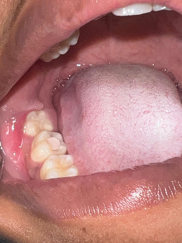
Preoperative intraoral view showing partially bony impacted right mandibular third molar.
Radiographical Evaluation
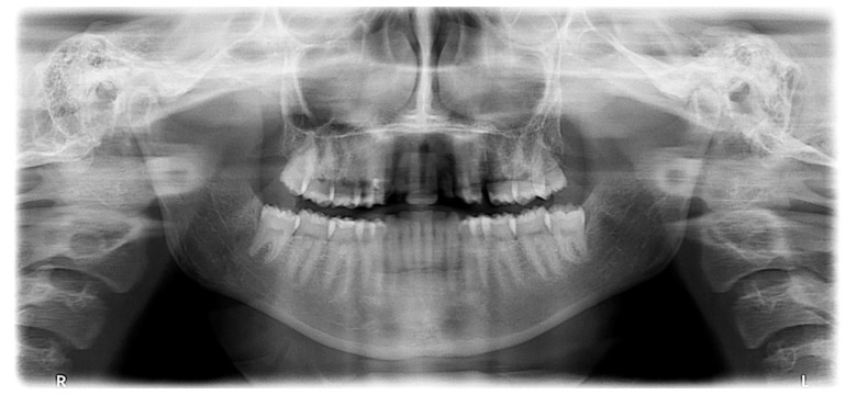
Panoramic radiographs were obtained for evaluation of the location and configuration of impacted mandibular third molar, surrounding bone, mandibular canal, and adjacent tooth.
The impacted mandibular third molars were categorized radiographically (classification, position, angulation) before surgery according to the Koerner index scale (Table 1) to reduce discrepancies in the degree of surgical difficulty (Figure 2).
Table 1.
Koerner difficulty index scale for removal of mandibular impacted third molar.
| Classification difficulty index | Value |
|---|---|
| Angulation | |
| Mesioangular | 1 |
| Horizontal | 2 |
| Vertical | 3 |
| Distoangular | 4 |
| Depth | |
| Level A | 1 |
| Level B | 2 |
| Level C | 3 |
| Ramus relationship | |
| Class I | 1 |
| Class II | 2 |
| Class III | 3 |
| Difficulty index | |
| Slightly difficult | 3–4 |
| Moderately difficult | 5–7 |
| Very difficult | 8–10 |
Figure 2.
Preoperative panoramic X-ray showing bilaterally vertically partially bony impacted right and left mandibular third molar, with moderate difficulty index (Koerner Index scale=5).
Surgical Procedures
The surgery was performed according to conventional surgical impacted third molar extraction, and aseptic, atraumatic, and non-heat producing techniques were considered to managing both soft and hard tissues.
Routine regional anesthesia was applied, including inferior alveolar nerve block together with buccal infiltration anesthesia by two 1.8-mL cartridges of a local anesthetic solution containing lidocaine hydrochloride 2% and epinephrine 1: 100 000 injection (manufactured for SEPTODONT, Louisville, CO 80027 by Novocol Pharmaceutical of Canada, Inc., Cambridge, Ontario, Canada, N1R 6X3).
In both groups, a 3-sided mucoperiosteal flap was reflected laterally, the tooth was extracted with the suitable instrument following adequate bone removal using the guttering technique on the buccal and the distal aspect of the tooth. Finally, the extraction socket was irrigated and debrided mechanically, and the flap was repositioned. In the study group (HA) (right side of the patients), hyaluronic acid sodium salt 1.5% (Hyalubrix 60mg/4ml; Fidia Farmaceutici S.P.A, Italy) was applied in the post-extraction socket before 3-0 Vicryl suture placement (Ethicon Vicryl Plus # 3-0 Absorbable, Braided, Polyglactin-Coated Violet Surgical Suture, Manufactured by Johanson & Johanson Pvt, Ltd, MIDC Area, Waluj, Aurangabad-MH, India).
In the control group (left side of the patients), the flaps were sutured with 3-0 Vicryl suture after a blood clot formed at the extraction site. In all patients, the right mandibular third molars were operated on first, then the second operation for removal of left mandibular third molars was performed at least 1 month later for objective evaluation (Figures 3–5).
Figure 3.
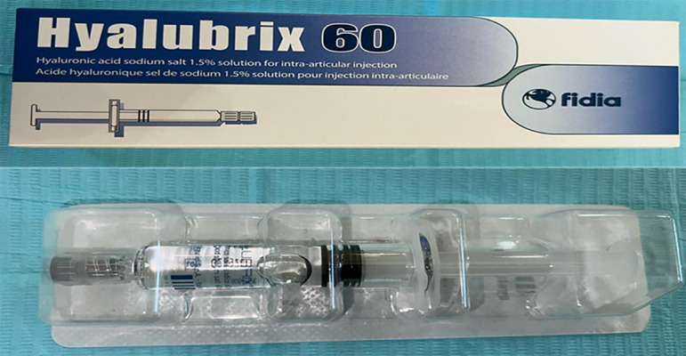
Hyaluronic acid sodium salt 1.5% (hyalubrix 60 mg/4 ml one prefilled syringe; Fidia Farmaceutici S.P.A, Italy).
Figure 4.
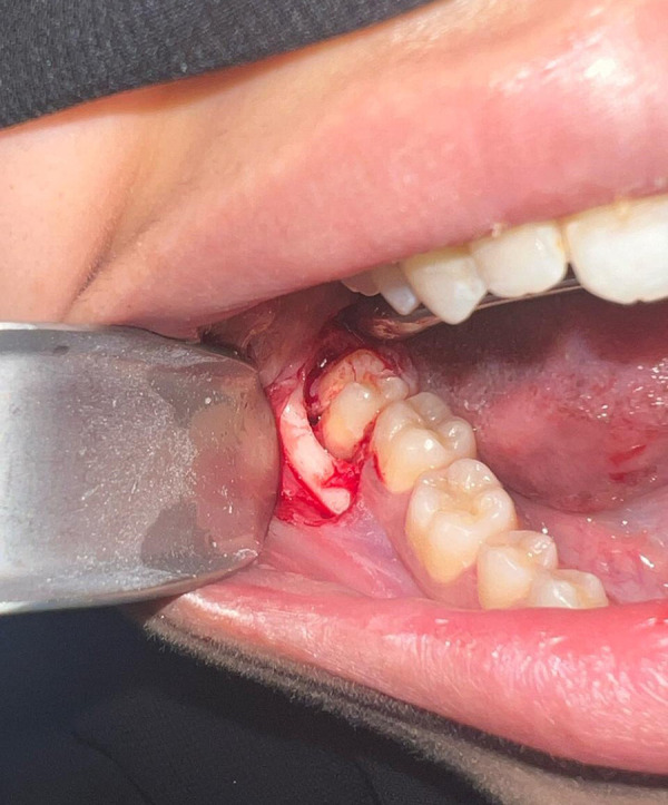
Intraoperative photograph showing surgical removal of partially impacted right mandibular third molar (study group), mucoperiosteal flap was retracted, the bone covering the tooth was removed, then the tooth was sectioned using a high-speed hand piece with a fissure bur.
Figure 5.
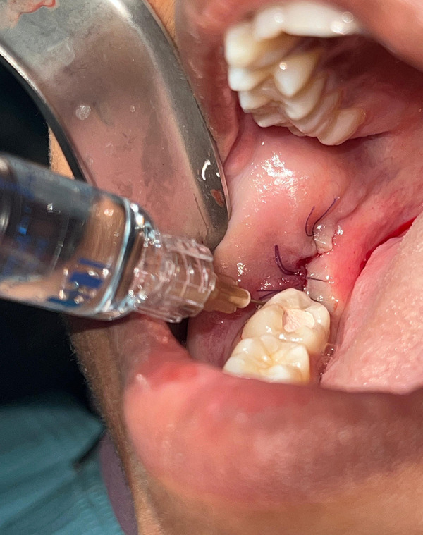
Intraoperative photograph showing a third molar tooth after hyaluronic acid (HA) was applied to the extraction socket.
Postoperative Care
Immediate postoperative care was started using ice packs 5 minutes every 20 minutes for 6 hours on the day of the surgery. Oral hygiene instructions were given, and patients were allowed to eat soft food for the first 2 days. Ibuprofen 600 mg (Manufactured by Tabuk pharmaceutical Co, Tabuk 47532, Saudi Arabia) was prescribed to be taken whenever needed, and the total analgesic dose taken in the postoperative period was recorded. The sutures were removed on the 7th postoperative day.
Postoperative Clinical Evaluation
All patients were evaluated postoperatively in the immediate postoperative 1st, 3rd, 7th days, and 1st month to assess the wound healing, pain, edema, and maximal jaw opening.
Outcome Measures
Soft Tissue Healing
The extraction sites were examined postoperatively to evaluate the soft tissue healing based on the criteria given by Huang et al [16] (score 1=uneventful healing with no gingival edema, erythema, suppuration, patient discomfort, or flap dehiscence; score 2=uneventful healing with slight gingival edema, erythema, patient discomfort, or flap dehiscence, but no suppuration; and score 3=poor wound healing with significant gingival edema, erythema, patient discomfort, flap dehiscence, or any suppuration).
Pain
The self-reported visual analog scale (VAS) [17] was used to assess the degree of pain postoperatively, starting from 0, which represents (no pain at all), and ending at 10, which represents (the most severe pain).
Edema
Assessment of postoperative edema was evaluated by using a vertical and horizontal guide with a tape on 4 reference points: canthus of the eye, angle of the mandible, tragus of the ear, and corner of the mouth. To assess the percentage of facial swelling, the difference between measurements of the postoperative and preoperative duration were divided by the value of the preoperative time [18].
Trismus
Maximal mouth openings were measured to determine the degree of trismus by the measurement of interincisal distance using calipers from the incisal edge of the upper central incisor to the incisal edge of the lower central incisor [19].
Statistical Analysis
All data were collected and statistically analyzed at the end of the study using IBM SPSS software version 20.0 (Armonk, NY: IBM Corp). Descriptive statistics, including mean values and standard deviations were determined for all variables in the test and control groups. The t test was used for normally distributed quantitative variables, to compare the 2 studied groups. The Friedman test for abnormally distributed quantitative variables was used to compare more than 2 periods or stages, and Dunn’s post hoc test was performed for pairwise comparisons. The level of significance was set at P<0.05.
Results
A total of 40 patients (16 males, 24 females) were included in this study. The age range was 18–29 years and the mean age was 21.16±2.97.
Clinical Results
Soft Tissue Healing
According to the healing index of Huang et al [12], at 1 month after surgery, normal extraction site healing with score 1 was achieved in 36 (90%) patients and 4 (10%) had slight gingival edema with score 2 in the right mandibular third molars extraction site (HA group). However, in the left mandibular third molars extraction site (control group) uneventful healing with score 1 was achieved in 28 (70%) patients, 8 (20%) had a score of 2, and 4 (10%) had a score of 3, indicating poor wound healing with significant gingival edema and flap dehiscence, which was treated by irrigation with warm normal saline, antiseptic mouthwash, and good oral hygiene.
Pain
Postoperative pain evaluation results using the visual analog scale (VAS) showed that the patients have lower VAS scores at all observation periods, the pain score 1 month postoperatively for study (HA) group ranged from 0 to 4 with a mean of 2.50±1.41 and for control group ranged from 4 to 7 with a mean of 5.50±0.93. There was statistically significant difference between groups at the last observational period (Table 2).
Table 2.
Comparison between the study (HA) and control group according to VAS values at the different observation periods.
| Groups | Pre-surgical Mean±SD |
Post-surgical Mean±SD |
|||
|---|---|---|---|---|---|
| 1st day | 3rd day | 7th day | 1st month | ||
| Study (HA) | 4.63±1.19 | 6.20±3.36 | 5.20±1.62 | 2.20±0.92 | 2.50±1.41 |
| Control | 4.88±1.13 | 6.80±2.62 | 5.90±1.79 | 4.50±1.18 | 5.50±0.93 |
| P-value | 0.672 | 0.912 | 0.372 | 0.350 | 0.001* |
HA – hyaluronic acid; VAS – Visual Analog Scale; SD – standard deviation.
Denotes significant difference at P<0.05.
Edema
Postoperative swelling reached peak intensity within 12–48 hours, resolving between the 5th and 7th days. In the study (HA) group, after 1 month, the edema score ranged from 9.85 to 11.53, with a mean of 10.81±0.74. However, in the control group, the edema score ranged from 11.05 to 11.81, with a mean of 11.29±0.45. There was a statistically significant difference between groups at follow-ups (Table 3).
Table 3.
Comparison between the study (HA) and control groups according to edema values at the different observation periods.
| Groups | Pre-surgical Mean±SD |
Post-surgical Mean±SD |
|||
|---|---|---|---|---|---|
| 1st day | 3rd day | 7th day | 1st month | ||
| Study (HA) | 11.09±0.59 | 11.41±0.52 | 10.50±0.63 | 11.10±0.61 | 10.81±0.74 |
| Control | 11.56±0.32 | 12.25±0.49 | 11.0±0.31 | 11.65±0.57 | 11.29±0.45 |
| P-value | 0.040* | 0.002* | 0.041* | 0.077 | 0.034* |
HA – hyaluronic acid; SD – standard deviation.
Denotes significant difference at P<0.05.
Trismus
The degree of mouth opening significantly decreased at postoperative days 1 and 3, there was no significant difference in pre- and postoperative measurements of the mean interincisal opening between the study (HA) and control groups. Adequate mouth opening was documented at 1-month follow-up in both groups. There were no statistically significant differences between the study (HA) and the control group at the different observation periods (P value >0.05) (Table 4).
Table 4.
Comparison of the mean maximal jaw opening in the study (HA) and control groups during different observation periods.
| Groups | Pre-surgical Mean±SD |
Post-surgical Mean±SD |
|||
|---|---|---|---|---|---|
| 1st day | 3rd day | 7th day | 1st month | ||
| Study (HA) | 43.63±1.92 | 24.25±2.76 | 20.63±2.50 | 35.75±2.19 | 41.3±6.5 |
| Control | 41.81±3.8 | 22.79±2.78 | 23.10±4.34 | 32.73±3.54 | 43.8±6.6 |
| P-value | 1.85 | 0.309 | 0.184 | 0.059 | 0.179 |
HA – hyaluronic acid; SD – standard deviation
Discussion
We found a significant decrease in pain scores after 24 hours, which continuously decreased until the end of the follow-up period in both groups. The pain score in the post-extraction site of the study (HA) group was significantly lower than in the control group at all observation points. This is in accordance with Gocemen et al [20], who found that topical application of HA after surgical removal of lower third molar surgery decreased postoperative pain by reducing leucocyte infiltration and increasing angiogenesis.
Postoperative edema after surgical removal of impacted third molars can occur due to inflammatory processes caused by surgical handling of soft tissues or bone cutting. In both groups there was a statistically significant increase of facial swelling after 24 and 72 hours, followed by a significant decrease until the end of the observational period. The facial swelling that occurs with surgical removal of the right mandibular impacted molar with the use of HA was significantly lower. These findings suggest that HA is effective in controlling postoperative swelling originating from the inflammatory process initiated by the surgical trauma, due to were prevention of excessive inflammation by the anti-inflammatory action of HA. This agrees with the results of Koray et al [21], who concluded that hyaluronic acid minimizes postoperative edema following impacted third molar surgery.
Limited mouth opening following impacted third molar surgery is mostly caused by the swelling associated with surgical trauma. The postoperative interincisal distance was decreased significantly in both groups by 24 hours after surgery, followed by a gradual increase to nearly the preoperative values in the HA-treated sockets group, while in the other group it was significantly lower than the preoperative value. This agrees with the results of Koray et al [21], who found that hyaluronic acid benefits management of trismus during the postoperative period after impacted third molar surgery.
Our study has some limitations. There have been few clinical studies assessing topical application of HA in the socket left behind after a lower impacted third molar tooth is extracted. Additionally, the study’s sample size was small, so more research should be planned with a larger sample size to assess the advantages of HA. Second, there is no recognized objective method for assessing pain; instead, the VAS is a subjective scale that is sensitive to individual variances such as pain threshold and psychological factors.
Conclusions
Intra-socket application of hyaluronic acid after surgical extraction of impacted mandibular third molars promote normal wound healing and reduces postoperative pain and swelling.
Statement
Patients were selected from the Dental Hospital of the College of Dentistry, Prince Sattam Bin Abdulaziz University.
Acknowledgment
The authors extend their appreciation to Prince Sattam bin Abdulaziz University for funding this research through project number 2024/01/99527.
Footnotes
Conflict of interest: None declared
Publisher’s note: All claims expressed in this article are solely those of the authors and do not necessarily represent those of their affiliated organizations, or those of the publisher, the editors and the reviewers. Any product that may be evaluated in this article, or claim that may be made by its manufacturer, is not guaranteed or endorsed by the publisher
Declaration of Figures’ Authenticity: All figures submitted have been created by the authors, who confirm that the images are original with no duplication and have not been previously published in whole or in part.
Financial support: Prince Sattam bin Abdulaziz University research project number 2024/01/99527
References
- 1.Kiencało A, Jamka-Kasprzyk M, Panaś M, Wyszyńska-Pawelec G. Analysis of complications after the removal of 339 third molars. Dent Med Probl. 2021;58(1):75–80. doi: 10.17219/dmp/127028. [DOI] [PubMed] [Google Scholar]
- 2.Carter K, Worthington S. Predictors of third molar impaction: A systematic review and meta-analysis. J Dent Res. 2016;95:267–76. doi: 10.1177/0022034515615857. [DOI] [PubMed] [Google Scholar]
- 3.Çanakçi FG, Er N, Duygu G, Karaca GT. Does saline irrigation at different temperatures affect pain, edema, and trismus after impacted third molar surgery: A clinical trial. J Oral Maxillofac Surg. 2023;81(1):88–94. doi: 10.1016/j.joms.2022.08.015. [DOI] [PubMed] [Google Scholar]
- 4.Yang H, Kim J, Kim J, et al. Non-inferiority study of the efficacy of two hyaluronic acid products in post-extraction sockets of impacted third molars. Maxillofac Plast Reconstr Surg. 2020;42(1):40. doi: 10.1186/s40902-020-00287-y. [DOI] [PMC free article] [PubMed] [Google Scholar]
- 5.Domic D, Bertl K, Lang T, et al. Hyaluronic acid in tooth extraction: A systematic review and meta-analysis of preclinical and clinical trials. Clin Oral Investig. 2023;27(12):7209–29. doi: 10.1007/s00784-023-05227-4. [DOI] [PMC free article] [PubMed] [Google Scholar]
- 6.Marques AFS, Silva NM, da Cruz M, Marques J, da Mata AD. Hyaluronic acid-based gels for oral application: Comparison of in vitro effects on gingival cells and bacteria. J Oral Biol Craniofac Res. 2024;14(3):238–44. doi: 10.1016/j.jobcr.2024.03.001. [DOI] [PMC free article] [PubMed] [Google Scholar]
- 7.Al-Saadi TS, Al-Quisi AF. The influence of the hyaluronic acid gel on the postoperative sequelae following surgical removal of the impacted mandibular third molar in comparison with the A-PRF: A randomized controlled trial. Int J Biometr. 2023;29:1883460. doi: 10.1155/2023/1883460. [DOI] [PMC free article] [PubMed] [Google Scholar]
- 8.Yilmaz N, Demirtas N, Kazancioglu HO, et al. The efficacy of hyaluronic acid in postextraction sockets of impacted third molars: A pilot study. Niger J Clin Pract. 2017;20:1626–31. doi: 10.4103/1119-3077.224131. [DOI] [PubMed] [Google Scholar]
- 9.Suchánek J, Ivančaková RK, Mottl R, et al. Hyaluronic acid-based medical device for treatment of alveolar osteitis-clinical study. Int J Environ Res Public Health. 2019;16:3698. doi: 10.3390/ijerph16193698. [DOI] [PMC free article] [PubMed] [Google Scholar]
- 10.Riccardo G, Egle P, Sergio M, et al. Effect on wound healing of a topical gel containing amino acid and sodium hyaluronate applied to the alveolar socket after mandibular third molar extraction: A double-blind randomized controlled trial. Quintessence In. 2018;49:831–40. doi: 10.3290/j.qi.a41157. [DOI] [PubMed] [Google Scholar]
- 11.Bouloux GF, Steed MB, Perciaccante VJ. Complications of third molar surgery. Oral Maxillofac Surg Clin North Am. 2007;19:117–28. doi: 10.1016/j.coms.2006.11.013. [DOI] [PubMed] [Google Scholar]
- 12.Yolcu U, Acar AH. Comparison of a new flap design with the routinely used triangular flap design in third molar surgery. Int J Oral Maxillofac Surg. 2015;44:1390–97. doi: 10.1016/j.ijom.2015.07.007. [DOI] [PubMed] [Google Scholar]
- 13.Pasqualini D, Cocero N, Castella A, et al. Primary and secondary closure of the surgical wound after removal of impacted mandibular third molars: A comparative study. Int J Oral Maxillofac Surg. 2005;34(1):52–57. doi: 10.1016/j.ijom.2004.01.023. [DOI] [PubMed] [Google Scholar]
- 14.Rodrigues ÉDR, Martins-de-Barros AV, Loureiro AMLC, et al. Comparison of two suture techniques on the inflammatory signs after third molars extraction – a randomized clinical trial. PLoS One. 2023;18(6):e0286413. doi: 10.1371/journal.pone.0286413. [DOI] [PMC free article] [PubMed] [Google Scholar]
- 15.Jeyashree T, Kumar MPS. Evaluation of difficulty index of impacted mandibular third molar extractions. J Adv Pharm Technol Res. 2022;13(Suppl 1):S98–S101. doi: 10.4103/japtr.japtr_362_22. [DOI] [PMC free article] [PubMed] [Google Scholar]
- 16.Huang LH, Neiva RE, Wang HL. Factors affecting the outcomes of coronally advanced flap root coverage procedure. J Periodontol. 2005;76:1729–34. doi: 10.1902/jop.2005.76.10.1729. [DOI] [PubMed] [Google Scholar]
- 17.Wewers ME, Lowe NK. A critical review of visual analogue scales in the measurement of clinical phenomena. Res Nurs Health. 1990;13:227–36. doi: 10.1002/nur.4770130405. [DOI] [PubMed] [Google Scholar]
- 18.Liu S, Zhao H, Wang Y, et al. oral bromelain for the control of facial swelling, trismus, and pain after mandibular third molar surgery: A systematic review and meta-analysis. J Oral Maxillofac Surg. 2019;77(8):1566–74. doi: 10.1016/j.joms.2019.02.044. [DOI] [PubMed] [Google Scholar]
- 19.Sayed N, Bakathir A, Pasha M, Al-Sudairy S. Complications of third molar extraction: A retrospective study from a tertiary healthcare centre in Oman. Sultan Qaboos Univ Med J. 2019;19:e230–e35. doi: 10.18295/squmj.2019.19.03.009. [DOI] [PMC free article] [PubMed] [Google Scholar]
- 20.Gocemen G, Gonul O, Oktay NS. The antioxidant and anti-inflammatory efficiency of hyaluronic acid after third molar extraction. J Craniomaxillofac Surg. 2015;43:1033–37. doi: 10.1016/j.jcms.2015.04.022. [DOI] [PubMed] [Google Scholar]
- 21.Koray M, Ofluoglu D, Onal EA. Efficacy of hyaluronic acid spray on swelling, pain, and trismus after surgical extraction of impacted mandibular third molars. Int J Oral Maxillofac Surg. 2014;43:1399–403. doi: 10.1016/j.ijom.2014.05.003. [DOI] [PubMed] [Google Scholar]


