Abstract
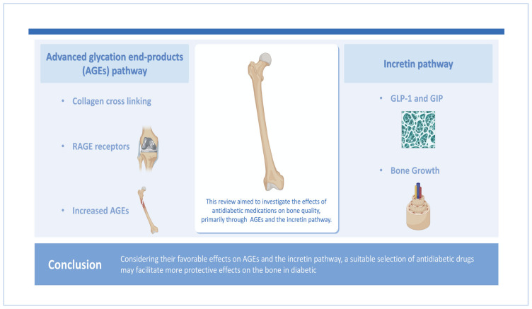
Diabetes mellitus is associated with inadequate bone health and quality and heightened susceptibility to fractures, even in patients with normal or elevated bone mineral density. Elevated advanced glycation end-products (AGEs) and a suppressed incretin pathway are among the mechanisms through which diabetes affects the bone. Accordingly, the present review aimed to investigate the effects of antidiabetic medications on bone quality, primarily through AGEs and the incretin pathway. Google Scholar, Cochrane Library, and PubMed were used to examine related studies until February 2024. Antidiabetic medications influence AGEs and the incretin pathway directly or indirectly. Certain antidiabetic drugs including metformin, glucagon-like peptide-1 receptor agonists (GLP-1RA), dipeptidyl-peptidase-4 (DDP-4) inhibitors, α-glucosidase inhibitors (AGIs), sodium-glucose co-transporter-2 inhibitors, and thiazolidinediones (TZDs), directly affect AGEs through multiple mechanisms. These mechanisms include decreasing the formation of AGEs and the expression of AGEs receptor (RAGE) in tissue and increasing serum soluble RAGE levels, resulting in the reduced action of AGEs. Similarly, metformin, GLP-1RA, DDP-4 inhibitors, AGIs, and TZDs may enhance incretin hormones directly by increasing their production or suppressing their metabolism. Additionally, these medications could influence AGEs and the incretin pathway indirectly by enhancing glycemic control. In contrast, sulfonylureas have not demonstrated any obvious effects on AGEs or the incretin pathway. Considering their favorable effects on AGEs and the incretin pathway, a suitable selection of antidiabetic drugs may facilitate more protective effects on the bone in diabetic patients.
Keywords: Antidiabetic agents, Bone, Diabetes Mellitus, Glycation, Incretin
GRAPHICAL ABSTRACT
INTRODUCTION
Skeletal fragility frequently coexists with type 1 and type 2 diabetes, where it is regarded as a pathological consequence of this condition. While low bone mass in type 1 diabetes mellitus (T1DM) can greatly increase the risk of fractures, individuals with type 2 DM (T2DM) also experience an increased incidence of fractures, even when normal or high bone mineral density (BMD) and a higher body mass index are present (factors that protect against fractures in non-diabetic individuals). Therefore, diabetes may be linked to a reduction in the strength of bone that is not accurately represented by the measurement of BMD. Rendering to the current definition, determining bone strength involves considering both bone density and quality, which include the structural and material characteristics of bone.[1] Historically, the most commonly used measurement for fracture risk prediction was BMD, a quantitative parameter measured by dual energy X-ray absorptiometry, however, this technique could incompletely expresses the fracture risk.[2] On the other hand, image-based noninvasive parameters, such as the trabecular bone score, quantitative computed tomography (QCT), and high-resolution peripheral QCT are newer approaches for assessing the bone quality.[3]
Generally, diabetes decreases the quality of bone instead of BMD. The process of collagen cross-linking is crucial for bone strength. Collagen cross-links can be further classified as enzymatic immature divalent cross-links, mature trivalent cross-links mediated by lysyl hydroxylase and lysyl oxidase, and non-enzymatic cross-links caused by oxidation or glycation (advanced glycation end-products [AGEs]) such as pentosidine (PEN), which is a surrogate marker of AGEs. The formation mechanisms and functionalities of these cross-link types differ from one another.[4] The presence of either reduced enzymatic cross-linking or an increase in non-enzymatic cross-links in bone collagen is believed to be a major contributing factor to the fragility of bones in aging, osteoporosis, and DM.[5]
Furthermore, the incretin hormone which includes glucose-dependent insulinotropic polypeptide (GIP), and glucagon-like peptide-1 (GLP-1) was demonstrated to have beneficial effects on bone metabolism, the effect of these hormones is reduced or even absent in diabetic patients.[6] The material qualities of bone tissue are controlled by cellular activity, the rate of bone tissue turnover, and the degrees of oxidative stress and glycation.[7]
Some antidiabetic drugs may decrease the risk of fracture and improve bone quality while others do not. As shown in Table 1, the mechanisms that promote these effects are multiple.[8] However, some antidiabetic medications, including sulfonylureas and insulin, have a greater tendency to cause hypoglycemia, which in turn increases the risk of fall-related fractures regardless of their effects on the bone.[9] This review will summarize the current knowledge about the impacts of antidiabetic medications on bone health, focusing on their effects on AGEs and incretin pathways in diabetic patients. This review article will discuss the effects of AGEs on the bone, the bone effects of incretin hormones including GIP, GLP-1, and GLP-2, the effects of antidiabetic medications on AGEs and incretin pathway, in addition to the clinical impacts of these medications on the bone.
Table 1.
Effects of antidiabetic drugs on the bone
| Drug | Effect on AGEs | Effects on incretin pathway | Effects on bone | References |
|---|---|---|---|---|
| Metformin | Decrease | Increase | Positive | Marx et al. [54], Kim [55] |
| Sulfonylureas | Decrease | Neutral | Neutral | Driessen et al. [60], Kanemoto et al. [91] |
| GLP-1 agonists | Decrease | Increase | Positive/neutral | Melton et al. [58], Alassaf et al. [92] |
| DPP-4 inhibitors | Decrease | Increase | Positive/neutral | Loh et al. [57], Vestergaard et al. [59] |
| AGIs | Decrease | Increase | Neutral | Bunck et al. [61] |
| SGLT2 inhibitorsa) | Decrease | Increase | Negative/neutral | Cheng et al. [62], Dabhi et al. [65], Bilezikian et al. [66] |
| TZDsb) | Decrease | Increase | Negative | Ljunggren et al. [71], Owusu et al. [93] |
Their effects on bone by mechanism not related to AGEs or incretin pathway, they cause alteration in calcium and phosphate homeostasis that can lead to bone loss.[67]
Their effects on bone is not related to AGEs or incretin pathway, but by stimulating PPAR-γ.[72]
AGEs, advanced glycation end-products; GLP-1, glucagon-like peptide-1; DPP-4, dipeptidyl peptidase 4; AGIs, α-glucosidase inhibitors; SGLT2, sodium glucose cotransporter 2; TZDs, thiazolidinediones; PPAR-γ, peroxisome proliferator-activated receptor-γ.
METHODS
This article is a narrative review that mainly discusses the impacts of antidiabetic medications on bone health, focusing on their effects on AGEs and the incretin pathway in T2DM. Using keywords related to the main subject of this study, a literature search was conducted between November 2023 and February 2024 on databases including Google Scholar, Cochrane Library, and PubMed. Antidiabetics, DM, diabetic complications, and bone diseases, were used separately and in combination to reveal articles related to the main topic until the date of drafting this review. Inclusion criteria included articles that discuss bone alterations in diabetic patients, and the effects of antidiabetic drugs on the bone particularly their effects on AGEs and incretin pathway, in addition to articles that discuss the clinical significance and/or non-significance of antidiabetics on the bone. The current narrative review goes into additional detail about these articles.
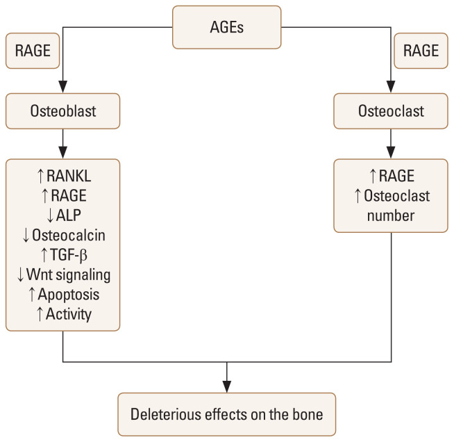
EFFECT OF AGES ON THE BONE (Fig. 1)
Fig. 1.
Effects of advanced glycation end-products (AGEs) on the bone.[10] AGEs exert direct effects on osteoblast and osteoclast, causing damaging effects on the bone. RAGE, receptor for AGEs; RANKL, receptor activator of nuclear factor-κB ligand; ALP, alkaline phosphatase; TGF-β, transforming growth factor-β.
Type I collagen is a crucial constituent of bone and acts as a structural scaffold that improves the strength of the skeleton through mineralization. Type I collagen is a fibrous protein arranged in a triple-helix pattern, which allows it to form organized fibrils that are made stronger through enzymatic cross-linking. Aside from enzymatic cross-linking, type I collagen can also undergo chemical cross-linking with “free-floating” sugars in the serum through the bonding of exposed amino acid residues. This can lead to post-translational modifications of collagen and ultimately the formation of AGEs. Individuals with DM are more likely to have AGEs accumulation in various organs, including bone.[4]
By interacting with the cell surface receptor for AGEs (RAGE), both non-cross-linking includes Nɛ-carboxymethyllysine and cross-linking forms of AGEs impair osteoblastic function as shown in Figure 1. In the bone AGE-RAGE binding primarily induces apoptosis and altered differentiation of osteoblasts. This is accompanied by an increased expression of the receptor activator of nuclear factor-κB ligand (RANKL), which promotes increased osteoclastogenesis and impedes bone mineralization.[10] Along with an increase in RANKL mRNA expression, these effects are at least in part mediated by osteocalcin mRNA and alkaline phosphatase (ALP) downregulation, as well as an increase in RAGE expression that promotes the AGE-RAGE pathway.[11] Additionally, transforming growth factor-β (TGF-β) is secreted and expressed more when AGE-RAGE binding occurs, which prevents osteoblastic cells from differentiation and mineralization.[12] AGEs also results in the inhibition of signaling pathways, include Wnt/β-catenin (the primary regulator of osteoblast development and activity), that are crucial for maintaining skeletal homeostasis.[13]
On the other hand, the effects of AGEs on bone resorption by osteoclasts are controversial. In response to RANKL stimulation, bone marrow macrophages release a non-histone nuclear protein known as high mobility group box 1 (HMGB1). Via RAGE, the extracellular HMGB1 regulates the remodeling, differentiation, and function of the osteoclastic actin cytoskeleton. Thus, it appears that the regulation of osteoclast activity is also influenced by the interaction between RAGE and other ligands.[14] Additionally, it appears that RANKL encourages the expression of RAGE and, as a result, osteoclast differentiation.[15]
Moreover, RAGE can be generated as a soluble “decoy” receptor that could prevent AGE–RAGE signaling axes by binding AGEs. It has been demonstrated that low serum concentrations of soluble RAGE and elevated serum concentrations of the AGE PEN are independent of BMD risk indicators for fractures in diabetes.[16] Furthermore, AGEs competitively block the production of enzymatic cross-links by binding to lysine residues, which are critical locations for enzymatic cross-linking in collagen molecules. Additionally, AGEs cross-link themselves decrease the strength of bone without causing cellular dysfunction. The overproduction of AGE cross-links in bone results in the brittleness of collagen fibers, leading to the buildup of microdamage. This microdamage then causes a decline in the post-yield properties and toughness of the bone.[17] The regulation of AGEs generation is dependent upon tissue life span, oxidative stress, and glycemic control.[18]
EFFECTS OF INCRETIN PATHWAY ON THE BONE
GLP-1 and GIP are the primary incretins generated by the intestine in response to dietary intake, and they function through signaling-mediated mechanisms by incretin receptors. Enteroendocrine cells in the intestinal mucosa secrete GLP-1, which binds to GLP-1 receptors (GLP-1Rs) found on various cell types, including islet β-cells. This interaction leads to metabolic consequences in multiple organs. Bone-related cells also express GLP-1Rs, which trigger a variety of effects. On the other hand, GIP receptors (GIPRs) are also expressed in human osteoblasts, indicating various stages of differentiation.[19]
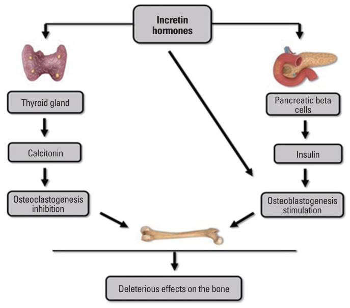
Bone turnover is defined as a dynamic process that includes the use of energy from meal consumption. Inadequate availability of essential nutrients, particularly during the night, might stimulate the breakdown of bone tissue to maintain a consistent balance of calcium in the body.[20] Following the intake of meals, bone resorption is subsequently suppressed. The occurrence of this event may be influenced by gastrointestinal hormones that are secreted following the intake of food, such as GLP-1 and GIP.[21] Consistent with this theory, the finding that intravenous feeding is linked to lowered bone density indicates that a deficiency in incretin hormones may contribute to changes in bone remodeling.[22] Notably, many investigations have suggested that the incretin hormones GLP-1 and GIP may have a beneficial effect on bone health related to their ability to reduce bone loss and promote bone growth, as shown in Figure 2. Suggests that antidiabetic medicines such as GLP-1R agonists (GLP-1RA) or dipeptidyl peptidase-4 (DPP-4) inhibitors may have a positive effect on bone metabolism.[23–25]
Fig. 2.
The impact of incretin hormones on the metabolic process of bone.[25] Incretin hormones have the dual ability to directly act on osteoblasts and indirectly stimulate osteoblastogenesis through an increase in insulin secretion. In addition, incretin hormones can suppress osteoclastogenesis by enhancing thyroid calcitonin synthesis.
GIP AND BONE METABOLISM
A functioning GIPR with high affinity that increases intracellular calcium levels via the cyclic adenosine 3′,5′-monophosphate pathway was found by Bollag et al. [19] in their study of GIPR expression in normal rat bone, osteoblasts, osteocytes, and osteoblast-like cell lines. Furthermore, in osteoblastic-like cells, GIP increases ALP activity and collagen type 1 expression, suggesting the anabolic effects of this incretin hormone.[26] These observations have been validated in ovariectomized rats that are associated with bone loss as compared with normal rats, intermittent administration of GIP in these models results in a significant rise in lumbar BMD specifically in ovariectomized rats. In addition, they confirmed that the activity of GIP is dependent on both dose and duration, where within three hours of treatment, high concentrations of GIP decreased GIPR expression in Saos-2 cells, whereas lower concentrations of GIP required a longer exposure period to reduce GIPR. Consequently, intermittent administration of GIP seems to have a beneficial effect on bone health, similar to parathyroid hormone, by promoting bone formation and preventing bone loss.[27] Furthermore, a study on animals with a deletion of the GIPR found a significant reduction in the degree of mineralization of bone matrix, the amount of mature cross-links of the collagen matrix, and lowered intrinsic material characteristics. In this study, the decline in collagen cross-link ratio was higher in trabecular bone (~42%) as compared with cortical bone (~16%). On the other hand, heterogeneity in BMD distribution was not significantly reduced in trabecular bone, while it was significantly increased in cortical bone. In other words, it appears that the alteration of collagen maturity was greater in the trabecular bone, but the alteration of mineral maturity was greater in the cortical bone.[28] The presence of GIPRs was also observed in osteoclastic-like cells of murine, where GIP seems to inhibit bone resorption triggered by RANKL.[29]
In addition, the process of bone formation and bone resorption was evaluated using two collagen-based markers, procollagen type N-terminal propeptide of type I collagen (P1NP) and C-terminal telopeptide (CTX). The administration of GIP infusion in this study led to an augmentation of the P1NP response, which is associated with bone formation, and a reduction in the CTX response, which is linked to bone resorption, during the hyperglycemic clamp. Thus, GIP has been shown to have advantageous impacts on bone quality.[30]
GLP-1, GLP-2 AND BONE METABOLISM
Examining the impact of GLP-1 administration on bone in three different glycometabolic condition revealed that participants with glucose metabolism exhibited a beneficial impact of GLP-1 on bone structure after 3 days treatment with either GLP-1 or a placebo.[31]
It was also shown that GLP-1 can interact with human osteoblasts through a receptor that is distinct from the GLP-1R receptor found in the pancreas. GLP-1 selectively attaches to the cell membrane of osteoblastic MC3T3-EI cells to trigger the rapid breakdown of glycosylphosphatidylinositols, resulting in the production of diacylglycerol and inositol phosphoglycans which function as secondary messengers. The interaction between GLP-1 and MC3T3-EI influenced gene expression, specifically increasing the expression of osteocalcin, which is the primary protein released by osteoblasts.[32] In addition, osteocytes release sclerostin (SOST), which is produced by the SOST gene and favors the inhibition of bone development. It has been discovered that GLP-1 decreases SOST mRNA levels, resulting in decreasing the harmful effects of SOST on the bone.[33]
Furthermore, Henriksen et al. [21] demonstrated a reduction in nocturnal bone breakdown following the delivery of GLP-2 at 10 PM. A total of 160 postmenopausal women were randomly grouped into four different groups. Each group received a daily dose of either 0.4, 1.6, or 3.2 mg of GLP-2, or a placebo. Additionally, all participants were given calcium and vitamin D supplements for 120 days. The results indicated that GLP-2 treatment did not have any harmful effects, and, notably, there was a substantial increase in BMD that was depending on the dosage. The 1.6 mg and 3.2 mg doses showed statistically significant improvements in BMD compared to the placebo. The administration of GLP-2 resulted in an instant and continuous reduction in bone resorption markers including CTX. However, the levels of bone production markers, such as osteocalcin, were unaffected.[21]
EFFECTS OF ANTIDIABETIC MEDICATIONS ON AGES AND INCRETIN PATHWAY
Metformin has been demonstrated to reduce AGE-RAGE pathway, beyond glycemic control, it led to inactivation of reactive carbonyl species, thus resulting in lower carbonyl stress and decreased formation of deleterious AGEs. Furthermore, it can effectively decrease the expression of RAGE in endothelial cells subjected to elevated levels of glucose or AGEs.[34] Additionally, metformin inhibited cell death caused by AGE and reduced the production of RAGE protein in cultured osteoblastic cells.[35] Moreover, metformin has been observed to increase plasma active GLP-1 levels with various mechanisms. It promotes the production of pro-glucagon (a precursor of GLP-1) in the large intestine and directly boosts GLP-1 release from entero-endocrine GLP-1-producing cells leading to an elevation in total GLP-1 concentrations.[36] Beyond this stimulating action, metformin has also been linked to the up-regulation of GLP-1R and GIPR expression in pancreatic beta cells through a peroxisome proliferator-activated receptor (PPAR) dependent pathway.[37] Also, it has been observed that metformin inhibits DPP-4 activity in T2DM patients.[38] On the other hand, sulfonylureas reduces AGEs indirectly through the enhancement in glycemic control.[39] Also, it demonstrated no significant effects on the incretin pathway.[40]
GLP-1RA directly activating the GLP-1R that potentiates the incretin pathway.[41] Regarding AGEs, GLP-1 agonists was suggested to suppress oxidative stress generation induced by AGEs-RAGE and reduce tissue expression of RAGE via activation of cyclic adenosine monophosphate pathways.[42]
DPP-4 inhibitors act by inhibiting the degradation of endogenous incretin by DDP-4 enzyme and so improve the action of incretin pathway.[43] It has been discovered that the production of oxidative stress caused by AGE-RAGE interaction leads to the release of DPP-4. This, in turn, enhances the harmful effects of AGEs, DPP-4 inhibitors provide a protection from these effects.[44,45] Additionally, it has been revealed that, vildagliptin, a DPP-4 inhibitor, is associated with decreased AGEs to some extents.[46]
α-glucosidase inhibitors (AGIs) including acarbose demonstrated to reduce the level of AGEs by acting as AGEs-inhibiting in patients with diabetes and decreases AGEs serum levels.[47] Additionally, by blocking α-glucosidase in the upper part of the small intestine, leads to the presence of significant quantities of undigested carbohydrates in the lower part of the small intestine. The ileum, a section of the small intestine, contains a high concentration of entero-endocrine L-cells. When these cells come into contact with foods, particularly carbohydrates they release GLP-1. Therefore, AGIs result in enhancement the secretion of GLP-1 from the intestine and potentiate the incretin pathway.[48]
The impact of sodium glucose cotransporter 2 (SGLT2) inhibitors on AGEs is not well understood however, it was suggested that improvements in glycemic control, as well as the restoration of renal and cardiovascular function, are anticipated mechanism by which SGLT2 inhibitors minimize AGE generation and accumulation.[49] Also, SGLT2 inhibitors have been demonstrated to decrease the formation of reactive oxygen species, therefore decreasing the buildup of AGEs in chronic diseases, including DM. Furthermore, due to enhancements in glomerular filtration resulting from SGLT2 inhibition, AGEs will likely be eliminated more efficiently.[50] These medications have shown to enhance the sensitivity of pancreatic beta cells to GLP-1 and GIP in diabetic patients, which may be related to improved glycemic control.[51] On the other hand, Administering canagliflozin to individuals after gastric bypass surgery did not decrease the 4-hr plasma GLP-1 responses, but it decreased the initial increase in GLP-1 and GIP hormones.[52]
Thiazolidinediones (TZDs) demonstrated to increase the level of total serum soluble RAGE that may lead to decrease the harmful effects of AGEs.[53] Furthermore, it reduce the tissue RAGE expression resulting in decreases proinflammatory effects of AGEs.[54] Additionally, TZDs was suggested to enhance GIP secretion and action that potentiates the incretin pathway in diabetic patients.[55]
CLINICAL IMPACT OF ANTIDIABETICS ON THE BONE
Many investigations demonstrated that metformin has beneficial effects on the bone.[56,57] A cohort study of 1,964 patients with T2DM has confirmed a decreased incidence of fractures with the use of metformin.[58] Furthermore, in Denmark, a comprehensive case-control study has shown that administering metformin could significantly be linked to a reduced risk of fracture.[59] GLP-1RA and DPP-4 inhibitors have been revealed to induce beneficial or neutral effects on the bone.[60,61] However, a meta-analysis of randomized controlled trials, showed that therapy with GLP-1RA decreased risk of fractures.[62] Additionally, Monami et al. [63] study reported that DPP-4 inhibitors usage may substantially be associated with lower fracture risk rate when compared with non-users. These favorable impacts on the bone by metformin, GLP-1RA, and DPP-4 inhibitors, may be attributed to the ability of these medications to suppress the actions of AGEs and improve the incretin pathway. On the other hand, sulfonylureas and AGIs, have exhibited neutral effects on the bone.[64,65] Concerning SGLT2 inhibitors, despite their beneficial effects on AGEs and incretin pathway, canagliflozin revealed to have detrimental impacts on bone density, bone resorption, and fracture risk. In September 2015, the U.S. Food and Drug Administration revised the drug’s label and added a new warning.[66,67] However, although the CANVAS data (canagliflozin cardiovascular assessment study) has shown that this drug has a harmful impact on the bone, there is still debate over the bone-related adverse effects of canagliflozin, as these effects have not been broadly verified in the subsequent trials.[68] While except for one study involving dapagliflozin,[69] there is no evidence to suggest any impact on BMD, bone markers, or the risk of fractures for empagliflozin and dapagliflozin. Therefore, it appears that both medications have a neutral effect on the metabolism of bones.[70,71] Nevertheless, the issues indicated by research on canagliflozin inevitably influence the entire class. However, SGLT2 inhibitors influence the bone by a mechanism unrelated to AGEs or incretin pathway. SGLT2 inhibitors affect bone by inhibiting the SGLT2 in the cells of the proximal tubule epithelium, where they can disrupt the balance of calcium and phosphate levels in the body. As a result, secondary hyperparathyroidism may be developed causing bone resorption.[72,73] Even though, the promising effects of TZDs in reducing AGEs and enhancing the incretin pathway, there is extensive evidence to support the fact that the clinically approved TZDs, including rosiglitazone and pioglitazone, have been shown to reduce bone density and increase the likelihood of fractures, particularly in women.[74,75] Furthermore, a longitudinal follow-up of patients in the action to control cardiovascular risk in T2DM (ACCORD) study revealed that women were more likely to experience non-spine fractures when taking TZDs, compared to men, and that the risk decreased once the TZDs were discontinued.[76] TZDs modify bone remodeling that results in increasing bone resorption and inhibiting bone formation through their action as full PPAR-γ agonists.[77,78]
According to the above-mentioned evidences, and beyond glycemic control, a proper selection criteria of certain antidiabetics, considering their effects on AGEs and incretin pathway, should be considered to result in a better bone profile and reduced risk of bone abnormality in diabetic individuals.[79] Metformin is recommended in such situation, GLP-1RA and DPP-4 inhibitors also could improve the bone health, while, sulfonylureas and AGIs have a less beneficial effect on the bone. On the other hand, TZDs and canagliflozin should be evaded as their detrimental effects are observed in clinical trials, whereas other SGLT2 inhibitors are less well-validated options. Additionally, when a combination of antidiabetics is the option, an augmentation in the favorable effects of such medications on AGEs and incretin pathway could occur.[80] The use of metformin and sitagliptin together may result in enhanced beneficial effects on bone when compared to metformin alone.[81] Similarly, the combination therapy of metformin and linagliptin has been demonstrated to have a better impact on the bone than metformin only.[82] Furthermore, the administration of a GIP together with GLP-1 resulted in a pronounced suppression of the bone resorption marker CTX than each hormone alone in overweight men without diabetes.[83] In the context of this combination, tirzepatide, which is a dual GIPR/GLP-1RA was recently added, however, more investigations are required about its impact on bone metabolism.[84] Additionally, a combination of GIP and GLP-2 was demonstrated to have a synergistic decrease in bone resorption that was greater than the reduction achieved by each hormone separately supporting a dual GIPR/GLP-2 receptor agonist as future osteoporosis management.[85]
On the other hand, glucagon has been shown to have a beneficial impact on bone metabolism in T2DM. It appears that glucagon may speed up skeletal remodeling, expedite osteogenesis, and enhance the creation of mature bone tissue. Simultaneously, the osteoclastic process was also increased, providing raw materials for osteogenesis and maintaining dynamic balance. Accordingly, the successful use of a drug that acts as a glucagon receptor (GcgR) agonist alone or in combination with other drugs may result in a reduction in osteoporosis in diabetic patients.[86] A novel long-acting GLP-1R/GIPR/GcgR triple agonist (HM15211) that is being developed to treat obesity, has been demonstrated to have bone protective effects despite the significant weight loss that is associated with this compound. Consistent with the bone protective effect in vivo, HM15211 led to significant increases in type 1 collagen-α1, -α2, and carboxylated osteocalcin expression.[87]
Moreover, not only antidiabetics could impact the AGEs and incretin pathways, other medications and dietary supplements had an effect.[88–90] Vitamin D3 has been observed to reduce the level of AGEs, and show synergistic protective effects on the bone when using with metformin.[91–95] Additionally, omega-3 fatty acids reported to have favorable effects on AGEs in diabetic patients, and accordingly its combination with metformin could be preferably used in the management of bone health in diabetic patients.[96–98] On the other hand, calcium supplementation has shown to enhance the incretin pathway and its combination with metformin could alleviate the osteoporosis induced by T2DM.[99,100]
CONCLUSIONS
Individuals with T2DM are more likely to experience fragility and fractures due to the damaging effects of diabetes on the bone including the negative effects of AGEs and reduction in the beneficial effects of incretin pathway. Antidiabetic medications could influence AGEs and incretin pathway either directly or indirectly, which could impact the bone. Metformin has been considered to have protective effects on the bone, GLP-1RA and DPP-4 inhibitors could also have beneficial effects on the bone, such effects may be related to the favorable effects of such medications on AGEs and incretin pathway. Sulfonylureas and AGIs reported to have neutral effects on the bone while canagliflozin and TZDs have been shown to have detrimental effects. However, combination therapy of antidiabetic could result in augmentation in the favorable effects of antidiabetics on AGEs and incretin pathway that result in more protective effects on the bone.
Acknowledgments
Our sincere gratitude goes to the University of Mosul and the College of Pharmacy for guidance and assistance.
Footnotes
Ethics approval and consent to participate
Not applicable.
Conflict of interest
No potential conflict of interest relevant to this article was reported.
REFERENCES
- 1.Schwartz AV. Diabetes, bone and glucose-lowering agents: clinical outcomes. Diabetologia. 2017;60:1170–9. doi: 10.1007/s00125-017-4283-6. [DOI] [PubMed] [Google Scholar]
- 2.Broy SB, Cauley JA, Lewiecki ME, et al. Fracture risk prediction by non-BMD DXA measures: the 2015 ISCD official positions part 1: Hip geometry. J Clin Densitom. 2015;18:287–308. doi: 10.1016/j.jocd.2015.06.005. [DOI] [PubMed] [Google Scholar]
- 3.Hunt HB, Donnelly E. Bone quality assessment techniques: geometric, compositional, and mechanical characterization from macroscale to nanoscale. Clin Rev Bone Miner Metab. 2016;14:133–49. doi: 10.1007/s12018-016-9222-4. [DOI] [PMC free article] [PubMed] [Google Scholar]
- 4.Saito M, Marumo K. Bone quality in diabetes. Front Endocrinol (Lausanne) 2013;4:72. doi: 10.3389/fendo.2013.00072. [DOI] [PMC free article] [PubMed] [Google Scholar]
- 5.Vestergaard P. Discrepancies in bone mineral density and fracture risk in patients with type 1 and type 2 diabetes--a meta-analysis. Osteoporos Int. 2007;18:427–44. doi: 10.1007/s00198-006-0253-4. [DOI] [PubMed] [Google Scholar]
- 6.Boer GA, Holst JJ. Incretin hormones and type 2 diabetes-mechanistic insights and therapeutic approaches. Biology (Basel) 2020;9:473. doi: 10.3390/biology9120473. [DOI] [PMC free article] [PubMed] [Google Scholar]
- 7.Saito M, Marumo K. Collagen cross-links as a determinant of bone quality: a possible explanation for bone fragility in aging, osteoporosis, and diabetes mellitus. Osteoporos Int. 2010;21:195–214. doi: 10.1007/s00198-009-1066-z. [DOI] [PubMed] [Google Scholar]
- 8.Deeba F, Younis S, Qureshi N, et al. Effect of diabetes mellitus and anti-diabetic drugs on bone health: a review. J Bioresour Manag. 2021;8:131–48. doi: 10.35691/JBM.1202.0187. [DOI] [Google Scholar]
- 9.Johnston SS, Conner C, Aagren M, et al. Association between hypoglycaemic events and fall-related fractures in Medicare-covered patients with type 2 diabetes. Diabetes Obes Metab. 2012;14:634–43. doi: 10.1111/j.1463-1326.2012.01583.x. [DOI] [PubMed] [Google Scholar]
- 10.Cavati G, Pirrotta F, Merlotti D, et al. Role of advanced glycation end-products and oxidative stress in type-2-diabetes-induced bone fragility and implications on fracture risk stratification. Antioxidants (Basel) 2023;12:928. doi: 10.3390/antiox12040928. [DOI] [PMC free article] [PubMed] [Google Scholar]
- 11.Taguchi K, Fukami K. RAGE signaling regulates the progression of diabetic complications. Front Pharmacol. 2023;14:1128872. doi: 10.3389/fphar.2023.1128872. [DOI] [PMC free article] [PubMed] [Google Scholar]
- 12.Kay AM, Simpson CL, Stewart JA., Jr The role of AGE/RAGE signaling in diabetes-mediated vascular calcification. J Diabetes Res. 2016;2016:6809703. doi: 10.1155/2016/6809703. [DOI] [PMC free article] [PubMed] [Google Scholar]
- 13.Li G, Xu J, Li Z. Receptor for advanced glycation end products inhibits proliferation in osteoblast through suppression of Wnt, PI3K and ERK signaling. Biochem Biophys Res Commun. 2012;423:684–9. doi: 10.1016/j.bbrc.2012.06.015. [DOI] [PubMed] [Google Scholar]
- 14.Zhou Z, Han JY, Xi CX, et al. HMGB1 regulates RANKL-induced osteoclastogenesis in a manner dependent on RAGE. J Bone Miner Res. 2008;23:1084–96. doi: 10.1359/jbmr.080234. [DOI] [PMC free article] [PubMed] [Google Scholar]
- 15.Zhou Z, Immel D, Xi CX, et al. Regulation of osteoclast function and bone mass by RAGE. J Exp Med. 2006;203:1067–80. doi: 10.1084/jem.20051947. [DOI] [PMC free article] [PubMed] [Google Scholar]
- 16.Schwartz AV, Garnero P, Hillier TA, et al. Pentosidine and increased fracture risk in older adults with type 2 diabetes. J Clin Endocrinol Metab. 2009;94:2380–6. doi: 10.1210/jc.2008-2498. [DOI] [PMC free article] [PubMed] [Google Scholar]
- 17.Saito M, Mori S, Mashiba T, et al. Collagen maturity, glycation induced-pentosidine, and mineralization are increased following 3-year treatment with incadronate in dogs. Osteoporos Int. 2008;19:1343–54. doi: 10.1007/s00198-008-0585-3. [DOI] [PubMed] [Google Scholar]
- 18.Saito M, Fujii K, Mori Y, et al. Role of collagen enzymatic and glycation induced cross-links as a determinant of bone quality in spontaneously diabetic WBN/Kob rats. Osteoporos Int. 2006;17:1514–23. doi: 10.1007/s00198-006-0155-5. [DOI] [PubMed] [Google Scholar]
- 19.Bollag RJ, Zhong Q, Phillips P, et al. Osteoblast-derived cells express functional glucose-dependent insulinotropic peptide receptors. Endocrinology. 2000;141:1228–35. doi: 10.1210/endo.141.3.7366. [DOI] [PubMed] [Google Scholar]
- 20.Abed MN, Alassaf F, Qazzaz ME, et al. Insights into the perspective correlation between vitamin D and regulation of hormones: thyroid and parathyroid hormones. Clin Rev Bone Miner Metab. 2020;18:87–93. doi: 10.1007/s12018-021-09279-6. [DOI] [Google Scholar]
- 21.Henriksen DB, Alexandersen P, Hartmann B, et al. Four-month treatment with GLP-2 significantly increases hip BMD: a randomized, placebo-controlled, dose-ranging study in postmenopausal women with low BMD. Bone. 2009;45:833–42. doi: 10.1016/j.bone.2009.07.008. [DOI] [PubMed] [Google Scholar]
- 22.Henriksen DB, Alexandersen P, Bjarnason NH, et al. Role of gastrointestinal hormones in postprandial reduction of bone resorption. J Bone Miner Res. 2003;18:2180–9. doi: 10.1359/jbmr.2003.18.12.2180. [DOI] [PubMed] [Google Scholar]
- 23.Kitaura H, Ogawa S, Ohori F, et al. Effects of incretin-related diabetes drugs on bone formation and bone resorption. Int J Mol Sci. 2021;22:6578. doi: 10.3390/ijms22126578. [DOI] [PMC free article] [PubMed] [Google Scholar]
- 24.Hansen MSS, Tencerova M, Frølich J, et al. Effects of gastric inhibitory polypeptide, glucagon-like peptide-1 and glucagon-like peptide-1 receptor agonists on Bone Cell Metabolism. Basic Clin Pharmacol Toxicol. 2018;122:25–37. doi: 10.1111/bcpt.12850. [DOI] [PubMed] [Google Scholar]
- 25.Ceccarelli E, Guarino EG, Merlotti D, et al. Beyond glycemic control in diabetes mellitus: effects of incretin-based therapies on bone metabolism. Front Endocrinol (Lausanne) 2013;4:73. doi: 10.3389/fendo.2013.00073. [DOI] [PMC free article] [PubMed] [Google Scholar]
- 26.Zhong Q, Ding KH, Mulloy AL, et al. Glucose-dependent insulinotropic peptide stimulates proliferation and TGF-beta release from MG-63 cells. Peptides. 2003;24:611–6. doi: 10.1016/s0196-9781(03)00103-7. [DOI] [PubMed] [Google Scholar]
- 27.Bollag RJ, Zhong Q, Ding KH, et al. Glucose-dependent insulinotropic peptide is an integrative hormone with osteotropic effects. Mol Cell Endocrinol. 2001;177:35–41. doi: 10.1016/s0303-7207(01)00405-1. [DOI] [PubMed] [Google Scholar]
- 28.Mieczkowska A, Irwin N, Flatt PR, et al. Glucose-dependent insulinotropic polypeptide (GIP) receptor deletion leads to reduced bone strength and quality. Bone. 2013;56:337–42. doi: 10.1016/j.bone.2013.07.003. [DOI] [PubMed] [Google Scholar]
- 29.Zhong Q, Itokawa T, Sridhar S, et al. Effects of glucose-dependent insulinotropic peptide on osteoclast function. Am J Physiol Endocrinol Metab. 2007;292:E543–E8. doi: 10.1152/ajpendo.00364.2006. [DOI] [PubMed] [Google Scholar]
- 30.Gasbjerg LS, Hartmann B, Christensen MB, et al. GIP’s effect on bone metabolism is reduced by the selective GIP receptor antagonist GIP(3-30)NH(2) Bone. 2020;130:115079. doi: 10.1016/j.bone.2019.115079. [DOI] [PubMed] [Google Scholar]
- 31.Nuche-Berenguer B, Moreno P, Portal-Nuñez S, et al. Exendin-4 exerts osteogenic actions in insulin-resistant and type 2 diabetic states. Regul Pept. 2010;159:61–6. doi: 10.1016/j.regpep.2009.06.010. [DOI] [PubMed] [Google Scholar]
- 32.Nuche-Berenguer B, Portal-Núñez S, Moreno P, et al. Presence of a functional receptor for GLP-1 in osteoblastic cells, independent of the cAMP-linked GLP-1 receptor. J Cell Physiol. 2010;225:585–92. doi: 10.1002/jcp.22243. [DOI] [PubMed] [Google Scholar]
- 33.Daniilopoulou I, Vlachou E, Lambrou GI, et al. The impact of GLP1 agonists on bone metabolism: a systematic review. Medicina (Kaunas) 2022;58:224. doi: 10.3390/medicina58020224. [DOI] [PMC free article] [PubMed] [Google Scholar]
- 34.Merhi Z. Advanced glycation end products and their relevance in female reproduction. Hum Reprod. 2014;29:135–45. doi: 10.1093/humrep/det383. [DOI] [PubMed] [Google Scholar]
- 35.Schurman L, McCarthy AD, Sedlinsky C, et al. Metformin reverts deleterious effects of advanced glycation end-products (AGEs) on osteoblastic cells. Exp Clin Endocrinol Diabetes. 2008;116:333–40. doi: 10.1055/s-2007-992786. [DOI] [PubMed] [Google Scholar]
- 36.Migoya EM, Bergeron R, Miller JL, et al. Dipeptidyl peptidase-4 inhibitors administered in combination with metformin result in an additive increase in the plasma concentration of active GLP-1. Clin Pharmacol Ther. 2010;88:801–8. doi: 10.1038/clpt.2010.184. [DOI] [PubMed] [Google Scholar]
- 37.Pan QR, Li WH, Wang H, et al. Glucose, metformin, and AICAR regulate the expression of G protein-coupled receptor members in INS-1 beta cell. Horm Metab Res. 2009;41:799–804. doi: 10.1055/s-0029-1234043. [DOI] [PubMed] [Google Scholar]
- 38.Cuthbertson J, Patterson S, O’Harte FP, et al. Addition of metformin to exogenous glucagon-like peptide-1 results in increased serum glucagon-like peptide-1 concentrations and greater glucose lowering in type 2 diabetes mellitus. Metabolism. 2011;60:52–6. doi: 10.1016/j.metabol.2010.01.001. [DOI] [PubMed] [Google Scholar]
- 39.Md Isa SH, Najihah I, Nazaimoon WM, et al. Improvement in C-reactive protein and advanced glycosylation end-products in poorly controlled diabetics is independent of glucose control. Diabetes Res Clin Pract. 2006;72:48–52. doi: 10.1016/j.diabres.2005.09.011. [DOI] [PubMed] [Google Scholar]
- 40.Cordiner RLM, Mari A, Tura A, et al. The impact of low-dose gliclazide on the incretin effect and indices of beta-cell function. J Clin Endocrinol Metab. 2021;106:2036–46. doi: 10.1210/clinem/dgab151. [DOI] [PMC free article] [PubMed] [Google Scholar]
- 41.Drucker DJ. Mechanisms of action and therapeutic application of glucagon-like peptide-1. Cell Metab. 2018;27:740–56. doi: 10.1016/j.cmet.2018.03.001. [DOI] [PubMed] [Google Scholar]
- 42.Yamagishi S, Nakamura N, Suematsu M, et al. Advanced glycation end products: a molecular target for vascular complications in diabetes. Mol Med. 2015;21(Suppl 1):S32–40. doi: 10.2119/molmed.2015.00067. [DOI] [PMC free article] [PubMed] [Google Scholar]
- 43.Vella A. Mechanism of action of DPP-4 inhibitors-new insights. J Clin Endocrinol Metab. 2012;97:2626–8. doi: 10.1210/jc.2012-2396. [DOI] [PMC free article] [PubMed] [Google Scholar]
- 44.Nakashima S, Matsui T, Takeuchi M, et al. Linagliptin blocks renal damage in type 1 diabetic rats by suppressing advanced glycation end products-receptor axis. Horm Metab Res. 2014;46:717–21. doi: 10.1055/s-0034-1371892. [DOI] [PubMed] [Google Scholar]
- 45.Ishibashi Y, Matsui T, Maeda S, et al. Advanced glycation end products evoke endothelial cell damage by stimulating soluble dipeptidyl peptidase-4 production and its interaction with mannose 6-phosphate/insulin-like growth factor II receptor. Cardiovasc Diabetol. 2013;12:125. doi: 10.1186/1475-2840-12-125. [DOI] [PMC free article] [PubMed] [Google Scholar]
- 46.Zhang L, Li P, Tang Z, et al. Effects of GLP-1 receptor analogue liraglutide and DPP-4 inhibitor vildagliptin on the bone metabolism in ApoE(−/−) mice. Ann Transl Med. 2019;7:369. doi: 10.21037/atm.2019.06.74. [DOI] [PMC free article] [PubMed] [Google Scholar]
- 47.Tsunosue M, Mashiko N, Ohta Y, et al. An alpha-glucosidase inhibitor, acarbose treatment decreases serum levels of glyceraldehyde-derived advanced glycation end products (AGEs) in patients with type 2 diabetes. Clin Exp Med. 2010;10:139–41. doi: 10.1007/s10238-009-0074-9. [DOI] [PubMed] [Google Scholar]
- 48.Kalra S. Incretin enhancement without hyperinsulinemia: α-glucosidase inhibitors. Expert Rev Endocrinol Metab. 2014;9:423–5. doi: 10.1586/17446651.2014.931807. [DOI] [PubMed] [Google Scholar]
- 49.Ahmed GM, Abed MN, Alassaf FA. An overview of the effects of sodium-glucose co-transporter-2 inhibitors on hematological parameters in diabetic patients. Iraqi J Pharm. 2023;20:65–71. [Google Scholar]
- 50.Tang L, Wu Y, Tian M, et al. Dapagliflozin slows the progression of the renal and liver fibrosis associated with type 2 diabetes. Am J Physiol Endocrinol Metab. 2017;313:E563–E76. doi: 10.1152/ajpendo.00086.2017. [DOI] [PubMed] [Google Scholar]
- 51.Ahn CH, Oh TJ, Min SH, et al. Incretin and pancreatic β-cell function in patients with type 2 diabetes. Endocrinol Metab (Seoul) 2023;38:1–9. doi: 10.3803/EnM.2023.103. [DOI] [PMC free article] [PubMed] [Google Scholar]
- 52.Martinussen C, Veedfald S, Dirksen C, et al. The effect of acute dual SGLT1/SGLT2 inhibition on incretin release and glucose metabolism after gastric bypass surgery. Am J Physiol Endocrinol Metab. 2020;318:E956–E64. doi: 10.1152/ajpendo.00023.2020. [DOI] [PubMed] [Google Scholar]
- 53.Tan KCB, Chow WS, Tso AWK, et al. Thiazolidinedione increases serum soluble receptor for advanced glycation end-products in type 2 diabetes. Diabetologia. 2007;50:1819–25. doi: 10.1007/s00125-007-0759-0. [DOI] [PubMed] [Google Scholar]
- 54.Marx N, Walcher D, Ivanova N, et al. Thiazolidinediones reduce endothelial expression of receptors for advanced glycation end products. Diabetes. 2004;53:2662–8. doi: 10.2337/diabetes.53.10.2662. [DOI] [PubMed] [Google Scholar]
- 55.Kim SW. Triple combination therapy using metformin, thiazolidinedione, and a GLP-1 analog or DPP-IV inhibitor in patients with type 2 diabetes mellitus. Korean Diabetes J. 2010;34:331–7. doi: 10.4093/kdj.2010.34.6.331. [DOI] [PMC free article] [PubMed] [Google Scholar]
- 56.Wang LX, Wang GY, Su N, et al. Effects of different doses of metformin on bone mineral density and bone metabolism in elderly male patients with type 2 diabetes mellitus. World J Clin Cases. 2020;8:4010–6. doi: 10.12998/wjcc.v8.i18.4010. [DOI] [PMC free article] [PubMed] [Google Scholar]
- 57.Loh DKW, Kadirvelu A, Pamidi N. Effects of metformin on bone mineral density and adiposity-associated pathways in animal models with type 2 diabetes mellitus: a systematic review. J Clin Med. 2022;11:4193. doi: 10.3390/jcm11144193. [DOI] [PMC free article] [PubMed] [Google Scholar]
- 58.Melton LJ, 3rd, Leibson CL, Achenbach SJ, et al. Fracture risk in type 2 diabetes: update of a population-based study. J Bone Miner Res. 2008;23:1334–42. doi: 10.1359/jbmr.080323. [DOI] [PMC free article] [PubMed] [Google Scholar]
- 59.Vestergaard P, Rejnmark L, Mosekilde L. Relative fracture risk in patients with diabetes mellitus, and the impact of insulin and oral antidiabetic medication on relative fracture risk. Diabetologia. 2005;48:1292–9. doi: 10.1007/s00125-005-1786-3. [DOI] [PubMed] [Google Scholar]
- 60.Driessen JH, Henry RM, van Onzenoort HA, et al. Bone fracture risk is not associated with the use of glucagon-like peptide-1 receptor agonists: a population-based cohort analysis. Calcif Tissue Int. 2015;97:104–12. doi: 10.1007/s00223-015-9993-5. [DOI] [PMC free article] [PubMed] [Google Scholar]
- 61.Bunck MC, Poelma M, Eekhoff EM, et al. Effects of vildagliptin on postprandial markers of bone resorption and calcium homeostasis in recently diagnosed, well-controlled type 2 diabetes patients. J Diabetes. 2012;4:181–5. doi: 10.1111/j.1753-0407.2011.00168.x. [DOI] [PubMed] [Google Scholar]
- 62.Cheng L, Hu Y, Li YY, et al. Glucagon-like peptide-1 receptor agonists and risk of bone fracture in patients with type 2 diabetes: a meta-analysis of randomized controlled trials. Diabetes Metab Res Rev. 2019;35:e3168. doi: 10.1002/dmrr.3168. [DOI] [PubMed] [Google Scholar]
- 63.Monami M, Dicembrini I, Antenore A, et al. Dipeptidyl peptidase-4 inhibitors and bone fractures: a meta-analysis of randomized clinical trials. Diabetes Care. 2011;34:2474–6. doi: 10.2337/dc11-1099. [DOI] [PMC free article] [PubMed] [Google Scholar]
- 64.Colhoun HM, Livingstone SJ, Looker HC, et al. Hospitalised hip fracture risk with rosiglitazone and pioglitazone use compared with other glucose-lowering drugs. Diabetologia. 2012;55:2929–37. doi: 10.1007/s00125-012-2668-0. [DOI] [PMC free article] [PubMed] [Google Scholar]
- 65.Dabhi AS, Bhatt NR, Shah MJ. Voglibose: an alpha glucosidase inhibitor. J Clin Diagn Res. 2013;7:3023–7. doi: 10.7860/jcdr/2013/6373.3838. [DOI] [PMC free article] [PubMed] [Google Scholar]
- 66.Bilezikian JP, Watts NB, Usiskin K, et al. Evaluation of bone mineral density and bone biomarkers in patients with type 2 diabetes treated with canagliflozin. J Clin Endocrinol Metab. 2016;101:44–51. doi: 10.1210/jc.2015-1860. [DOI] [PMC free article] [PubMed] [Google Scholar]
- 67.Watts NB, Bilezikian JP, Usiskin K, et al. Effects of canagliflozin on fracture risk in patients with type 2 diabetes mellitus. J Clin Endocrinol Metab. 2016;101:157–66. doi: 10.1210/jc.2015-3167. [DOI] [PMC free article] [PubMed] [Google Scholar]
- 68.Wikarek A, Grabarczyk M, Klimek K, et al. Effect of drugs used in pharmacotherapy of type 2 diabetes on bone density and risk of bone fractures. Medicina (Kaunas) 2024;60:393. doi: 10.3390/medicina60030393. [DOI] [PMC free article] [PubMed] [Google Scholar]
- 69.Kohan DE, Fioretto P, Tang W, et al. Long-term study of patients with type 2 diabetes and moderate renal impairment shows that dapagliflozin reduces weight and blood pressure but does not improve glycemic control. Kidney Int. 2014;85:962–71. doi: 10.1038/ki.2013.356. [DOI] [PMC free article] [PubMed] [Google Scholar]
- 70.Ptaszynska A, Johnsson KM, Parikh SJ, et al. Safety profile of dapagliflozin for type 2 diabetes: pooled analysis of clinical studies for overall safety and rare events. Drug Saf. 2014;37:815–29. doi: 10.1007/s40264-014-0213-4. [DOI] [PubMed] [Google Scholar]
- 71.Ljunggren Ö, Bolinder J, Johansson L, et al. Dapagliflozin has no effect on markers of bone formation and resorption or bone mineral density in patients with inadequately controlled type 2 diabetes mellitus on metformin. Diabetes Obes Metab. 2012;14:990–9. doi: 10.1111/j.1463-1326.2012.01630.x. [DOI] [PubMed] [Google Scholar]
- 72.Meier C, Schwartz AV, Egger A, et al. Effects of diabetes drugs on the skeleton. Bone. 2016;82:93–100. doi: 10.1016/j.bone.2015.04.026. [DOI] [PubMed] [Google Scholar]
- 73.Taylor SI, Blau JE, Rother KI. Possible adverse effects of SGLT2 inhibitors on bone. Lancet Diabetes Endocrinol. 2015;3:8–10. doi: 10.1016/s2213-8587(14)70227-x. [DOI] [PMC free article] [PubMed] [Google Scholar]
- 74.Kaku K, Hashiramoto M. Thiazolidinediones and bone fractures. J Diabetes Investig. 2011;2:354–5. doi: 10.1111/j.2040-1124.2011.00142.x. [DOI] [PMC free article] [PubMed] [Google Scholar]
- 75.Aubert RE, Herrera V, Chen W, et al. Rosiglitazone and pioglitazone increase fracture risk in women and men with type 2 diabetes. Diabetes Obes Metab. 2010;12:716–21. doi: 10.1111/j.1463-1326.2010.01225.x. [DOI] [PubMed] [Google Scholar]
- 76.Paschou SA, Dede AD, Anagnostis PG, et al. Type 2 diabetes and osteoporosis: a guide to optimal management. J Clin Endocrinol Metab. 2017;102:3621–34. doi: 10.1210/jc.2017-00042. [DOI] [PubMed] [Google Scholar]
- 77.Lazarenko OP, Rzonca SO, Hogue WR, et al. Rosiglitazone induces decreases in bone mass and strength that are reminiscent of aged bone. Endocrinology. 2007;148:2669–80. doi: 10.1210/en.2006-1587. [DOI] [PMC free article] [PubMed] [Google Scholar]
- 78.Kanda J, Izumo N, Kobayashi Y, et al. Effect of the antidiabetic agent pioglitazone on bone metabolism in rats. J Pharmacol Sci. 2017;135:22–8. doi: 10.1016/j.jphs.2017.08.004. [DOI] [PubMed] [Google Scholar]
- 79.Ahmed GM, Abed MN, Alassaf FA. Impact of calcium channel blockers and angiotensin receptor blockers on hematological parameters in type 2 diabetic patients. Naunyn Schmiedebergs Arch Pharmacol. 2024;397:1817–28. doi: 10.1007/s00210-023-02731-y. [DOI] [PubMed] [Google Scholar]
- 80.Ahmed GM, Abed MN, Alassaf FA. The diabetic-anemia nexus: implications for clinical practice. Mil Med Sci Lett. 2023;92:1–11. doi: 10.31482/mmsl.2023.042. [DOI] [Google Scholar]
- 81.Liu K, Ahemaiti A, Tuernisaguli K, et al. [Effect of metformin combined with DPP-4 inhibitor on alveolar bone density in patients with type 2 diabetes mellitus and chronic periodontitis]. Shanghai Kou Qiang Yi Xue. 2023;32:410–6. [PubMed] [Google Scholar]
- 82.Nirwan N, Vohora D. Linagliptin in combination with metformin ameliorates diabetic osteoporosis through modulating BMP-2 and sclerostin in the high-fat diet fed C57BL/6 mice. Front Endocrinol (Lausanne) 2022;13:944323. doi: 10.3389/fendo.2022.944323. [DOI] [PMC free article] [PubMed] [Google Scholar]
- 83.Bergmann NC, Lund A, Gasbjerg LS, et al. Separate and combined effects of GIP and GLP-1 infusions on bone metabolism in overweight men without diabetes. J Clin Endocrinol Metab. 2019;104:2953–60. doi: 10.1210/jc.2019-00008. [DOI] [PubMed] [Google Scholar]
- 84.Nauck MA, D’Alessio DA. Tirzepatide, a dual GIP/GLP-1 receptor co-agonist for the treatment of type 2 diabetes with unmatched effectiveness regrading glycaemic control and body weight reduction. Cardiovasc Diabetol. 2022;21:169. doi: 10.1186/s12933-022-01604-7. [DOI] [PMC free article] [PubMed] [Google Scholar]
- 85.Gabe MBN, Skov-Jeppesen K, Gasbjerg LS, et al. GIP and GLP-2 together improve bone turnover in humans supporting GIPR-GLP-2R co-agonists as future osteoporosis treatment. Pharmacol Res. 2022;176:106058. doi: 10.1016/j.phrs.2022.106058. [DOI] [PubMed] [Google Scholar]
- 86.Guo H, Sui C, Ge S, et al. Positive association of glucagon with bone turnover markers in type 2 diabetes: a cross-sectional study. Diabetes Metab Res Rev. 2022;38:e3550. doi: 10.1002/dmrr.3550. [DOI] [PubMed] [Google Scholar]
- 87.Lee S, Kim YY, Lee JS, et al. Bone protective effect of a novel long-acting GLP-1/GIP/Glucagon triple agonist (HM15211) in an animal model. Diabetes. 2018;67(Suppl 1):1105-P. doi: 10.2337/db18-1105-P. [DOI] [Google Scholar]
- 88.Kim B, Kim YJ, Kim JH, et al. Melatonin protects bone microarchitecture against deterioration due to high-fat diet-induced obesity. J Bone Metab. 2023;30:69–75. doi: 10.11005/jbm.2023.30.1.69. [DOI] [PMC free article] [PubMed] [Google Scholar]
- 89.Alassaf FA, Jasim MHM, Alfahad M, et al. Effects of bee propolis on FBG, HbA1c, and insulin resistance in healthy volunteers. Turk J Pharm Sci. 2021;18:405–9. doi: 10.4274/tjps.galenos.2020.50024. [DOI] [PMC free article] [PubMed] [Google Scholar]
- 90.Al-Dabbagh BMA, Abed MN, Mahmood NM, et al. Anti-inflammatory, antioxidant and hepatoprotective potential of milk thistle in albino rats. Lat Am J Pharm. 2022;41:1832–41. [Google Scholar]
- 91.Kanemoto Y, Iwaki M, Sawada T, et al. Advances in the administration of vitamin D analogues to support bone health and treat chronic diseases. J Bone Metab. 2023;30:219–29. doi: 10.11005/jbm.2023.30.3.219. [DOI] [PMC free article] [PubMed] [Google Scholar]
- 92.Alassaf FA, Alfahad M, Jasim MHM, et al. Insights into the perspective correlation between vitamin D and regulation of hormones: adrenal hormones and vasopressin. Lat Am J Pharm. 2022;41:2242–7. [Google Scholar]
- 93.Owusu J, Huffman F, Liuzzi J, et al. Effect of 4000 IU vitamin D3 supplements on advanced glycation end products (AGEs) among adults with type 2 diabetes and hypovitaminosis. D Curr Dev Nutr. 2020;4(Suppl 2):nzaa067_56. doi: 10.1093/cdn/nzaa067_05. [DOI] [Google Scholar]
- 94.Ha NN, Huynh TKT, Phan NUP, et al. Synergistic effect of metformin and vitamin D(3) on osteogenic differentiation of human adipose tissue-derived mesenchymal stem cells under high d-glucose conditions. Regen Ther. 2024;25:147–56. doi: 10.1016/j.reth.2023.12.003. [DOI] [PMC free article] [PubMed] [Google Scholar]
- 95.Qazzaz ME, Abed MN, Alassaf FA, et al. Insights into the perspective correlation between vitamin D and regulation of hormones: sex hormones and prolactin. Curr Issues Pharm Med Sci. 2021;34:192–200. [Google Scholar]
- 96.Mirhashemi SM, Rahimi F, Soleimani A, et al. Effects of omega-3 fatty acid supplementation on inflammatory cytokines and advanced glycation end products in patients with diabetic nephropathy: a randomized controlled trial. Iran J Kidney Dis. 2016;10:197–204. [PubMed] [Google Scholar]
- 97.Adeyemi WJ, Olayaki LA, Abdussalam TA, et al. Co-administration of omega-3 fatty acids and metformin showed more desirable effects than the single therapy on indices of bone mineralisation but not gluco-regulatory and antioxidant markers in diabetic rats. Biomed Pharmacother. 2020;121:109631. doi: 10.1016/j.biopha.2019.109631. [DOI] [PubMed] [Google Scholar]
- 98.Thanoon IAJ, Jasim MHM, Abed MN, et al. Effects of omega-3 on renal function tests and uric acid level in healthy volunteers. Lat Am J Pharm. 2021;40:2319–23. [Google Scholar]
- 99.Gonzalez JT, Stevenson EJ. Calcium co-ingestion augments postprandial glucose-dependent insulinotropic peptide(1–42), glucagon-like peptide-1 and insulin concentrations in humans. Eur J Nutr. 2014;53:375–85. doi: 10.1007/s00394-013-0532-8. [DOI] [PubMed] [Google Scholar]
- 100.Li XY, He R, Jiang YZ. Effect of metformin combined with calcium on the metabolism of bone in elderly type 2 diabetes patients. Shiyong Pharm Clin Remedies. 2014;17:1638–41. [Google Scholar]


