Abstract
OBJECTIVE
Continuous glucose monitoring (CGM) can detect early dysglycemia in older children and adults with presymptomatic type 1 diabetes (T1D) and predict risk of progression to clinical onset. However, CGM data for very young children at greatest risk of disease progression are lacking. This study aimed to investigate the use of CGM data measured in children being longitudinally observed in the Australian Environmental Determinants of Islet Autoimmunity (ENDIA) study from birth to age 10 years.
RESEARCH DESIGN AND METHODS
Between January 2021 and June 2023, 31 ENDIA children with persistent multiple islet autoimmunity (PM Ab+) and 24 age-matched control children underwent CGM assessment alongside standard clinical monitoring. The CGM metrics of glucose SD (SDSGL), coefficient of variation (CEV), mean sensor glucose (SGL), and percentage of time >7.8 mmol/L (>140 mg/dL) were determined and examined for between-group differences.
RESULTS
The mean (SD) ages of PM Ab+ and Ab− children were 4.4 (1.8) and 4.7 (1.9) years, respectively. Eighty-six percent of eligible PM Ab+ children consented to CGM wear, achieving a median (quartile 1 [Q1], Q3) sensor wear period of 12.5 (9.0, 15.0) days. PM Ab+ children had higher median (Q1, Q3) SDSGL (1.1 [0.9, 1.3] vs. 0.9 [0.8, 1.0] mmol/L; P < 0.001) and CEV (17.3% [16.0, 20.9] vs. 14.7% [12.9, 16.6]; P < 0.001). Percentage of time >7.8 mmol/L was greater in PM Ab+ children (median [Q1, Q3] 8.0% [4.4, 13.0] compared with 3.3% [1.4, 5.3] in Ab− children; P = 0.005). Mean SGL did not differ significantly between groups (P = 0.10).
CONCLUSIONS
CGM is feasible and well tolerated in very young children at risk of T1D. Very young PM Ab+ children have increased SDSGL, CEV, and percentage of time >7.8 mmol/L, consistent with prior studies involving older participants.
Graphical Abstract
Introduction
Prospective longitudinal studies of individuals at increased genetic risk have significantly advanced the understanding of the natural history of type 1 diabetes (T1D) and characterized stages of disease progression (1–3). Prospective studies focusing on at-risk birth cohorts, such as the TEDDY, DAISY, Finish DIPP, DPT-1, and German and Australian BABYDIAB cohorts, have shown that the peak incidence of islet autoantibody positivity occurs between ages 9 and 30 months (4). In addition, those who develop persistent multiple islet autoantibodies (PM Ab+) have a 70% 10-year risk and ∼100% lifetime risk of progressing to clinical insulin-requiring T1D (4). Consequently, a staging system was developed to define the progression of individuals at risk of T1D, spanning from presymptomatic early stages to symptomatic T1D and eventual clinical diagnosis. Stage 1 T1D is characterized by multiple islet autoantibodies and normoglycemia; stage 2 T1D by islet autoimmunity and evidence of dysglycemia, without clinical symptoms or signs of T1D (2,3); and stage 3, or clinical, T1D by overt hyperglycemia resulting in clinical presentation and commencement of insulin replacement (1–3).
Increased urgency for T1D screening and monitoring for disease progression (5–7) has followed the approval of teplizumab (Tzield) to delay disease progression from stage 2 to 3 T1D (8). Increasing numbers of genetic and/or islet autoantibody screening programs are underway globally to identify and monitor individuals with persistent islet autoimmunity (1,9), thereby preventing diabetic ketoacidosis and optimizing timely access to disease-modifying therapy (1,3). Current approaches to glycemic monitoring and staging of individuals with stage 1 T1D include serial measurements of HbA1c and oral glucose tolerance tests (OGTTs) (1,3). Longitudinal measures of HbA1c have been investigated in presymptomatic T1D (10), and a change of ≥10% in HbA1c, even within the normal range, has been shown to predict progression to stage 3 T1D (11). OGTTs and random and fasting plasma glucose levels have been shown to provide useful measures of glycemic progression in the early stages of T1D (12–14). However, OGTTs have minimal feasibility in children in whom the tests are poorly accepted, and blood draws for fasting/random plasma glucose levels are also less acceptable than less invasive testing in young children (1,15).
The availability of continuous glucose monitoring (CGM) devices with sufficient accuracy to provide 24-h continuous measurements of interstitial glucose values provides an alternative method for glycemic monitoring, but its role in glycemic staging remains unclear (16). CGM metrics have been shown to detect early dysglycemia in children and young adults with presymptomatic T1D and predict progression to clinical (stage 3) T1D (15–19). Of note, CGM has also been shown to identify dysglycemia in individuals at risk of T1D who have normal findings on OGTTs (20,21). Importantly, minimal CGM data are available for young children, who likely have a more rapidly progressive phenotype and age-specific feasibility and acceptability requirements to consider (1,15).
Therefore, this study aimed to investigate early dysglycemia using CGM in very young children with, and without, persistent islet autoimmunity being longitudinally observed in the Australia-wide Environmental Determinants of Islet Autoimmunity (ENDIA) study (22).
Research Design and Methods
Study Population
The ENDIA pregnancy-childhood cohort study commenced in 2013 and is observing 1,473 children who have a first-degree relative with T1D. It collects health and lifestyle data alongside biologic sampling across pregnancy and early life to determine environmental factors that trigger islet autoimmunity in early life (22). The primary outcome of ENDIA is persistent islet autoimmunity, defined as one or more islet autoantibodies detected on consecutive blood tests taken at least 3 months apart. Serum autoantibody testing of insulin, glutamic acid decarboxylase, IA2, and zinc transporter 8 is conducted every 3 months from birth to age 2 years, and every 6 months thereafter, to age 10 years (22).
Since 1 January 2021, all ENDIA children with persistent islet autoimmunity have been invited to wear blinded CGM devices every 3 to 6 months. Between 1 January 2021 and 30 June 2023, 36 PM Ab+ children were invited to wear CGM devices, and 31 (86%) agreed to participate; 48 age- and sex-matched Ab− children were invited to act as control participants, of whom 24 (50%) agreed to participate. Ab− status was defined as having undetectable islet autoantibodies within a 45- to 90-day window of the PM Ab+ child’s date of seroconversion.
At the time of their first CGM, 28 of the 31 Ab+ children had an HbA1c measurement within a 6-month window of the CGM period. All these children had HbA1c <5.7% (39 mmol/mol), and none had a ≥10% increase in HbA1c over the previous 12 months. No data were available for fasting plasma glucose or postglucose load, because OGTTs are not conducted as part of the ENDIA study protocol. Based on the current HbA1c diagnostic criteria for stage 2 T1D, the Ab+ children included in this study were at stage 1 (3).
CGM Substudy Protocol
Members of the ENDIA Study Consumer and Community Engagement group were consulted at multiple stages in the development of this protocol. Input was sought on key aspects, including the type of sensor used, blinding of real-time sensor glucose levels, strategies to improve acceptability for young children, and preferred mode of feedback of CGM data to families.
The Dexcom G6 CGM device used in this study is a factory-calibrated system that has been demonstrated to measure interstitial glucose values reliably and accurately for up to 10 days of continuous wear in children (23). For this substudy, children underwent blinded CGM wear for a minimum of 14 consecutive days, wearing two sensors consecutively (24). CGM data were uploaded to a secure-access study-specific Dexcom CLARITY clinic. Parents/carers were asked to continue their child’s usual activities and eating habits during the CGM period.
Outcomes and Sample Size
The primary outcome was SD of sensor glucose levels (SDSGL), a summary measure of glycemic variability (amplitude, frequency, and duration of fluctuations in glucose measurements). Because of a paucity of data from a comparable population of children with an affected first-degree relative, power calculations were based on findings in older children (19). A sample size of 30 in each group was estimated to achieve a power of at least 0.8 (α = 0.05) to detect a difference in SDSGL of 0.31 mmol/L.
Secondary outcomes included the coefficient of variation (CEV) of sensor glucose levels (CEV = SDSGL/mean) and percentage of CGM time with sensor glucose levels within specified target ranges (time in range 3.9–7.8 mmol/L [70–140 mg/dL]), time in hypoglycemia (<3.9 mmol/L [<70 mg/dL]; <3.5 mmol/L [<63 mg/dL]; <3.0 mmol/L [<54 mg/dL]), and time in hyperglycemia (>7.8 mmol/L [>140 mg/dL]; >8.9 mmol/L [>160 mg/dL]; >10.0 mmol/L [>180 mg/dL]; >11.1 mmol/L [>200 mg/dL]) (15,24).
Statistical Analysis
CGM data for all CGM periods conducted between 1 January 2021 and 30 June 2023 were extracted as individual CSV files for each participant from the study-specific Dexcom CLARITY clinic. CGM data management, validation, and analysis were conducted using R (version 4.3.1) and R Studio (version 2023.6.1.524), including the open access packages cgmanalysis and iglu (25,26). Only CGM records with >70% CGM data for at least 4 days were included, resulting in one PM Ab+ and one Ab− child being excluded from analysis.
CGM metrics were calculated for each study participant’s CGM period over 24 h, as well as stratified into day- (6 a.m.–10 p.m.) and nighttime (10 p.m.–6 a.m.). CGM metrics are presented from the first CGM period for the PM Ab+ ENDIA children, and a single CGM period conducted for the age- and sex-matched Ab− ENDIA children. Data are presented as mean (SD), median (quartile 1 [Q1], Q3), and n (%) unless otherwise stated. Between-group comparisons were conducted using linear mixed models including a random intercept for matched pair. Where heteroscedasticity in model residuals could not be adequately addressed through log transformation, Wilcoxon rank sum tests were conducted.
Ethics
Ethics approval was obtained nationally for the ENDIA CGM substudy from the Women and Children’s Hospital in Adelaide (South Australia, Australia; 2020/HRE01400) and Child and Adolescent Health Service in Western Australia (Nedlands, Western Australia, Australia; HREC RGS 0000002402). The study is also registered on the Australia New Zealand Clinical Trials Registry (ACTRN12620000947909). Written informed consent was provided by each child’s parent/caregiver.
Data and Resource Availability
Deidentified participant data will be made available after completion of the ENDIA CGM substudy to investigators whose proposed use of the data has been approved by an independent review committee (i.e., learned intermediary) identified for this purpose. Requests for data can be made by e-mail to the ENDIA Study Chief Operating Officer at endia@adelaide.edu.au.
Results
Characteristics of the PM Ab+ and Ab− groups are presented in Table 1. The mean age at time of persistent islet autoantibody detection was 2.0 (1.5) years. The median age at first CGM was 4.3 (1.5, 5.9) years, with 15 (48%) of the 31 PM Ab+ children age ≤3 years and 11 (35%) age between 4 and 6 years. A similar proportion of PM Ab+ and Ab− children had a father or mother as their only affected first-degree relative. A greater proportion of the PM Ab+ children had the DR34, DR4X, and DR44 haplotypes, as expected, compared with the Ab− children, a majority of whom had the DRXX haplotype.
Table 1.
Demographic and clinical characteristics of PM Ab+ and Ab− children
| PM Ab+ | Ab− | |
|---|---|---|
| Total n | 31 | 24 |
| Boys, n (%) | 19 (61) | 16 (67) |
| Age at first CGM session, years | ||
| Mean (SD) | 4.4 (1.8) | 4.7 (1.9) |
| Median (Q1, Q3) | 4.3 (3.4, 5.9) | 4.7 (3.6, 6.7) |
| Age at time of autoantibody detection, years | ||
| Mean (SD) | 2.5 (1.5) | NA |
| Median (Q1, Q3) | 2.2 (1.3, 3.5) | NA |
| Duration of PM Ab+ at time of CGM session, years | ||
| Mean (SD) | 2.2 (1.7) | NA |
| Median (Q1, Q3) | 1.8 (0.9, 3.2) | NA |
| Median HbA1c (Q1, Q3) | ||
| % | 5.3 (5.0, 5.4) | |
| mmol/mol | 33.7 (31.5, 35.5) | |
| HLA haplotype, n (%) | ||
| DR34 | 7 (23) | 2 (8) |
| DR4X | 9 (29) | 3 (13) |
| DR44 | 8 (26) | 2 (8) |
| DR3X | 2 (6) | 6 (25) |
| DR33 | — | 1 (4) |
| DRXX | 3 (10) | 9 (38) |
| Pending | 2 (6) | 1 (4) |
| T1D-affected relative, n (%) | ||
| Father | 9 (29) | 8 (33) |
| Mother | 15 (48) | 12(50) |
| Sibling | 3 (10) | 4 (17) |
| Father and mother | 1 (3) | 0 (0) |
| Father and sibling | 2 (7) | 0 (0) |
| Mother and sibling | 1 (3) | 0 (0) |
NA, not applicable.
Sensor insertions were well tolerated by children, with no topical anesthetic used or requested, and no families declined the second sensor insertion during the CGM period. The median duration of sensor wear data was similar in both groups (PM Ab+ 12.5 [9.0, 15.0] and Ab− 13.0 [12.5, 15.0] days).
CGM Metrics in PM Ab+ and Ab− Children
CGM data were analyzed for 30 of 31 PM Ab+ and 23 of 24 Ab− children who had >70% CGM data for 4 consecutive days.
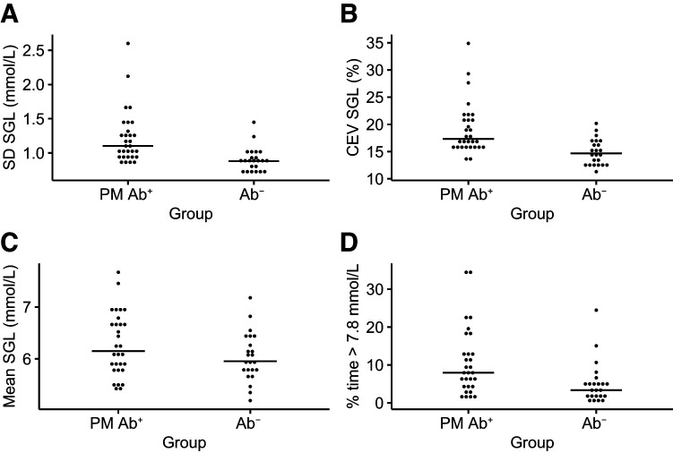
Higher SDSGL and CEV were observed in PM Ab+ compared with Ab− children (Table 2 and Fig. 1A and B), with no difference observed in mean sensor glucose level between the groups (Table 2 and Fig. 1C). PM Ab+ children spent a median (Q1, Q3) 90.7% (83.0, 95.0) of time with sensor glucose values between 3.9 and 7.8 mmol/L (70–140 mg/dL), compared with 95.5% (93.6, 97.1) in the Ab− group (Table 2 and Fig. 1D).
Table 2.
CGM metrics at time of first CGM for PM Ab+ and Ab− children
| PM Ab+ | Ab− | P | |
|---|---|---|---|
| Total n | 30 | 23 | |
| No. of days of sensor wear | 12.5 (9.0, 15.0) | 13.0 (12.5, 14.5) | |
| SDSGL | |||
| mmol/L | 1.1 (0.9, 1.3) | 0.9 (0.8, 1.0) | <0.001* |
| mg/dL | 19.8 (16.2, 23.4) | 15.8 (14.4, 18.0) | |
| CEV, % | 17.3 (16.0, 20.9) | 14.7 (12.9, 16.6) | <0.001* |
| Mean sensor glucose level | |||
| mmol/L | 6.1 (5.8, 6.7) | 6.0 (5.8, 6.3) | 0.10* |
| mg/dL | 109.8 (104.4, 120.6) | 108.0 (104.4, 113.4) | |
| Percentage of CGM time spent, mmol/L (mg/dL) | |||
| 3.9–7.8 (70–140) | 90.7 (83.0, 95.0) | 95.5 (93.6, 97.1) | 0.002† |
| >7.8 (>140) | 8.0 (4.4, 13.0) | 3.3 (1.4, 5.3) | 0.005† |
| >8.9 (>160) | 2.1 (0.9, 4.8) | 0.6 (0.2, 1.2) | 0.002† |
| >10.0 (>180) | 0.7 (0.1, 2.0) | 0.1 (0, 0.3) | 0.006† |
| >11.1 (>200) | 0.2 (0, 0.9) | 0 (0, 0.1) | 0.003† |
| <3.9 (<70) | 0.4 (0.1, 1.5) | 0.1 (0, 0.8) | 0.163† |
| <3.5 (<63) | 0.1 (0, 0.3) | 0 (0, 0.2) | 0.206† |
| <3.0 (<54) | 0 (0, 0.1) | 0 (0, 0.1) | 0.463† |
| Mean amplitude of glycemic excursion | 2.8 (2.3, 3.2) | 2.3 (2.0, 2.4) | 0.008* |
| Continuous overlapping net glycemic action | 1.3 (1.1, 1.6) | 1.1 (0.9, 1.2) | <0.001* |
Data are median (Q1, Q3). *Linear mixed model with log normal transformation.
Wilcoxon rank sum test.
Figure 1.
Mean (SD) sensor glucose (A), mean CEV sensor glucose (B), mean sensor glucose (C), and mean percentage of time spent >7.8 mmol/L (>140 mg/dL) (D) at time of first CGM in PM Ab+ and age- and sex-matched (assigned at birth) Ab− children. Solid line represents median value in each group for metric shown in figure. SGL, sensor glucose level.
The median percentage of time spent >7.8 mmol/L (>140 mg/dL) during the first CGM period in PM Ab+ children was 8.0% (4.4, 13.0) compared with 3.3% (1.4, 5.3) in Ab− children (P = 0.005) (Fig. 1D), with PM Ab+ children spending a statistically significantly higher percentage of time above higher cutoffs of 8.9 mmol/L (160 mg/dL), 10.0 mmol/L (180 mg/dL), and 11.1 mmol/L (200 mg/dL) (Table 2).
Because the ENDIA CGM substudy commenced in 2021, 8 years after the ENDIA study commenced, the first CGM period for PM Ab+ children occurred at varying durations of islet autoimmunity. Interindividual variability in CGM metrics by age at first islet autoantibody detection and duration of islet autoimmunity at time of first CGM was observed (Supplementary Fig. 1). Some PM Ab+ children who were younger at time of islet autoantibody detection who had their first CGM at a shorter duration of islet autoimmunity had higher CGM metrics (SDSGL, CEV, and percentage of time >7.8 mmol/mol [>140 mg/dL]), whereas others who had their first CGM at longer duration of islet autoimmunity did not (Supplementary Fig. 1). Conversely, some children who were older at time of first islet autoantibody had higher CGM metrics within a shorter duration of islet autoimmunity.
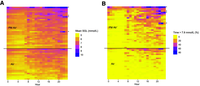
When CGM data were stratified into day and night periods, similar differences were observed; SDSGL and CEV remained significantly higher in PM Ab+ compared with Ab− children during both day- and nighttime (Supplementary Table 1); no difference was observed between groups in mean sensor glucose during the day- or nighttime period. PM Ab+ and Ab− children spent a median of 88.3% and 94.0% of time with sensor glucose values between 3.9 and 7.8 mmol/L (70–140 mg/dL) during the daytime and 95.9% and 98.2% during the nighttime periods, respectively (Supplementary Table 1). The mean sensor glucose and percentage of CGM time spent >7.8 mmol/L (>140 mg/dL) for each hour across 24 h during the CGM period for each study participant included in this analysis are shown in Fig. 2.
Figure 2.
Mean sensor glucose (mmol/L) (A) and percentage of CGM time spent >7.8 mmol/L (>140 mg/dL) (B) by hour of day at time of first CGM period in PM Ab+ and Ab− children. Each row represents values for an individual study participant. SGL, sensor glucose level. *Child later diagnosed with clinical T1D.
No difference was observed in percentage of time spent <3.9, <3.5, or <3.0 mmol/L (<70, <63, or <64 mg/dL) in PM Ab+ compared with Ab− children over 24 h (Table 2) or with day- and nighttime sensor readings analyzed separately (Supplementary Table 1).
Conclusions
This study provides unique CGM data from the Australian population–based ENDIA study cohort of very young children at risk of T1D who are being observed longitudinally from mother’s pregnancy to age 10 years (22). The cross-sectional analysis, comparing CGM metrics in PM Ab+ children with age- and sex-matched Ab− children, found higher glycemic variability measured as SDSGL and CEV. No significant difference was observed in the median sensor glucose between the groups. However, children with PM Ab+ spent significantly less time in the range 3.9–7.8 mmol/L (70–140 mg/dL) and higher percentage of CGM time with sensor glucose values >7.8 mmol/L (>140 mg/dL).
Although differences in the point estimates remained between the groups in SDSGL, CEV, and percentage of CGM time >7.8 mmol/L (>140 mg/dL) during both the day- and nighttime periods, these differences were greater during the daytime, consistent with observations in older children and young adults with early-stage T1D (17).
A key strength of this study is the use of the same CGM device for all participants, with blinded wear to minimize the effect of changes in dietary intake or other daily activities in response to sensor glucose readings. In addition, this study reports summary CGM data collected over a median duration of ∼12 days, longer than most previously published studies in children at risk (17,19).
Because CGM monitoring of PM Ab+ children commenced in January 2021, and the ENDIA study commenced in 2013, a limitation to interpreting the differences observed in CGM metrics between PM Ab+ children and their matched control participants is the variable duration between time of islet autoantibody detection at time of first CGM for PM Ab+ children. In addition, comparison between the groups was restricted to unmatched analyses because unique age- and sex-matched Ab− ENDIA children were not available for each PM Ab+ child.
Importantly, this study contributes additional CGM data on very young children at risk of T1D, who may have a more rapidly progressive phenotype of T1D, and in whom CGM data are limited to a few studies with a minimal number of participants age <5 years (19). The higher SD and CEV of sensor glucose values observed at time of first CGM in this young group of PM Ab+ children with a median age of <5 years are consistent with observations in older children (17,19). Notably, the median percentage of CGM time spent >7.8 mmol/L (>140 mg/dL) in this young cohort of children with stage 1 T1D at time of first CGM was significantly higher than the rate observed in Ab− children and children age 1–6 years without T1D (27). Recently reported data from the Autoantibody Screening for Kids (ASK) study in the U.S. found that for individuals with >10% CGM time spent >7.8 mmol/L (>140 mg/dL), the risk of progression to clinical (stage 3) T1D was 80% in the following 12 months (19). Currently, the role of CGM metrics in the prediction and/or definition of clinical T1D is an important area of investigation, especially for young children age <6 years, in whom OGTTs have low feasibility and acceptability, and relying on a change in HbA1c in this young population may miss those rapidly progressing (1,28).
Longer-term follow-up of these ENDIA children is underway and will focus on characterizing within-person variability in serial CGM periods to increase understanding of how and when dysglycemia develops in this cohort of children with young-onset persistent islet autoimmunity. These unique data for young children will contribute to global efforts investigating the role of CGM in disease staging and monitoring. The need for feasible, acceptable, accurate, equitable, and cost-effective clinical pathways for families with children identified as having presymptomatic T1D has become increasingly urgent as islet autoantibody screening programs are increasingly rolled out.
This article contains supplementary material online at https://doi.org/10.2337/figshare.26240234.
Article Information
Funding. This study was supported by grants from Diabetes Research Western Australia, the Women and Children’s Hospital Research Foundation, the Australasian Paediatric Endocrinology Group, and the Leona M. and Harry B. Helmsley Charitable Trust (grant key 2205-05241). ENDIA follow-up was supported by grants from JDRF Australia, a Commonwealth of Australia grant for Accelerated Research under the Medical Research Future Fund, and the Leona M. and Harry B. Helmsley Charitable Trust (grant keys 3-SRA-2023-1374-M-N, 3-SRA-2020-966-M-N, 1-SRA-2019-871-M-B, and 4-SRA-2015-127-M-B). A.H. was supported to lead this study by a JDRF postdoctoral fellowship (grant key 3-PDF-2020-939-A-N) and Raine Medical Research Foundation priming grant. Dexcom, Inc., provided CGM products via an investigator-initiated research agreement (Steeprock ID OUS-2019-029).
Dexcom, Inc., was not involved in study design, data analysis, or interpretation of findings.
Duality of Interest. No potential conflicts of interest relevant to this article were reported.
Author Contributions. A.H. drafted the initial manuscript, which all authors critically reviewed and revised. A.H., A.T., G.J.S., M.A.S.P., and H.O. were responsible for data curation and/or analysis. A.H., A.T., J.J.C., and E.A.D. conceptualized the study design and methodology. A.H. and G.J.S. directly accessed and verified the underlying data reported in this manuscript. A.H., M.A.S.P., M.E.C., J.M.W., T.H., J.J.C., and E.A.D. were responsible for funding acquisition. A.T. and A.J.A. were responsible for ethics and governance-related matters. M.E.C., J.M.W., T.H., P.G.C., G.S., J.J.C., and E.A.D. provided supervision of study personnel and clinical oversight of study participants, including interpretation of CGM data for providing feedback to families and interpretation of grouped CGM data and study findings. K.J.M. provided supervision of ENDIA research coordinators and led consultation with the ENDIA Parent Feedback Group. All authors had full access to all the data in the study, approved the final version submitted for publication, and agree to be responsible for all aspects of the work. A.H. is the guarantor of this work and, as such, had full access to all the data in the study and takes responsibility for the integrity of the data and the accuracy of the data analysis.
Prior Presentation. Preliminary findings based on a subset of the data included in this manuscript were presented at the 58th Annual Scientific Meeting of the European Association for the Study of Diabetes, Stockholm, Sweden, 19–23 September 2022.
Handling Editors. The journal editors responsible for overseeing the review of the manuscript were John B. Buse and Jeremy Pettus.
Appendix
The ENDIA Study Group. Investigators: Simon C. Barry, Maria E. Craig, Peter G. Colman, Jennifer J. Couper, Elizabeth A. Davis, Emma Hamilton-Williams, Leonard C. Harrison, Aveni Haynes, Tony Huynh, Ki Wook Kim, Grant Morahan, Helena Oakey, Megan A.S. Penno, William D. Rawlinson, Richard O. Sinnott, Georgia Soldatos, Rebecca L. Thomson, Jason Tye-Din, Peter J. Vuillermin, and John M. Wentworth. Associate investigators: Fergus Cameron, Andrew Day, and Prudence Lopez. Project, data, and biospecimen managers: Amanda J. Anderson, Pat Ashwood, James D. Brown, William Hu, Dao Huynh, and Kelly J. McGorm. Clinical scientist: Kelly Watson. Coordinators: Sarah Beresford, Debra Bezuidenhout, Susan Brandrick, Carlie Butterworth, Jacki Catteau, Helen Griffiths, Alison Gwiazdzinski, Candice Hall, Amanda Hulley, Lee Henneken, Renee Kludas, Ying Mateevici, Benjamin Ramoso, Alison Roberts, Alexandra Tully, and Rosemary Wood. Research officers: Sabrina Binkowski, Minh Bui, Abbey Gilbert, Dexing Huang, Ana Karceva, Brydie-Rose Mellor, Gaetano Naselli, Katrina Ngui, Trung Nguyen, Bina Patel, Vanessa Prajitno, Natalie Stone, Thao Tran, Sapphire Vaega, Emily Ward, Yan Xu, and Cynthia Yau. Dietitian: Rachel Battersby. Postdoctoral fellows: Bek Brittain, Charles Foster, Christopher Hope, Preston Leung, Kylie-Ann Mallitt, Alexandra Roth-Schulze, Tim Sadlon, Bree Tillett, Gregory Walker, Ying Wong, and Enrique Zozaya-Valdes. Administrator: Leanne Cavenett.
Funding Statement
This study was supported by grants from Diabetes Research Western Australia, the Women and Children’s Hospital Research Foundation, the Australasian Paediatric Endocrinology Group, and the Leona M. and Harry B. Helmsley Charitable Trust (grant key 2205-05241). ENDIA follow-up was supported by grants from JDRF Australia, a Commonwealth of Australia grant for Accelerated Research under the Medical Research Future Fund, and the Leona M. and Harry B. Helmsley Charitable Trust (grant keys 3-SRA-2023-1374-M-N, 3-SRA-2020-966-M-N, 1-SRA-2019-871-M-B, and 4-SRA-2015-127-M-B). A.H. was supported to lead this study by a JDRF postdoctoral fellowship (grant key 3-PDF-2020-939-A-N) and Raine Medical Research Foundation priming grant. Dexcom, Inc., provided CGM products via an investigator-initiated research agreement (Steeprock ID OUS-2019-029).
Footnotes
J.J.C. and E.A.D. are joint senior authors.
A complete list of ENDIA Study Group members can be found in the Appendix.
Clinical trial reg. no. ACTRN12620000947909, www.anzctr.org.au
Contributor Information
ENDIA Study Group:
Simon C. Barry, Maria E. Craig, Peter G. Colman, Jennifer J. Couper, Elizabeth A. Davis, Emma Hamilton-Williams, Leonard C. Harrison, Aveni Haynes, Tony Huynh, Ki Wook Kim, Grant Morahan, Helena Oakey, Megan A.S. Penno, William D. Rawlinson, Richard O. Sinnott, Georgia Soldatos, Rebecca L. Thomson, Jason Tye-Din, Peter J. Vuillermin, John M. Wentworth, Fergus Cameron, Andrew Day, Prudence Lopez, Amanda J. Anderson, Pat Ashwood, James D. Brown, William Hu, Dao Huynh, Kelly J. McGorm, Kelly Watson, Sarah Beresford, Debra Bezuidenhout, Susan Brandrick, Carlie Butterworth, Jacki Catteau, Helen Griffiths, Alison Gwiazdzinski, Candice Hall, Amanda Hulley, Lee Henneken, Renee Kludas, Ying Mateevici, Benjamin Ramoso, Alison Roberts, Alexandra Tully, Rosemary Wood, Sabrina Binkowski, Minh Bui, Abbey Gilbert, Dexing Huang, Ana Karceva, Brydie-Rose Mellor, Gaetano Naselli, Katrina Ngui, Trung Nguyen, Bina Patel, Vanessa Prajitno, Natalie Stone, Thao Tran, Sapphire Vaega, Emily Ward, Yan Xu, Cynthia Yau, Rachel Battersby, Bek Brittain, Charles Foster, Christopher Hope, Preston Leung, Kylie-Ann Mallitt, Alexandra Roth-Schulze, Tim Sadlon, Bree Tillett, Gregory Walker, Ying Wong, Enrique Zozaya-Valdes, and Leanne Cavenett
References
- 1. Besser REJ, Bell KJ, Couper JJ, et al. ISPAD clinical practice consensus guidelines 2022: stages of type 1 diabetes in children and adolescents. Pediatr Diabetes 2022;23:1175–1187 [DOI] [PubMed] [Google Scholar]
- 2. Insel RA, Dunne JL, Atkinson MA, et al. Staging presymptomatic type 1 diabetes: a scientific statement of JDRF, the Endocrine Society, and the American Diabetes Association. Diabetes Care 2015;38:1964–1974 [DOI] [PMC free article] [PubMed] [Google Scholar]
- 3. American Diabetes Association Professional Practice Committee . 2. Diagnosis and classification of diabetes: Standards of Care in Diabetes—2024. Diabetes Care 2024;47(Suppl. 1):S20–S42 [DOI] [PMC free article] [PubMed] [Google Scholar]
- 4. Ziegler AG, Rewers M, Simell O, et al. Seroconversion to multiple islet autoantibodies and risk of progression to diabetes in children. JAMA 2013;309:2473–2479 [DOI] [PMC free article] [PubMed] [Google Scholar]
- 5. Tatovic D, Narendran P, Dayan CM. Author Correction: A perspective on treating type 1 diabetes mellitus before insulin is needed. Nat Rev Endocrinol 2023;19:371–370 [DOI] [PubMed] [Google Scholar]
- 6. Jacobsen LM, Haller MJ, Schatz DA. Understanding pre-type 1 diabetes: the key to prevention. Front Endocrinol (Lausanne) 2018;9:70. [DOI] [PMC free article] [PubMed] [Google Scholar]
- 7. Wherrett DK, Chiang JL, Delamater AM, et al. ; Type 1 Diabetes TrialNet Study Group . Defining pathways for development of disease-modifying therapies in children with type 1 diabetes: a consensus report. Diabetes Care 2015;38:1975–1985 [DOI] [PMC free article] [PubMed] [Google Scholar]
- 8. Hirsch JS. FDA approves teplizumab: a milestone in type 1 diabetes. Lancet Diabetes Endocrinol 2023;11:18. [DOI] [PubMed] [Google Scholar]
- 9. Sims EK, Besser REJ, Dayan C, et al. ; NIDDK Type 1 Diabetes TrialNet Study Group . Screening for type 1 diabetes in the general population: a status report and perspective. Diabetes 2022;71:610–623 [DOI] [PMC free article] [PubMed] [Google Scholar]
- 10. Helminen O, Aspholm S, Pokka T, et al. HbA1c predicts time to diagnosis of type 1 diabetes in children at risk. Diabetes 2015;64:1719–1727 [DOI] [PubMed] [Google Scholar]
- 11. Vehik K, Boulware D, Killian M, et al. ; TEDDY Study Group . Rising hemoglobin A1c in the nondiabetic range predicts progression of type 1 diabetes as well as oral glucose tolerance tests. Diabetes Care 2022;45:2342–2349 [DOI] [PMC free article] [PubMed] [Google Scholar]
- 12. Veijola R, Koskinen M, Helminen O, et al. Dysregulation of glucose metabolism in preclinical type 1 diabetes. Pediatr Diabetes 2016;17(Suppl. 22):25–30 [DOI] [PubMed] [Google Scholar]
- 13. Simmons KM, Sosenko JM, Warnock M, et al. One-hour oral glucose tolerance tests for the prediction and diagnostic surveillance of type 1 diabetes. J Clin Endocrinol Metab 2020;105:e4094–e4101 [DOI] [PMC free article] [PubMed] [Google Scholar]
- 14. Helminen O, Aspholm S, Pokka T, et al. OGTT and random plasma glucose in the prediction of type 1 diabetes and time to diagnosis. Diabetologia 2015;58:1787–1796 [DOI] [PubMed] [Google Scholar]
- 15. Driscoll KA, Tamura R, Johnson SB, et al. ; TEDDY Study Group . Adherence to oral glucose tolerance testing in children in stage 1 of type 1 diabetes: the TEDDY study. Pediatr Diabetes 2021;22:360–368 [DOI] [PMC free article] [PubMed] [Google Scholar]
- 16. Kontola H, Alanko I, Koskenniemi JJ, et al. Exploring minimally invasive approach to define stages of type 1 diabetes remotely. Diabetes Technol Ther 2022;24:655–665 [DOI] [PubMed] [Google Scholar]
- 17. Steck AK, Dong F, Taki I, Hoffman M, Klingensmith GJ, Rewers MJ. Early hyperglycemia detected by continuous glucose monitoring in children at risk for type 1 diabetes. Diabetes Care 2014;37:2031–2033 [DOI] [PMC free article] [PubMed] [Google Scholar]
- 18. Galderisi A, Evans-Molina C, Martino M, Caprio S, Cobelli C, Moran A. β-Cell function and insulin sensitivity in youth with early type 1 diabetes from a 2-hour 7-sample OGTT. J Clin Endocrinol Metab 2023;108:1376–1386 [DOI] [PMC free article] [PubMed] [Google Scholar]
- 19. Steck AK, Dong F, Geno Rasmussen C, et al. ; ASK Study Group . CGM metrics predict imminent progression to type 1 diabetes: Autoimmunity Screening for Kids (ASK) study. Diabetes Care 2022;45:365–371 [DOI] [PubMed] [Google Scholar]
- 20. Wilson DM, Pietropaolo SL, Acevedo-Calado M, et al. ; Type 1 Diabetes TrialNet Study Group . CGM metrics identify dysglycemic states in participants from the TrialNet Pathway to Prevention study. Diabetes Care 2023;46:526–534 [DOI] [PMC free article] [PubMed] [Google Scholar]
- 21. Helminen O, Pokka T, Tossavainen P, et al. Continuous glucose monitoring and HbA1c in the evaluation of glucose metabolism in children at high risk for type 1 diabetes mellitus. Diabetes Res Clin Pract 2016;120:89–96 [DOI] [PubMed] [Google Scholar]
- 22. Penno MAS, Couper JJ, Craig ME, et al. ; ENDIA Study Group . Environmental determinants of islet autoimmunity (ENDIA): a pregnancy to early life cohort study in children at-risk of type 1 diabetes. BMC Pediatr 2013;13:124. [DOI] [PMC free article] [PubMed] [Google Scholar]
- 23. Welsh JB, Zhang X, Puhr SA, et al. Performance of a factory-calibrated, real-time continuous glucose monitoring system in pediatric participants with type 1 diabetes. J Diabetes Sci Technol 2019;13:254–258 [DOI] [PMC free article] [PubMed] [Google Scholar]
- 24. Battelino T, Alexander CM, Amiel SA, et al. Continuous glucose monitoring and metrics for clinical trials: an international consensus statement. Lancet Diabetes Endocrinol 2023;11:42–57 [DOI] [PubMed] [Google Scholar]
- 25. Broll S, Urbanek J, Buchanan D, et al. Interpreting blood GLUcose data with R package iglu. PLoS One 2021;16:e0248560. [DOI] [PMC free article] [PubMed] [Google Scholar]
- 26. Vigers T, Chan CL, Snell-Bergeon J, et al. cgmanalysis: an R package for descriptive analysis of continuous glucose monitor data. PLoS One 2019;14:e0216851. [DOI] [PMC free article] [PubMed] [Google Scholar]
- 27. DuBose SN, Kanapka LG, Bradfield B, et al. Continuous glucose monitoring profiles in healthy, nondiabetic young children. J Endocr Soc 2022;6:bvac060. [DOI] [PMC free article] [PubMed] [Google Scholar]
- 28. Ylescupidez A, Speake C, Pietropaolo SL, et al. OGTT metrics surpass continuous glucose monitoring data for T1D prediction in multiple-autoantibody-positive individuals. J Clin Endocrinol Metab 2023;109:57–67 [DOI] [PMC free article] [PubMed] [Google Scholar]


