

Abstract
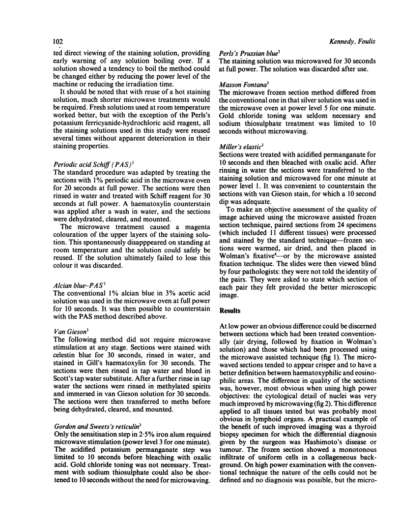
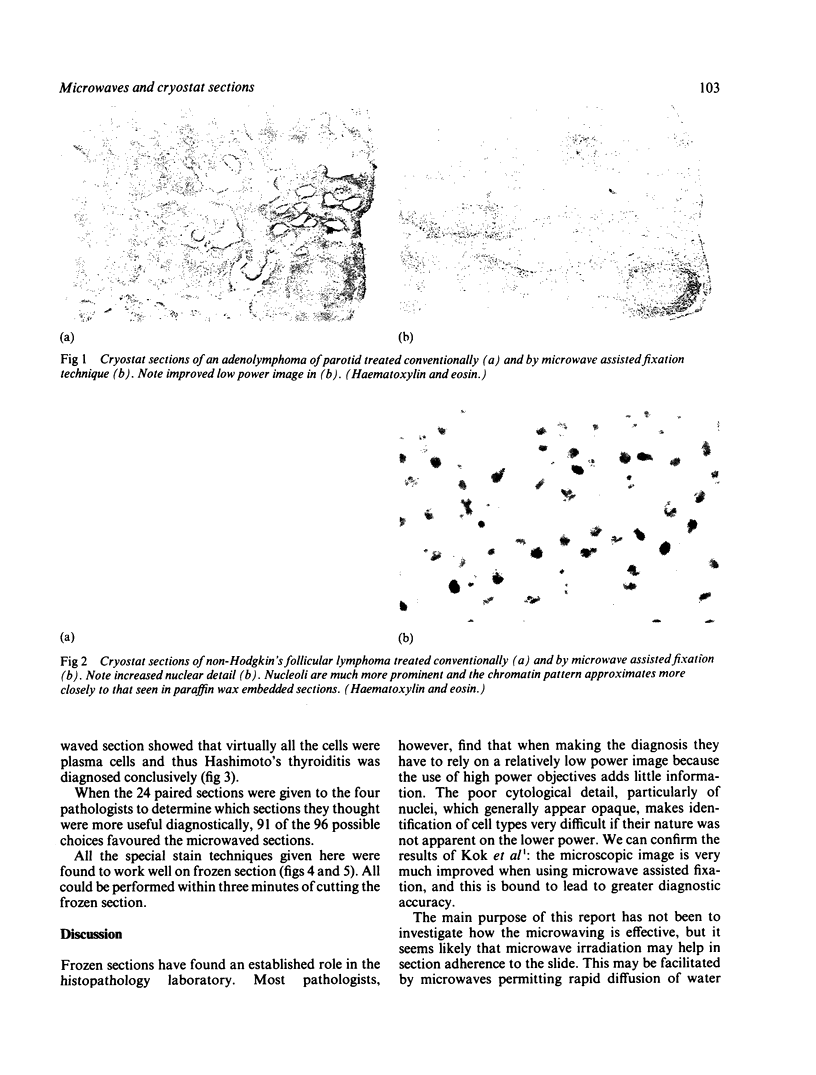
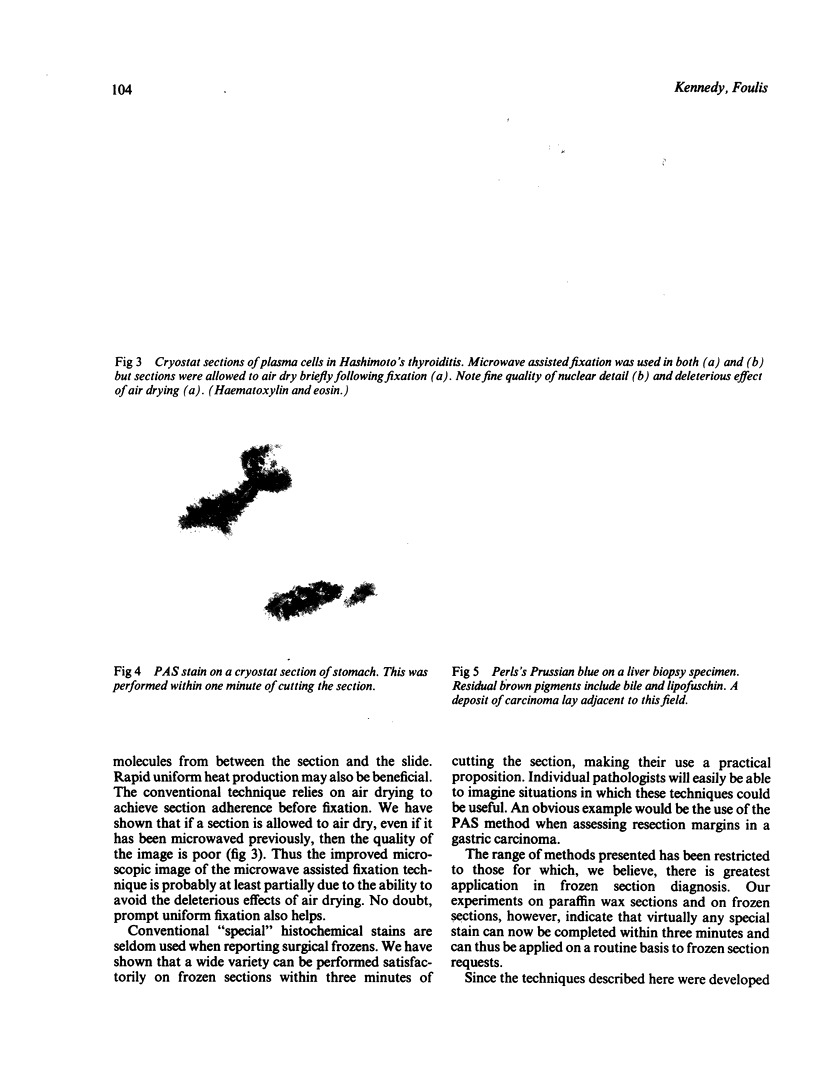
The quality of microscopic image of cryostat sections that had been subjected to microwave assisted fixation was compared with that resulting from conventional air drying of the sections. The role of microwaves in producing rapid special stains on cryostat sections was also assessed. Methods used permitted stains such as periodic acid Schiff, alcian blue, Gordon and Sweets's reticulin, Masson Fontana, Elastica, Prussian blue and Van Gieson to be performed within three minutes of cutting a cryostat section. The cytological detail of nuclei was much clearer using the microwave technique, allowing more accurate determination of cell type. The microwave oven seems to have major potential in improving the diagnostic accuracy of surgical frozen sections.
Full text
PDF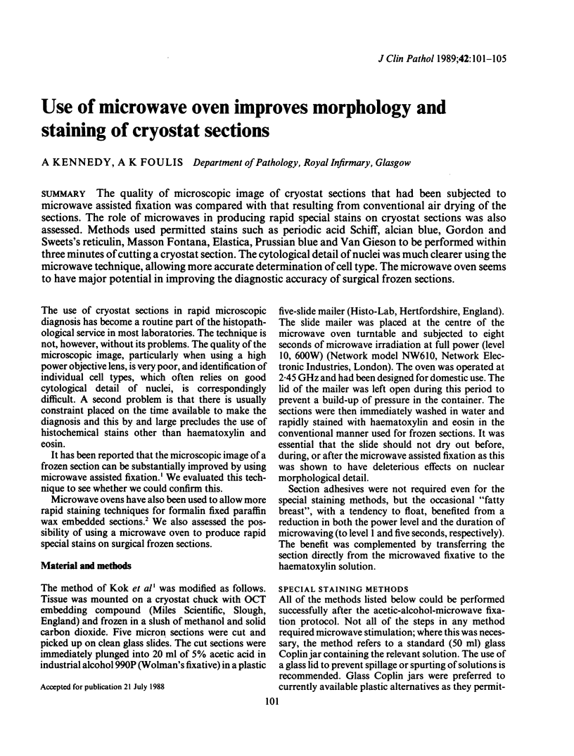
Images in this article
Selected References
These references are in PubMed. This may not be the complete list of references from this article.
- Kok L. P., Boon M. E., Suurmeijer A. J. Major improvement in microscopic-image quality of cryostat sections. Combining freezing and microwave-stimulated fixation. Am J Clin Pathol. 1987 Nov;88(5):620–623. doi: 10.1093/ajcp/88.5.620. [DOI] [PubMed] [Google Scholar]