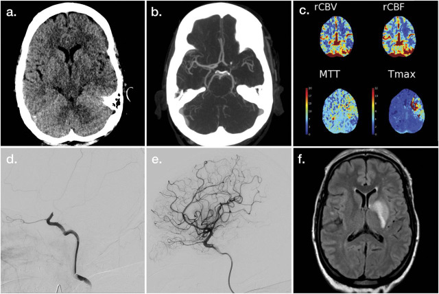
Figure 2. Illustrative Example of Neuroimaging Applications for Large Vessel Occlusion Acute Ischemic Stroke.
Noncontrast head CT (A), maximum intensity projection CT angiography (CTA) (B), postprocessed CT perfusion maps (C), and left carotid artery injection digital subtraction angiography (DSA) (D) demonstrate findings associated with acute left internal carotid artery terminus occlusion. Postrecanalization images are provided as left internal carotid artery injection DSA (E) and fluid-attenuated inversion recovery MRI (F). MTT = mean transit time; rCBF = relative cerebral blood flow; rCBV = relative cerebral blood volume; Tmax = time to maximum of residue function.

