Abstract
Objectives
To measure the expression level of the vacuolar protein sorting 13 (VPS13) gene and stimulator of interferon genes (STING) in patients with SLE with and without reported neuropsychiatric symptoms to establish their possible role in the pathogenesis of neuropsychiatric SLE (NPSLE).
Methods
This study included 100 subjects: 50 patients diagnosed with SLE and 50 age-matched and sex-matched healthy participants as the control group. The patients with SLE were further subdivided into NPSLE and non-NPSLE groups. All the subjects underwent rheumatological, neurological and psychological evaluation, MRI, VPS13C gene and STING expression assessment via quantitative real-time PCR.
Results
Seventy-eight per cent of the SLE group were classified as non-NPSLE, and 22% were classified as NPSLE. Positive MRI results were found in 55% of the patients with NPSLE and 7.7% of the patients without NPSLE.
VPS13C expression levels were decreased in the patients with SLE compared with the control (p<0.001), while STING expression levels showed higher levels in the patients in comparison with the control (p<0.001). Both markers showed significant differences between the MRI-positive and MRI-negative groups.
At a cut-off value of 0.225 for the VPS13C assessment and a cut-off value of 3.15 for STING expression, both markers were able to distinguish patients with NPSLE from those who were non-NPSLE; however, VPS13C performed better.
Conclusion
The VPS13C expression levels were decreased in patients with NPSLE compared with patients without NPSLE, while STING expression levels showed higher levels in NPSLE. Both were associated with the MRI findings. To distinguish patients with NPSLE from those without it, the VPS13C assessment performed better.
Keywords: lupus erythematosus, systemic; magnetic resonance imaging; autoimmune diseases
WHAT IS ALREADY KNOWN ON THIS TOPIC
Neuropsychiatric SLE (NPSLE) is still considered to be a mystery in many aspects.
WHAT THIS STUDY ADDS
Inflammation and neurodegeneration are now known to be a shared pathogenesis in NPSLE and neurodegenerative diseases.
HOW THIS STUDY MIGHT AFFECT RESEARCH, PRACTICE OR POLICY
The stimulator of interferon genes pathway, a new approach to unpuzzle the NPSLE dilemma, may add to our armamentarium of drugs used to treat SLE.
Introduction
Lupus cerebritis, now known as neuropsychiatric SLE (NPSLE), is a serious complication of SLE characterised by several neurological and psychiatric manifestations.1
Since the American College of Rheumatology (ACR) proposed, in 1999, a classification and nomenclature system that included 19 neurological manifestations, 12 of the central and 7 of the peripheral nervous system, the identification and diagnosis of NPSLE has become less puzzling.2 Nonetheless, despite the major advances in clinical research, none of the neuroimaging and laboratory biomarkers have been proven reliable in diagnosing NPSLE accurately. This may be because it differs from other aspects of SLE due to its development without serological changes.3
The culprit mechanisms in NPSLE pathophysiology are yet to be fully understood. Recently, two mechanisms have been proposed to be incriminated, mostly in conjunction but maybe in separate, in the development of NPSLE. The first is the autoimmune or inflammatory pathway, and the second is the ischaemic or thrombotic pathway.3
With the recent awareness of the major contribution of the stimulator of interferon genes (STING) pathway in SLE disease pathophysiology, particularly in sensing mitochondrial DNA that has failed to be cleared by autophagy4 5 and double-stranded (ds)DNA present in apoptotic-derived membrane vesicles.6
Furthermore, defective mitochondrial DNA clearance seems to be a shared pathogenic triggering factor in both SLE and neurodegenerative diseases like Parkinson’s disease (PD), Alzheimer’s disease, amyotrophic lateral sclerosis and others.7
These findings have led us to believe that the vacuolar protein sorting 13 (VPS13C) gene (a member of the family that encodes lipid transfer proteins) is localised to various autonomous contact sites between membranous organelles (eg, endoplasmic reticulum (ER)).8 Its subsequent activation of the cyclic GMP–AMP synthase (cGAS)-STING pathway (one of the most recently researched topics regarding the pathogenesis of neurodegenerative disorders) has been found to mediate both neurodegeneration with functional decline and low-grade inflammation8 and may be involved more deeply in NPSLE pathogenesis. Due to the presence of VPS13C in mitochondria-associated membrane fractions in ER, the event of mitochondrial dysfunction in the case of VPS13C mutation or absence has been reported.9
So, we assume that NPSLE with the evidence of STING pathway expression might also have VPS13C mutation, like most of the previously mentioned neurodegenerative conditions with their shared precedence of microglial activation, neurodegeneration and a state of low-grade inflammation. To investigate our hypothesis, we measured the expression level of the VPS13C gene and STING in patients with SLE with and without reported neuropsychiatric (NP) symptoms, trying to test the accuracy of our assumption.
Subjects and methods
Study design and participants
The studied participants were recruited from the follow-up unit of the Rheumatology and Rehabilitation Department at Zagazig University. An analysis of the samples was performed at the Clinical Pathology Department. The time frame for conducting this investigation was from November 2023 to April 2024. It was estimated that 50 patients with SLE would be required for this case-control study; 50 healthy participants of comparable age and gender were also included. The sample size was calculated at a 95% CI and 80% power using Epi Software V.6 (Atlanta, Georgia, USA), assuming a mean difference expression of 0.2 and SD of 0.3 and 0.4 for controls and cases, respectively.10
All the patients who met the revised ACR classification criteria for SLE by the Systemic Lupus International Collaborating Clinics (SLICC) were diagnosed with SLE.11 They were recruited independently of NP events or disease activity. However, during the analysis of our patients’ medical histories, we came across multiple different scenarios of their NP disease development: some of our participants experienced fits as an initial presentation of both NPSLE and SLE itself, while others developed their symptoms during different stages of the disease. We chose patients between the ages of 18 and 55 years to decrease the likelihood of age-related abnormalities.
The patients with SLE were divided into two groups with and without NPSLE according to the diagnostic criteria for NPSLE proposed by the ACR in 1999.2 The ACR defined 19 neurological syndromes (12 central and 7 peripheral NP); we included only the 12 central syndromes in the present study and excluded the peripheral neurological lesions because of the substantial differences in anatomy, function and clinical characteristics between the central and peripheral nervous systems.
We assumed that the examined patients with SLE had primary NP manifestations after ruling out the probability of the presence of secondary lesions of the nervous system related to antiphospholipid antibodies, infections, electrolyte disturbances, drug intake (neuroleptics, L-Dopa), concurrent syndromes like chronic renal failure or diseases like hyperthyroidism. Subjects with a history of overlapping SLE with other systemic diseases of the connective tissue, illiteracy, chronic alcoholism and drug abuse were not included in the study.
Based on the medical records, all 50 patients with SLE were treated with variable doses of corticosteroids with or without steroid pulse, along with other immune-modulating or immunosuppressive agents such as cyclophosphamide and/or rituximab according to their manifestations and disease activity level.
Antipsychotics or anticonvulsants were used according to the patient’s clinical condition and after consultation with the psychiatrist and the neurologist.
After the initial interview, obtaining their consent and a thorough history and clinical examination (rheumatological, neurological and psychological), the participants were sent to the radiologist in our team who was blinded to the clinical status of the recruit, whether a patient or a control subject. Finally, a laboratory investigation and gene expression were carried out.
Clinical examination
Every patient filled out a standard medical history form, and they all underwent examinations that included rheumatological and neurological examinations. A psychiatric evaluation was employed when needed. The SLE Disease Activity Index-2K (SLEDAI-2K) score was used to quantify the activity of SLE.12 To assess the severity of the disease, we used the SLICC/ACR Damage Index for SLE.13
Psychiatric assessment
When psychiatric manifestations were suspected during the initial evaluation, a thorough psychiatric history and mental status examination were performed. The diagnosis of psychiatric disorders was assessed clinically using the Diagnostic and Statistical Manual of Mental Disorders Fifth Edition (DSM-5) criteria14 and confirmed using psychometric measures when needed. Cognitive dysfunction, depression and anxiety disorders were confirmed using Arabic versions of the Montreal Cognitive Assessment (MoCA) for cognitive dysfunction15 16 and the Hospital Anxiety and Depression Scale (HADS)17 18 for depression and anxiety disorders, respectively. An acute confusional state was diagnosed with the equivalent DSM-5 criteria for delirium.
MRI acquisition and sequences acquired
All the subjects underwent an MRI scan with a 1.5 T magnet device (Philips Medical Systems) using a head-phased-array coil with eight channels within a week of enrolment and after the initial rheumatological and neurological evaluation. The scans were aligned parallel to the axial plane through the anterior-to-posterior commissure and covered the entire brain in all sequences. T1-weighted, T2-weighted and fluid-attenuated inversion recovery (FLAIR) images, diffusion-weighted imaging and apparent diffusion coefficient maps were acquired from all the participants. All the MRI images, as well as the grey matter, white matter, WMH and cerebral-spinal fluid maps, were carefully inspected by a radiologist who had >10 years of experience in neuroradiology. She was also blinded to the clinical data of both groups. The MRI images with large cerebral infarcts were excluded. After the white matter hyperintensity (WMHI) volume with associated significant volumetric brain changes was considered, a positive finding related to our study was found as it was mostly attributed to inflammatory changes in patients with SLE.19
Laboratory test
One millilitre of whole blood was donated by each participant, and it was collected in an EDTA tube for the study of gene expression. Three millilitres of each patient’s entire blood were collected and placed in a plain tube. The tube was centrifuged at 1200 × g speed for 10 min to separate the serum. In the erythrocyte sedimentation rate (ESR) tube, 1.6 mL of whole blood was drawn. Becton Dickinson vacutainers (Franklin Lakes, New Jersey, USA) were used in this study.
The ESR was determined using the Vision B automated analyzer (YHLO Biotech, Shenzhen, China). Serum was used to measure the levels of C reactive protein (CRP), C3 and C4 on the Cobas 6000/c501 analyzer (Roche Diagnostic, Mannheim, Germany). The assay of indirect immunofluorescence was used to identify antinuclear and dsDNA antibodies present in the serum. The ANAFLUOR test system from DiaSorin (Stillwater, Minnesota, USA) was used to measure ANAs. INOVA Diagnostic, located in San Diego, California, evaluated anti-dsDNA antibodies. Using the human antineuronal nuclear antibody (ANNA) ELISA Kit (catalogue no.: MBS765001). ANNA was quantified. Every stage of the kit was used in compliance with the guidelines provided by the manufacturer, MyBiosource, in Southern California, San Diego, USA. The intra-assay and inter-assay precision of this kit were indicated by a coefficient of variation <8% and <10%, respectively.
The reference interval of ANNA was 8.2–16.4 ng/mL. The reference intervals of C3 and C4 were 0.9–1.8 g/L and 0.1–0.4 g/L, respectively.
Quantitative real-time PCR was used to assess the gene expression levels in the peripheral blood. The total RNA was isolated from the plasma using the QIAGEN, Hilden, Germany, miRNeasy Serum/Plasma Kit, following the manufacturer’s instructions. The concentration and purity of the isolated RNA were assessed using Thermo Scientific’s NanoDrop-2000 spectrophotometer (USA). The miScript RT II kit (QIAGEN) was then used to reverse-transcribe the total RNA into complementary DNA (cDNA).
Following the manufacturer’s instructions, the miScript SYBR Green PCR kit was used to perform real-time RT-PCR on the cDNA templates. As directed by the manufacturer, a 20 µL of PCR reaction mix was generated using 5 µL of cDNA. The degree of gene expression was assessed using the StepOne System real-time PCR apparatus (Applied Biosystems, Foster City, California, USA). There were 40 cycles, each lasting 10 s at 94°C and 1 min at 60°C, following a 15 min initial denaturation period at 95°C. To identify the specific amplification, a melting curve analysis was performed. To normalise the expression level of genes, β-actin expression was used.
β-Actin (forward: 5′-GGACTTCGAGCAAGAGATGG-3′, reverse: 5′-AGCACTGTGTTGGCGTACAG-3′), VPS13C (forward: 5′-TTGGAAAAGGGCTTGTGGGT-3′, reverse: 5′-GGGGACGGAGGCTAGATACT-3′) and STING (forward: 5′-CATTGGGTACTTGCGGTT-3′, reverse: 5′-CTGAGCATGTTGTTATGTAGC-3′) primers were used. Calculations were made to determine the genes’ relative expression level by the 2−ΔΔCT method.
Statistical analysis
SPSS V.26.0 was used to tabulate and statistically analyse all the data (SPSS, Chicago, Illinois, USA). The data were confirmed to be non-parametric using the Shapiro-Wilk test. Whereas categorical data were shown as absolute values and percentages, quantitative variables were shown as the median and range. To compare the variables, the Mann-Whitney U test and χ2 test were used. An examination of the receiver operating characteristic (ROC) curve was used to evaluate the marker’s predictive power. Performance was evaluated using the area under the ROC curve (AUC) and its 95% CI. The link between the various study variables was evaluated using the Spearman’s correlation test. Logistic regression analysis was used to determine the independent predictive factors by estimating the OR and its 95% CI. A p value of <0.05 indicates statistical significance.
Results
In all, 100 individuals participated in the study: half of them with SLE, 7 men and 42 women with a median age of 35 (19–55) years; the other half were a control group (9 men and 41 women, with a median age of 39.5 (25–55) years. There was no noticeable difference in the demographic data between the two groups as both the patients’ and control group’s ages (p=0.16) and sexes (p=0.66) were matched. The laboratory results of the control group were negative for ANA and anti-dsDNA. The median ANNA level was 10 (range: 4–22 ng/mL). The CRP was 2.3 (range: 0.3–4.1 mg/L), ESR was 12 (range: 7–15 mm/hour), the C3 was 1.1 (range: 0.9–1.5 g/L) and the C4 was 0.23 (range: 0.1–0.36 g/L).
Among the 50 studied patients with SLE, based on the medical history and NP examinations by rheumatologists, an experienced neurologist and a psychiatrist, and supported by conventional laboratory tests and appropriate complementary tests, 39 patients (78%) were classified as non-NPSLE, while 11 patients (22%) were classified as NPSLE. The patient groups’ demographic, clinical and laboratory results are shown in table 1.
Table 1. Patients’ demographics, clinical and laboratory characteristics.
| Parameters | NPSLE group (no.: 11) | Non-NPSLE group (no.: 39) | P value |
| Age (years) | 34 (19–55) | 35 (20–55) | 0.19 |
| Sex (male/female) | 3/8 (27.2/72.7) | 4/35 (10.3/89.7) | 0.48 |
| Family history of SLE | 2 (18.2) | 4 (10.3) | 0.47 |
| Duration (years) | 4 (0–13) | 6 (0–20) | 0.19 |
| Clinical features | |||
| Malar rash/discoid rash | 4 (36.4) | 17 (43.6) | 0.67 |
| Oral or nasal ulcers | 1 (9.1) | 6 (15.4) | 0.59 |
| Arthritis | 3 (27.3) | 11 (28.2) | 0.95 |
| Serositis | 1 (9.1) | 5 (12.8) | 0.73 |
| Renal disorder | 5 (45.5) | 15 (38.6) | 0.66 |
| SLEDAI-2K activity score | 12 (4–44) | 6 (1–21) | 0.017* |
| SLICC/ACR Damage Index | 3.8 (2–5) | 1.6 (1–4) | <0.001* |
| MRI-positive | 6 (54.5) | 3 (7.7) | <0.001* |
| Laboratory tests | |||
| ANA | 10 (90.9) | 36 (92.3) | 0.88 |
| Anti-dsDNA antibody | 7 (63.6) | 22 (56.4) | 0.6 |
| ANNA (ng/mL) | 25 (10–85) | 12 (5–35) | <0.001* |
| CRP (mg/L) | 5.2 (0.6–74.1) | 4.7 (0.3–64.2) | 0.88 |
| ESR (mm/hour) | 22 (14–50) | 25 (10–105) | 0.11 |
| C3 (g/L) | 0.62 (0.3–1.47) | 0.73 (0.2–1.4) | 0.47 |
| C4 (g/L) | 0.2 (0.06–0.36) | 0.15 (0.1–0.34) | 0.47 |
Data are expressed as median [(Mminimum-M–maximum)] or number (%).
: Significant.
ANNAantineuronal nuclear antibodyCcomplementCRPC reactive proteindsDNAdouble-stranded DNAESRerythrocyte sedimentation rateNPSLEneuropsychiatric SLESLEDAISLE Disease Activity IndexSLICC/ACRSystemic Lupus International Collaborating Clinics/American College of Rheumatology
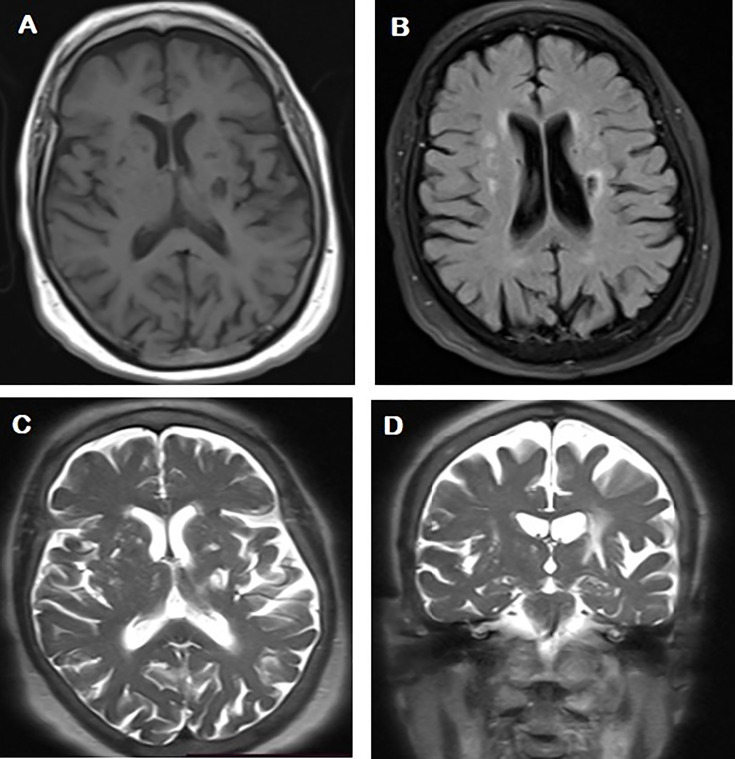
Both the SLEDAI and SLICC scores were considerably greater in patients with NPSLE (0.017 and p<0.001, respectively). Positive MRI results were found in around 55% of the patients with NPSLE, but some patients without NPSLE also had positive MRI findings (7.7%). Among the patients with NPSLE, eight patients (72.7%) had white matter lesions, six (54.5%) had cerebral atrophy and only two (18.2%) had grey matter lesions (figure 1). Acute phase reactants, CRP, ESR and complements C3 and C4, did not differ significantly between the patient groups (p>0.05). Patients with NPSLE had higher levels of ANNA than those without it (p<0.001). Table 2 shows how positive MRI results were found in around 55% of patients with NPSLE, and some patients without NPSLE had positive MRI findings (7.7%).
Figure 1. Abnormal MRI signals in a female patient with SLE aged 55 years presented with convulsions. (A) Axial T1-weighted imaging, showing multiple bilateral periventricular foci of low signal intensity (SI). (B) Axial fluid-attenuated inversion recovery (FLAIR), (C) and (D) axial and coronal T2-weighted imaging (T2WI) images: showing (1) multiple bilateral white matter hyperintensity lesions seen at a periventricular location and frontal subcortical white matter, as well as corona radiata high SI lesions seen on T2WI and FLAIR. (2) Mild atrophic changes in the form of mild dilated ventricles with prominent cortical sulci and Sylvian fissures. Peri-ventricular linear sheets of abnormal SI display high SI on T2WI and FLAIR, consistent with leukoencephalopathy (additional two cases illustrated in online supplemental figure 1 and 2).
Table 2. SLE neuropsychiatric symptoms and MRI positivity in each symptom.
| Parameter | NPSLE group(no.: 11) | MRI-positive in each symptom |
| Neurological disorders | ||
| Aseptic meningitis | 0 (0) | 0 (0) |
| Headache | 4 (36.4%) | 3 (75) |
| Myelopathy | 0 (0) | 0 (0) |
| Cerebrovascular disease | 0 (0) | 0 (0) |
| Demyelinating syndrome | 2 (18.2%) | 1 (50%) |
| Seizures | 4 (36.4%) | 2 (50%) |
| Movement disorders | 0 (0) | 0 (0) |
| Psychiatric disorders | ||
| Acute confusion | 3 (27.3%) | 1 (33.3%) |
| Anxiety disorder | 2 (18.2) | 0 (0) |
| Cognitive dysfunction | 3 (27.3%) | 1 (33.3%) |
| Mood disorders | 2 (18.2%) | 0 (0) |
| Psychosis | 1 (9.1%) | 0 (0) |
MRI considered to be postive when changes attributed to inflammatory process are resent, eg, white matter hyperintensities (WMHI) and or associated significant volumetric brain changes.
Data are expressed as number (%).
NPSLEneuropsychiatric SLE
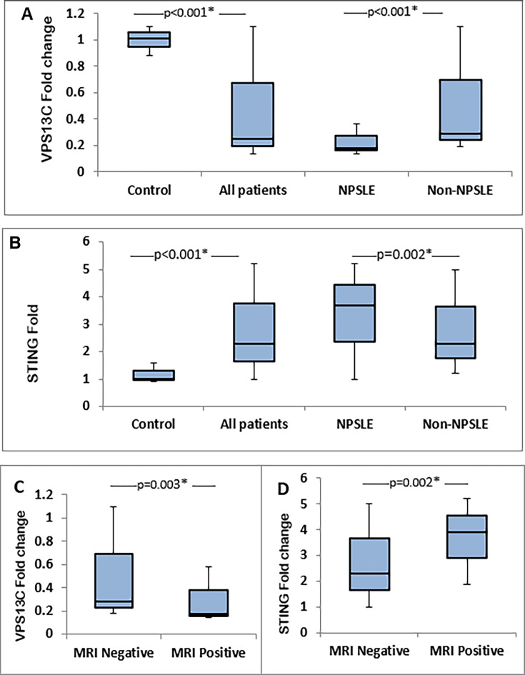
The VPS13C expression levels were decreased in the patients with SLE compared with the control group (p<0.001) (figure 2A). On the other hand, the STING expression levels were higher in the patients (p<0.001) (figure 2B). The VPS13C expression levels were lower in patients with NPSLE compared with those without NPSLE. Meanwhile, the STING expression levels were higher in the patients with NPSLE. The relationship between the studied markers and MRI status is illustrated in figure 2C,D. Both markers showed significant differences between the MRI-positive and MRI-negative groups.
Figure 2. (A) Expression levels of vacuolar protein sorting 13 (VPS13C) and (B) expression levels of stimulator of interferon gene (STING) in patients with SLE and healthy controls. (C) Relationship between VPS13C and MRI. (D) Relationship between STING expression levels and MRI. NPSLE, neuropsychiatric SLE.
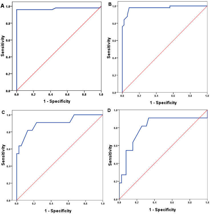
On conducting a ROC analysis to discriminate between the patients with SLE and the healthy control subjects (figure 3), the STING expression showed the highest sensitivity (98%) and specificity (92%) at a cut-off value of 1.2 with an AUC of 0.979. Also, the VPS13C gene expression at a cut-off value of 0.88 showed (100%) sensitivity and (96%) specificity with an AUC of 0.971.
Figure 3. Receiver operating characteristic curves of (A) vacuolar protein sorting 13 (VPS13C) and (B) stimulator of interferon gene (STING) for SLE diagnosis. Receiver operating characteristic curves of (C) VPS13C and (D) STING for neuropsychiatric SLE diagnosis.

A ROC curve analysis was used to evaluate the significance of the markers in the diagnosis of NPSLE (figure 3). The ROC-AUC values for the VPS13C and STING were 0.904 and 0.805, respectively. The cut-off value for VPS13C was 0.225 (Youden’s index=0.69), resulting in 81.8% sensitivity and 87.2% specificity. At the cut-off of 3.15 (Youden’s index=0.56), the STING exhibited 81.8% sensitivity and 74.4% specificity. To distinguish patients with NPSLE from those without it, the VPS13C performed better.
We looked into the relationships between the characteristics of patients and the levels of markers. The VPS13C levels were negatively correlated with SLEDAI, SLICC/ACR, CRP, ESR, ANNA and STING (online supplemental figure 3). However, there was a positive correlation with C3 and C4. However, the STING levels showed the opposite pattern of correlation (online supplemental figure 4) (table 3).
Table 3. The correlation of VPS13C and STING expression levels with patients' characteristics.
| Parameters | VPS13C | STING | ||
| rs | P value | rs | P value | |
| Age | 0.09 | 0.35 | −0.11 | 0.27 |
| Duration | 0.08 | 0.57 | 0.07 | 0.63 |
| SLEDAI | −0.36 | 0.01* | 0.34 | 0.017* |
| SLICC/ACR | −0.52 | <0.001* | 0.65 | <0.001* |
| CRP | −0.35 | <0.001* | 0.39 | <0.001* |
| ESR | −0.61 | <0.001* | 0.69 | <0.001* |
| C3 | 0.38 | <0.001* | −0.47 | <0.001* |
| C4 | 0.44 | <0.001* | −0.48 | <0.001* |
| ANNA | −0.49 | <0.001* | 0.37 | 0.009* |
| STING | −0.72 | <0.001* | 1 | … |
: Significant.
ANNAantineuronal nuclear antibodyCcomplementCRPC reactive proteinESRerythrocyte sedimentation rateSLEDAISLE Disease Activity IndexSLICC/ACRSystemic Lupus International Collaborating Clinics/American College of RheumatologySTINGstimulator of interferon genesVPS13Cvacuolar protein sorting 13
In the univariate analysis for NPSLE prediction, the VPS13C and STING expressions were associated with an OR of 18.1 and 13, respectively. In addition, SLICC, ANNA and MRI were risk factors for NPSLE (table 4). Using the factors listed in table 4 in the multivariate analysis, the VPS13C expression was still significantly associated with NPSLE. The VPS13C expression seems to be an independent predictor of NPSLE. It showed an adjusted OR of 15.2 (95% CI 0.3 to 0.99) (p=0.03).
Table 4. Logistic regression analysis of risk factors for NPSLE.
| Covariate | Univariate analysis | Multivariate analysis | ||
| OR (95% CI) | P value | AOR (95% CI) | P value | |
| VPS13C | 18.1 (0.57 to 0.92) | <0.001* | 15.2 (0.3 to 0.99) | 0.03* |
| STING | 13 (2.4 to 70.89) | 0.003* | 7.03 (0.56 to 88.99) | 0.13 |
| ANNA | 1.15 (1.04 to 1.27) | 0.007* | 1.21 (0.98 to 1.47) | 0.07 |
| SLEDAI | 1.01 (0.98 to 4.54) | 0.09 | 1.23 (0.80 to 4.26) | 0.56 |
| SLICC/ACR | 1.14 (1.02 to 1.27) | 0.022* | 1.1 (0.87 to 1.39) | 0.43 |
| MRI | 14.4 (2.71 to 76.65) | 0.002* | 4.58 (0.11 to 182.33) | 0.42 |
: Significant.
ANNA, antineuronal nuclear antibody; AOR, adjusted OR; SLEDAISLE Disease Activity IndexSLICC/ACRSystemic Lupus International Collaborating Clinics/American College of RheumatologySTING, stimulator of interferon genes; VPS13C, vacuolar protein sorting 13
Discussion
NPSLE is a serious and potentially life-threatening manifestation of SLE. Its prevalence rates vary widely; it was estimated to be between 12% and 95%, which may be due to the lack of consistency of NPSLE definitions, differences in study designs, the variability of study populations and differences in ethnicities included, among other factors.20
Many factors hinder the identification and diagnosis of NPSLE, including the multifarious neurological symptoms, the absence of standardised assessment and the traditional markers of SLE being unreliable in terms of diagnosis and NPSLE disease monitoring.21
Even with the 2019 EULAR/ACR classification criteria (for the classification of SLE) and the SLEDAI (for stratification of disease activity), which incorporated several serological criteria,2 13 both are frequently deemed ineffective in predicting the course of NP disease activity in the absence of concurrent systemic inflammation.22
Our results support this notion because our NPSLE group had considerably higher disease activity using SLEDAI than the non-NPSLE group. It was demonstrated that patients with SLE with diffuse NPSLE manifestations, but not focal, had higher disease activity.23 This was further proved by many others and a recent systematic review even has set a SLEDAI score of >10 to be considered a prerequisite for diagnosis for NPSLE.24
Our current data support the widespread belief that NPSLE activity is different from other aspects of SLE disease activity because it may occur separately and in the absence of serological activity, as shown by our tested groups which indicated insignificant differences in the levels of CRP, ESR, C3 and C4. Moreover, it can happen solely without any other organ involvement.25
The old-known fact that anti-dsDNA antibodies possess less value when it comes to isolated NP involvement is supported by our existing results because both our NPSLE and non-NPSLE groups had more positive than negative anti-dsDNA, but in the end, there was no significant difference between them (p value=0.6).23
Anti-dsDNA antibodies are famous for being highly specific for SLE and correlating closely with SLE disease activity.26 Up to 70% of patients with NPSLE may have these antibodies, but their levels do not seem to be correlated with the activity of NP diseases.23 27
SLE was found to carry a type I interferon (IFN) signature. It was even classified among the α interferonopathy group of diseases.28 It was suggested that IFNα has a leading role in NPSLE pathogenesis as it has in SLE itself. The evidence presented by Shiozawa et al29 shows that IFNα is produced in the central nervous system (CNS), especially in NPSLE manifestations like psychosis and headache.30 These anecdotal pieces of evidence gave solid grounds for our presumption that the complex relationship between type I IFN, VP13C mutation and STING pathway activation might have a more crucial role in NPSLE pathophysiology than what is currently known. IFN expression via the cGAS-STING cytosolic DNA-sensing pathway was detected, with the resulting activation of JAK-STAT signalling and IFN-stimulated gene expression. Such activation culminates in IFN production.31
The local microglia in the CNS are known to be potent cytokine producers; they were found to be activated by elevated levels of type I IFNs (both in serum and the hippocampus), leading to erratic pruning of synaptic neurons in NPSLE mouse models.32
A recent highly regarded study by Gulen et al revealed that the activation of STING triggers reactive microglial transcriptional states, neurodegeneration and cognitive decline.7 Moreover, in multiple earlier studies, activated microglia were established to be a feature of several mouse models of lupus.33 34 Even their inhibition was found to attenuate the phenotype of NPSLE in these mice.35 36
In addition, Gkirtzimanaki et al proposed that sustained IFN signalling leads to anti-DNA autoimmunity by damaging mitochondrial metabolism. This, in turn, leads to oxidative stress that impairs lysosomal degradation and the obstruction of autophagic clearance. An antiviral-like response against self-DNA is initiated when the non-degraded mitochondrial DNA (mtDNA) escapes to the cytoplasm and is sensed.5
A much similar pattern was proposed in PD more recently by Hancock-Cerutti et al, who demonstrated that depleting VPS13C in HeLa cells causes an accumulation of lysosomes with an altered lipid profile. These cells have an activated DNA-sensing cGAS-STING pathway which results from a combination of increased cytosolic mtDNA and a malfunction in the degradation of activated STING, becoming a lysosome-dependent mechanism.37
These findings imply a connection between lysosome lipid transfer and innate immune activation in a model human cell line and put VPS13C and STING in a shared pathway relevant to PD pathogenesis and, by extension, NPSLE pathogenesis.
Indeed, we detected an elevated expression level of STING in both NPSLE and non-NPSLE groups more than in the control group, but the level of STING expression in the NPSLE group was slightly more evident than in the non-NPSLE group. Moreover, VPS13C expression levels decreased in patients with SLE compared with the control group, and even less in the NPSLE group.
The already-established link between VPS13C-dependent cGAS-STING in neurodegenerative disorders and its induced immune sensing of DNA has proved to be a critical driver of chronic inflammation and functional decline during ageing.38 39 Leading the way, these non-precedent results open a new chapter to a more in-depth search in the pathogenesis of NPSLE and may unveil a hidden territory that has not been broached before.
Low-grade inflammation mediated by an innate immune response and neurodegeneration are two mechanisms that have been proposed in the pathogenesis of NPSLE. Functional studies of neuronal networks, although performed on patients with rheumatoid arthritis and not on patients with SLE, have shown changes in network association patterns. These changes correlate with the degree of inflammation as well as pain and fatigue.40 The results suggest that the ongoing inflammatory process leads to functional changes in brain connectivity and function, which could explain some manifestations of NPSLE or contribute to their severity.41
The previous finding goes hand in hand with the positive correlation we detected between STING expression levels and markers of inflammation and activity such as ESR, CRP and SLEDAI, and the exact opposite with VPS13C expression levels.
Previous gene expression investigations in patients with SLE have generally found a cross-sectional correlation between disease activity and different types of genes, including type I IFN-induced gene expression.42 This was contradicted by Jin et al, who stated that in their sample type, the type I IFN patterns were independent of both disease activity and medical treatment, and they attributed the high disease activity (SLEDAI ≥10) to another immunoregulatory event.43 Perhaps this is also the case in our sample: the underexpression level of VPS13C and overly expressed STING are the cause of the higher disease activity we encountered in our study and, thus, both can be used as biomarkers to diagnose and monitor both SLE activity in general and NPSLE activity in particular. This might open a window for the newly proposed STING inhibitors to be tried for the more resistant cases of NPSLE.44
Although STING and VPS13C expression levels could differentiate between patients with SLE and healthy controls, VPS13C proved to be superior to STING in distinguishing between patients with NPSLE and patients without NPSLE.
The dysregulation of the cGAS-STING pathway in SLE, and its relevance to triggering adaptive immunity by promoting the development of autoreactive and dysfunctional B and T cells,45 as well as its involvement in innate immunity and INF-producing dendritic cell-related pathways, was not discovered until a few years ago.6 46
In the NPSLE group, headache was among our most encountered NP manifestations (36.4%), and both seizures and acute confusion came second (27.3%), followed equally by demyelinating syndrome, anxiety and mood disorders (18.2%). These findings were not different from multiple previous reports offering similar results with a minimal percentage difference.24 41 47
Conventional MRI is known to be an important tool in the evaluation of patients with NPSLE. It possesses the ability to detect structural brain abnormalities because of the excellent soft tissue contrast and the ability to acquire multiplanar images.48
To date, neuroimaging has been helpful in ruling out other possible causes of NP symptoms but cannot be the sole referee for diagnosing NPSLE, which still primarily relies on clinical expertise.41
In the current study, the MRI findings varied significantly between patients with NPSLE and patients without NPSLE (p<0.001). We had a minuscule number of patients without NPSLE showing variable MRI-positive signs (7.7%), which is not uncommon. Zaky et al detected MRI abnormalities in 20% of their non-NPSLE group compared with 46% of the NPSLE group.49 A noted cohort stated that 25%–50% of patients with SLE without CNS involvement had MRI abnormalities, mostly in the form of WMHIs that were also found in subjects without SLE. They explained that WMHIs represent the major histopathological changes observed in postmortem brain analyses of patients with SLE, implying that brain damage progresses over time in patients with SLE, regardless of NP clinical involvement, due to both SLE-related and non-SLE-related risk factors.50
Conversely, previous data have suggested that up to 40%–50% of patients with NPSLE may produce a normal MRI scan, especially in diffuse syndromes such as headache, mood disorder and psychiatric disease.51 52
Such a discrepancy between the MRI findings and the clinical presentation urged the EULAR task force to recommend the use of advanced imaging, such as the MRI methods of magnetisation transfer imaging, diffusion-weighted imaging and diffusion tensor imaging, and methods that use radioactive tracers, such as positron emission tomography and single-photon emission CT, in cases of normal MRI findings in patients with NPSLE.53
Our results have shown that MRI findings correlated with both markers (positively with STING expression and negatively with VPS13C expression). This was impressive considering various studies have not found a correlation with multiple variables, neither clinical, such as disease activity by SLEDAI-2K or damage by SLICC,54 55 nor laboratory-based, such as variable autoantibodies examined in an MRI cohort, where the results have shown that none of the autoantibodies they tested were associated with MRI findings, except for lupus anticoagulant, which was significantly associated with ischaemic lesions and cerebral atrophy.56
Study limitations
One limitation of our study is the number of cases. It would have been preferable to employ a larger sample size of patients with SLE in general or, specifically, patients with NPSLE covering all phenotypes to give a more definitive conclusion about the significance of our results in NPSLE, although it is appreciably within the range as previous studies on NPSLE.
Conclusions
VPS13C expression levels were decreased in patients with NPSLE compared with patients without NPSLE. While STING expression levels showed higher levels in NPSLE, both were associated with MRI findings. To distinguish patients with NPSLE from those without it, VPS13C performed better. VPS13C expression seems to be independently associated with NPSLE.
supplementary material
Footnotes
Funding: The authors have not declared a specific grant for this research from any funding agency in the public, commercial or not-for-profit sectors.
Provenance and peer review: Not commissioned; externally peer reviewed.
Patient consent for publication: Consent obtained directly from patient(s).
Ethics approval: The research protocol was approved by the research ethical committee of the Faculty of Medicine, Zagazig University, and the Institutional Research Board (IRB), number #11302. The Declaration of Helsinki, issued by the World Medical Association to ensure the protection of individuals participating in medical research, was strictly adhered to during this study. Participants gave informed consent to participate in the study before taking part.
Data availability free text: Data are available from corresponding author on reasonable request.
Patient and public involvement: Patients and/or the public were not involved in the design, or conduct, or reporting, or dissemination plans of this research.
Contributor Information
Amany M Ebaid, Email: AMMouhmmed@medicine.zu.edu.eg.
Mohammad A Zakaria, Email: dr.m.zakaria@gmail.com.
Enas M Mekkawy, Email: emmekaway@medicine.zu.edu.eg.
Rania S Nageeb, Email: rnsanad@yahoo.com.
Rabab M Elfwakhry, Email: rooobymahmoud@gmail.com.
Dina A Seleem, Email: Dinaaseleem@gmail.com.
Marwa A Shabana, Email: marwa_shabana@yahoo.com.
Marwa M Esawy, Email: dr.marwaesawy@ymail.com.
Data availability statement
Data are available on reasonable request.
References
- 1.Fujieda Y. Diversity of neuropsychiatric manifestations in systemic lupus erythematosus. Immunol Med. 2020;43:135–41. doi: 10.1080/25785826.2020.1770947. [DOI] [PubMed] [Google Scholar]
- 2.The American College of Rheumatology nomenclature and case definitions for neuropsychiatric lupus syndromes. Arthritis Rheum. 1999;42:599–608. doi: 10.1002/1529-0131(199904)42:4<599::AID-ANR2>3.0.CO;2-F. [DOI] [PubMed] [Google Scholar]
- 3.Sarwar S, Mohamed AS, Rogers S, et al. Neuropsychiatric Systemic Lupus Erythematosus: A 2021 Update on Diagnosis, Management, and Current Challenges. Cureus. 2021;13:e17969. doi: 10.7759/cureus.17969. [DOI] [PMC free article] [PubMed] [Google Scholar]
- 4.Caielli S, Cardenas J, de Jesus AA, et al. Erythroid mitochondrial retention triggers myeloid-dependent type I interferon in human SLE. Cell. 2021;184:4464–79. doi: 10.1016/j.cell.2021.07.021. [DOI] [PMC free article] [PubMed] [Google Scholar]
- 5.Gkirtzimanaki K, Kabrani E, Nikoleri D, et al. IFNα Impairs Autophagic Degradation of mtDNA Promoting Autoreactivity of SLE Monocytes in a STING-Dependent Fashion. Cell Rep. 2018;25:921–33. doi: 10.1016/j.celrep.2018.09.001. [DOI] [PMC free article] [PubMed] [Google Scholar]
- 6.Kato Y, Park J, Takamatsu H, et al. Apoptosis-derived membrane vesicles drive the cGAS-STING pathway and enhance type I IFN production in systemic lupus erythematosus. Ann Rheum Dis. 2018;77:1507–15. doi: 10.1136/annrheumdis-2018-212988. [DOI] [PMC free article] [PubMed] [Google Scholar]
- 7.Gulen MF, Samson N, Keller A, et al. cGAS-STING drives ageing-related inflammation and neurodegeneration. Nature New Biol. 2023;620:374–80. doi: 10.1038/s41586-023-06373-1. [DOI] [PMC free article] [PubMed] [Google Scholar]
- 8.Ugur B, Hancock-Cerutti W, Leonzino M, et al. Role of VPS13, a protein with similarity to ATG2, in physiology and disease. Curr Opin Genet Dev. 2020;65:61–8. doi: 10.1016/j.gde.2020.05.027. [DOI] [PMC free article] [PubMed] [Google Scholar]
- 9.Lesage S, Drouet V, Majounie E, et al. Loss of VPS13C Function in Autosomal-Recessive Parkinsonism Causes Mitochondrial Dysfunction and Increases PINK1/Parkin-Dependent Mitophagy. Am J Hum Genet. 2016;98:500–13. doi: 10.1016/j.ajhg.2016.01.014. [DOI] [PMC free article] [PubMed] [Google Scholar]
- 10.Safaralizadeh T, Jamshidi J, Esmaili Shandiz E, et al. SIPA1L2, MIR4697, GCH1 and VPS13C loci and risk of Parkinson’s diseases in Iranian population: A case-control study. J Neurol Sci. 2016;369:1–4. doi: 10.1016/j.jns.2016.08.001. [DOI] [PubMed] [Google Scholar]
- 11.Petri M, Orbai A-M, Alarcón GS, et al. Derivation and validation of the Systemic Lupus International Collaborating Clinics classification criteria for systemic lupus erythematosus. Arthritis Rheum. 2012;64:2677–86. doi: 10.1002/art.34473. [DOI] [PMC free article] [PubMed] [Google Scholar]
- 12.Bombardier C, Gladman DD, Urowitz MB, et al. Derivation of the SLEDAI. A disease activity index for lupus patients. The Committee on Prognosis Studies in SLE. Arthritis Rheum. 1992;35:630–40. doi: 10.1002/art.1780350606. [DOI] [PubMed] [Google Scholar]
- 13.Gladman D, Ginzler E, Goldsmith C, et al. The development and initial validation of the Systemic Lupus International Collaborating Clinics/American College of Rheumatology damage index for systemic lupus erythematosus. Arthritis Rheum. 1996;39:363–9. doi: 10.1002/art.1780390303. [DOI] [PubMed] [Google Scholar]
- 14.American Psychiatric Association . Diagnostic and statistical manual of mental disorders: DSM-5. Washington, DC: American psychiatric association; 2013. [Google Scholar]
- 15.Nasreddine ZS, Phillips NA, Bédirian V, et al. The Montreal Cognitive Assessment, MoCA: a brief screening tool for mild cognitive impairment. J Am Geriatr Soc. 2005;53:695–9. doi: 10.1111/j.1532-5415.2005.53221.x. [DOI] [PubMed] [Google Scholar]
- 16.Rahman TTA, El Gaafary MM. Montreal Cognitive Assessment Arabic version: reliability and validity prevalence of mild cognitive impairment among elderly attending geriatric clubs in Cairo. Geriatr Gerontol Int. 2009;9:54–61. doi: 10.1111/j.1447-0594.2008.00509.x. [DOI] [PubMed] [Google Scholar]
- 17.Zigmond AS, Snaith RP. The hospital anxiety and depression scale. Acta Psychiatr Scand. 1983;67:361–70. doi: 10.1111/j.1600-0447.1983.tb09716.x. [DOI] [PubMed] [Google Scholar]
- 18.Terkawi AS, Tsang S, AlKahtani GJ, et al. Development and validation of Arabic version of the Hospital Anxiety and Depression Scale. Saudi J Anaesth. 2017;11:S11–8. doi: 10.4103/sja.SJA_43_17. [DOI] [PMC free article] [PubMed] [Google Scholar]
- 19.Inglese F, Kant IMJ, Monahan RC, et al. Different phenotypes of neuropsychiatric systemic lupus erythematosus are related to a distinct pattern of structural changes on brain MRI. Eur Radiol. 2021;31:8208–17. doi: 10.1007/s00330-021-07970-2. [DOI] [PMC free article] [PubMed] [Google Scholar]
- 20.Unterman A, Nolte JES, Boaz M, et al. Neuropsychiatric syndromes in systemic lupus erythematosus: a meta-analysis. Semin Arthritis Rheum. 2011;41:1–11. doi: 10.1016/j.semarthrit.2010.08.001. [DOI] [PubMed] [Google Scholar]
- 21.Emerson JS, Gruenewald SM, Gomes L, et al. The conundrum of neuropsychiatric systemic lupus erythematosus: Current and novel approaches to diagnosis. Front Neurol. 2023;14:1111769. doi: 10.3389/fneur.2023.1111769. [DOI] [PMC free article] [PubMed] [Google Scholar]
- 22.Shimojima Y, Matsuda M, Gono T, et al. Relationship between clinical factors and neuropsychiatric manifestations in systemic lupus erythematosus. Clin Rheumatol. 2005;24:469–75. doi: 10.1007/s10067-004-1060-y. [DOI] [PubMed] [Google Scholar]
- 23.Morrison E, Carpentier S, Shaw E, et al. Neuropsychiatric systemic lupus erythematosus: association with global disease activity. Lupus (Los Angel) 2014;23:370–7. doi: 10.1177/0961203314520843. [DOI] [PubMed] [Google Scholar]
- 24.Zhang Y, Han H, Chu L. Neuropsychiatric Lupus Erythematosus: Future Directions and Challenges; a Systematic Review and Survey. Clinics (Sao Paulo) 2020;75:e1515. doi: 10.6061/clinics/2020/e1515. [DOI] [PMC free article] [PubMed] [Google Scholar]
- 25.Winfield JB, Brunner CM, Koffler D. Serologic studies in patients with systemic lupus erythematosus and central nervous system dysfunction. Arthritis Rheum. 1978;21:289–94. doi: 10.1002/art.1780210301. [DOI] [PubMed] [Google Scholar]
- 26.Arriens C, Wren JD, Munroe ME, et al. Systemic lupus erythematosus biomarkers: the challenging quest. Rheumatology (Oxford) 2017;56:i32–45. doi: 10.1093/rheumatology/kew407. [DOI] [PMC free article] [PubMed] [Google Scholar]
- 27.Joseph FG, Lammie GA, Scolding NJ. CNS lupus: a study of 41 patients. Neurology (ECronicon) 2007;69:644–54. doi: 10.1212/01.wnl.0000267320.48939.d0. [DOI] [PubMed] [Google Scholar]
- 28.Bengtsson AA, Sturfelt G, Truedsson L, et al. Activation of type I interferon system in systemic lupus erythematosus correlates with disease activity but not with antiretroviral antibodies. Lupus (Los Angel) 2000;9:664–71. doi: 10.1191/096120300674499064. [DOI] [PubMed] [Google Scholar]
- 29.Shiozawa S, Kuroki Y, Kim M, et al. Interferon-alpha in lupus psychosis. Arthritis Rheum. 1992;35:417–22. doi: 10.1002/art.1780350410. [DOI] [PubMed] [Google Scholar]
- 30.Fragoso-Loyo H, Atisha-Fregoso Y, Llorente L, et al. Inflammatory profile in cerebrospinal fluid of patients with headache as a manifestation of neuropsychiatric systemic lupus erythematosus. Rheumatology (Oxford) 2013;52:2218–22. doi: 10.1093/rheumatology/ket294. [DOI] [PubMed] [Google Scholar]
- 31.Ding S, Diep J, Feng N, et al. STAG2 deficiency induces interferon responses via cGAS-STING pathway and restricts virus infection. Nat Commun. 2018;9:1485. doi: 10.1038/s41467-018-03782-z. [DOI] [PMC free article] [PubMed] [Google Scholar]
- 32.Bialas AR, Presumey J, Das A, et al. Microglia-dependent synapse loss in type I interferon-mediated lupus. Nat New Biol. 2017;546:539–43. doi: 10.1038/nature22821. [DOI] [PubMed] [Google Scholar]
- 33.Crupi R, Cambiaghi M, Spatz L, et al. Reduced adult neurogenesis and altered emotional behaviors in autoimmune-prone B-cell activating factor transgenic mice. Biol Psychiatry. 2010;67:558–66. doi: 10.1016/j.biopsych.2009.12.008. [DOI] [PubMed] [Google Scholar]
- 34.Mondal TK, Saha SK, Miller VM, et al. Autoantibody-mediated neuroinflammation: pathogenesis of neuropsychiatric systemic lupus erythematosus in the NZM88 murine model. Brain Behav Immun. 2008;22:949–59. doi: 10.1016/j.bbi.2008.01.013. [DOI] [PubMed] [Google Scholar]
- 35.Nestor J, Arinuma Y, Huerta TS, et al. Lupus antibodies induce behavioral changes mediated by microglia and blocked by ACE inhibitors. J Exp Med. 2018;215:2554–66. doi: 10.1084/jem.20180776. [DOI] [PMC free article] [PubMed] [Google Scholar]
- 36.Chalmers SA, Wen J, Doerner J, et al. Highly selective inhibition of Bruton’s tyrosine kinase attenuates skin and brain disease in murine lupus. Arthritis Res Ther. 2018;20:10. doi: 10.1186/s13075-017-1500-0. [DOI] [PMC free article] [PubMed] [Google Scholar]
- 37.Hancock-Cerutti W, Wu Z, Xu P, et al. ER-lysosome lipid transfer protein VPS13C/PARK23 prevents aberrant mtDNA-dependent STING signaling. J Cell Biol. 2022;221:e202106046. doi: 10.1083/jcb.202106046. [DOI] [PMC free article] [PubMed] [Google Scholar]
- 38.Glück S, Guey B, Gulen MF, et al. Innate immune sensing of cytosolic chromatin fragments through cGAS promotes senescence. Nat Cell Biol. 2017;19:1061–70. doi: 10.1038/ncb3586. [DOI] [PMC free article] [PubMed] [Google Scholar]
- 39.van Deursen JM. The role of senescent cells in ageing. Nature New Biol. 2014;509:439–46. doi: 10.1038/nature13193. [DOI] [PMC free article] [PubMed] [Google Scholar]
- 40.Schrepf A, Kaplan CM, Ichesco E, et al. A multi-modal MRI study of the central response to inflammation in rheumatoid arthritis. Nat Commun. 2018;9:2243. doi: 10.1038/s41467-018-04648-0. [DOI] [PMC free article] [PubMed] [Google Scholar]
- 41.Schwartz N, Stock AD, Putterman C. Neuropsychiatric lupus: new mechanistic insights and future treatment directions. Nat Rev Rheumatol. 2019;15:137–52. doi: 10.1038/s41584-018-0156-8. [DOI] [PMC free article] [PubMed] [Google Scholar]
- 42.Baechler EC, Batliwalla FM, Karypis G, et al. Interferon-inducible gene expression signature in peripheral blood cells of patients with severe lupus. Proc Natl Acad Sci U S A. 2003;100:2610–5. doi: 10.1073/pnas.0337679100. [DOI] [PMC free article] [PubMed] [Google Scholar]
- 43.Jin Z, Fan W, Jensen MA, et al. Single-cell gene expression patterns in lupus monocytes independently indicate disease activity, interferon and therapy. Lupus Sci Med. 2017;4:e000202. doi: 10.1136/lupus-2016-000202. [DOI] [PMC free article] [PubMed] [Google Scholar]
- 44.Zhang S, Zheng R, Pan Y, et al. Potential Therapeutic Value of the STING Inhibitors. Molecules. 2023;28:3127. doi: 10.3390/molecules28073127. [DOI] [PMC free article] [PubMed] [Google Scholar]
- 45.Wobma H, Shin DS, Chou J, et al. Dysregulation of the cGAS-STING Pathway in Monogenic Autoinflammation and Lupus. Front Immunol. 2022;13:905109. doi: 10.3389/fimmu.2022.905109. [DOI] [PMC free article] [PubMed] [Google Scholar]
- 46.Thim-Uam A, Prabakaran T, Tansakul M, et al. STING Mediates Lupus via the Activation of Conventional Dendritic Cell Maturation and Plasmacytoid Dendritic Cell Differentiation. iScience. 2020;23:101530. doi: 10.1016/j.isci.2020.101530. [DOI] [PMC free article] [PubMed] [Google Scholar]
- 47.Memon W, Aijaz Z, Afzal MS, et al. Primary Psychiatric Disorder Masking the Diagnosis of Lupus Cerebritis. Cureus. 2020;12:e11643. doi: 10.7759/cureus.11643. [DOI] [PMC free article] [PubMed] [Google Scholar]
- 48.Postal M, Lapa AT, Reis F, et al. Magnetic resonance imaging in neuropsychiatric systemic lupus erythematosus: current state of the art and novel approaches. Lupus (Los Angel) 2017;26:517–21. doi: 10.1177/0961203317691373. [DOI] [PubMed] [Google Scholar]
- 49.Zaky MR, Shaat RM, El-Bassiony SR, et al. Magnetic resonance imaging (MRI) brain abnormalities of neuropsychiatric systemic lupus erythematosus patients in Mansoura city: Relation to disease activity. The Egyp Rheum. 2015;37:S7–11. doi: 10.1016/j.ejr.2015.09.004. [DOI] [Google Scholar]
- 50.Piga M, Peltz MT, Montaldo C, et al. Twenty-year brain magnetic resonance imaging follow-up study in Systemic Lupus Erythematosus: Factors associated with accrual of damage and central nervous system involvement. Autoimmun Rev. 2015;14:510–6. doi: 10.1016/j.autrev.2015.01.010. [DOI] [PubMed] [Google Scholar]
- 51.Sarbu N, Alobeidi F, Toledano P, et al. Brain abnormalities in newly diagnosed neuropsychiatric lupus: systematic MRI approach and correlation with clinical and laboratory data in a large multicenter cohort. Autoimmun Rev. 2015;14:153–9. doi: 10.1016/j.autrev.2014.11.001. [DOI] [PubMed] [Google Scholar]
- 52.Sarbu N, Bargalló N, Cervera R. Advanced and Conventional Magnetic Resonance Imaging in Neuropsychiatric Lupus. F1000Res. 2015;4:162. doi: 10.12688/f1000research.6522.2. [DOI] [PMC free article] [PubMed] [Google Scholar]
- 53.Bertsias GK, Ioannidis JPA, Aringer M, et al. EULAR recommendations for the management of systemic lupus erythematosus with neuropsychiatric manifestations: report of a task force of the EULAR standing committee for clinical affairs. Ann Rheum Dis. 2010;69:2074–82. doi: 10.1136/ard.2010.130476. [DOI] [PubMed] [Google Scholar]
- 54.Jeong HW, Her M, Bae JS, et al. Brain MRI in neuropsychiatric lupus: associations with the 1999 ACR case definitions. Rheumatol Int. 2015;35:861–9. doi: 10.1007/s00296-014-3150-8. [DOI] [PubMed] [Google Scholar]
- 55.Nystedt J, Nilsson M, Jönsen A, et al. Altered white matter microstructure in lupus patients: a diffusion tensor imaging study. Arthritis Res Ther. 2018;20:21. doi: 10.1186/s13075-018-1516-0. [DOI] [PMC free article] [PubMed] [Google Scholar]
- 56.Magro-Checa C, Kumar S, Ramiro S, et al. Are serum autoantibodies associated with brain changes in systemic lupus erythematosus? MRI data from the Leiden NP-SLE cohort. Lupus (Los Angel) 2019;28:94–103. doi: 10.1177/0961203318816819. [DOI] [PMC free article] [PubMed] [Google Scholar]
Associated Data
This section collects any data citations, data availability statements, or supplementary materials included in this article.
Supplementary Materials
Data Availability Statement
Data are available on reasonable request.


