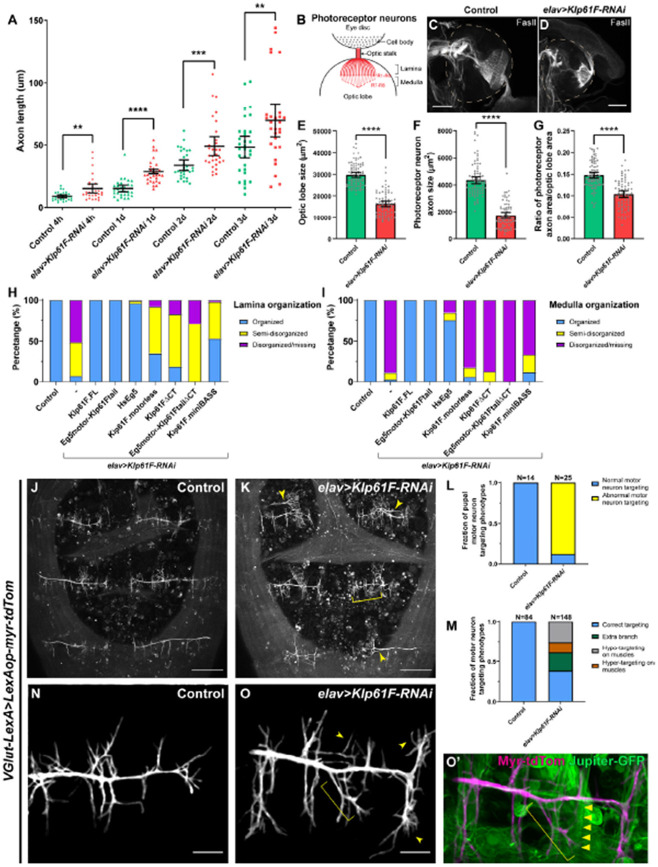
Figure 3. Kinesin-5/Klp61F regulates axonal growth in culture and in vivo.
(A) Axon length measurement in cultured Drosophila larval brain neurons of control and neuronal Klp61F-RNAi. Neurons were fixed and stained with both anti-α tubulin (DM1α) and anti-Elav antibodies. The length of the longest neurite of each neuron (Elav-positive) was measured as the axon length. Scatter plots with average ± 95 % confidence intervals are shown. Unpaired t-tests with Welch's correction were performed between control and neuronal Klp61F-RNAi samples: 4 hours, p= 0.0018 (**); 1 day, p <0.0001 (****); 2 days, p= 0.0008 (***); 3 days, p=0.0080 (**). (B) A schematic illustration of the photoreceptor neurons of Drosophila 3rd instar larval eye disc and optic lobe. (C-D) Representative examples of photoreceptor neuron axonal targeting in the optic lobes of control (C) and neuronal Klp61F depletion (D). Photoreceptor neuron axons were labeled with anti-Fasciclin II antibody. Dashed lines indicate the position of the optic lobes. Scale bars, 50 μm. (E-G) Optic lobe size (E), photoreceptor neuron axon size (F), and the ratio of photoreceptor neuron axon size to the optic lobe size (G) in control and neuronal Klp61F depletion. Scatter plots with average ± 95% confidence intervals are shown. Unpaired t-tests with Welch's correction were performed between control and neuronal Klp61F-RNAi samples: optic lobe size, p <0.0001 (****); photoreceptor neuron axon size, p <0.0001 (****); the ratio of photoreceptor neuron axon size to the optic lobe size, p <0.0001 (****). (H-I) Summary of lamina (H) and medulla (I) organization phenotypes in control, neuronal Klp61F-RNAi, and rescue with RNAi-resistant full-length Drosophila Klp61F (Klp61F.FL), chimeric Eg5motor-Klp61Ftail, full-length human Eg5 (HsEg5), and Klp61F.motorless, Klp61FΔCT, chimeric Eg5motor-Klp61FtailΔCT, and Klp61F.miniBASS. Sample sizes for each genotype: control, N=85; Klp61F-RNAi, N=72; Klp61F-RNAi + Klp61F.FL, N=84; Klp61F-RNAi + Eg5motor-Klp61Ftail, N=89; Klp61F-RNAi + HsEg5, N=97; Klp61F-RNAi + Klp61F.motorless, N=86; Klp61F-RNAi + Klp61FΔCT, N=97; Klp61F-RNAi + Eg5motor-Klp61FtailΔCT, N=24; Klp61F-RNAi + Klp61F.miniBASS, N=51. All samples carried one copy of elavP-Gal4 and were stained with anti-Fasciclin II antibody to label the photoreceptor neuron axons. (J-K) Pupal motor neuron targeting in dorsal abdomen segment A5-A7 in control (J) and in elav>Klp61F-RNAi (K) 45-50 hours after pupal formation (APF). Extra branching and hyper-targeting of motor neurons in elav>Klp61F-RNAi (K) are shown with arrowheads and a bracket, respectively. Scale bars, 100 μm. (L-M) Summary of motor neuron targeting phenotypes, by the numbers of pupae (L) and the numbers of motor neuron groups (M). (N-O’) Pupal motor neuron targeting 30 hours APF in control (N) and in elav>Klp61F-RNAi (O). Extra (arrowheads) and mistargeting (bracket) neurites were seen in elav>Klp61F-RNAi compared to control. (O’) In the overlay image of the motor neuron membrane (magenta) and muscles (green, labeled with Jupiter-GFP), an axonal branch (bracket) showed aberrant targeting, deviating from its expected perpendicular orientation relative to the main axon; instead, it originated from an incorrect position and extended obliquely, eventually innervating a distant muscle (indicated by triangles). Scale bars, 20 μm. (J-K) and (N-O’) Motor neurons were labeled with membrane-targeted tdTomato driven by a motor neuron-specific driver (VGlut-LexA, LexAop-myr-tdTom); knockdown of Klp61F by RNAi was driven independently by the UAS-Gal4 system (elavP-Gal4, UASp-Klp61F-RNAi).

