Abstract
Patient: Female, 23-year-old
Final Diagnosis: Choledochal cyst and pancreatic divisum
Symptoms: Pancreatitis
Clinical Procedure: Pancreaticoduodenectomy
Specialty: Gastroenterology and Hepatology • Surgery
Objective:
Rare disease
Background:
A choledochal cyst (CC), or biliary cyst, is a congenital or acquired anomaly of the biliary tree. Pancreas divisum (PD) is a rare congenital anomaly due to incomplete fusion of pancreatic ducts, which can complicate the clinical course of choledochal cysts. This rare combination is a surgical management challenge. This report presents the diagnosis and management of a 23-year-old woman with a combined choledochal cyst and pancreas divisum treated with pancreaticoduodenectomy.
Case Report:
This article presents the case of a 23-year-old woman who presented with severe, stabbing abdominal pain radiating to the back and epigastric tenderness and was diagnosed with pancreatitis. Initial imaging revealed a choledochal cyst, prompting further investigation with ERCP that showed concomitant PD. She was treated via pancreaticoduodenectomy. During the following 9 years, she was hospitalized over 2 dozen times for recurrent pancreatitis.
Conclusions:
This report presents a complex case of a combined choledochal cyst and pancreas divisum, which was surgically managed by pancreaticoduodenectomy. The association of CC with PD should be suspected in patients with recurrent acute pancreatitis and/or cholangitis with no identifiable cause. Surgical treatment of CC with PD depends on the classification of the CC, and complications can include recurrent pancreatitis, although the prognosis is often favorable. The purpose of this report is to emphasize that pancreaticoduodenectomy is unlikely to provide favorable outcomes for CC with PD, especially considering there is evidence that less extensive surgical interventions produce better outcomes.
Key words: Choledochal Cyst, Pancreatitis
Introduction
Choledochal cysts (CCs) are uncommon and pancreatic divisum (PD) is relatively common, but these conditions are exceedingly rare concomitantly, with less than 15 documented cases in the literature [1,2]. CCs are congenital abnormalities of the bile duct, defined as dilations of the intra- and/or extrahepatic segments of the biliary tree [3]. A popular hypothesis is that CCs are the result of an abnormally formed pancreaticobiliary junction, generating a channel that bypasses the sphincter of Oddi. Such anatomy allows for backflow and mixing of pancreatic and biliary secretions, with subsequent activation of pancreatic enzymes [3]. This results in increased pressure, leading to dilation, epithelial damage, and inflammation, and increased risk of malignancy [3], necessitating surgical removal [4]. There exists a regional variation in the prevalence of CCs, as they most often occur in Asian populations, and two-thirds of cases within Asia occur in Japan, where prevalence is estimated to be as high as 1: 1000 live births [5]. However, the prevalence in Western populations overall ranges from 1: 100 000 to 1: 150 000. Interestingly, the prevalence is 1: 13 500 in the United States, and 1: 15 000 in Australia [6]. Choledochal cysts occur predominantly in females, at a rate of 3–4: 1 compared to males [7]. The genetic associations of CCs depend on the classification (discussed further below), with Type I and IV being associated with mutations in the HNF1B gene, Type II with a deletion of EHBP1, and Type V with translocations of the APC gene [8]. The criterion standard for diagnosis of CCs is endoscopic retrograde cholangiopancreatography (ERCP), although magnetic resonance cholangiopancreatography (MRCP) has been shown to have higher sensitivity [9]). The management of CCs varies by type, discussed further below, but most require surgical removal [4].
In contrast, PD is the most common congenital pancreatic anomaly, with an overall prevalence estimated to be 1: 10, with roughly equal distribution between males and females [10]. PD occurs when the dorsal and ventral duct systems of the pancreas fail to fuse during embryogenesis, resulting in the main pancreatic duct draining through the minor papilla, with the ventral duct being the only pancreatic contribution to the major papilla [11]. PD is associated with genetic variations in PRSS1 and SPINK1, but most commonly with mutations in CFTR, all of which pre-dispose to pancreatitis, with CFTR mutations having the strongest association [12]. Like CCs, PD is best diagnosed via ERCP, although MCRP is also a valuable, noninvasive modality and is preferred as a first-line imaging test [13]. Symptomatic cases can be treated endoscopically with sphincterotomy, and if that fails, with surgical sphincteroplasty, duodenopancreatectomy, or duodenum-preserving pancreatic head resection [13].
The association between CCs and PD is not well established; with few cases of these congenital anomalies occurring concomitantly in the literature, it is difficult to determine an association. Further studies will be required to determine what factors contribute to the coexistence of CC and PD. A previously reported case was managed by hepaticoduodenostomy, but the patient developed chronic pancreatitis followed by several bouts of acute cholangitis in the years following surgery [1]. Other cases have been treated with more conservative producers like CC excision, with a case series of 3 of 4 patients treated with this technique experiencing long-term remission of symptoms [2]. This report presents the diagnosis and management of a 23-year-old woman with a combined choledochal cyst and pancreas divisum treated with pancreaticoduodenectomy.
Case Report
A diaphoretic 23-year-old woman presented to the Emergency Department (ED) with a chief concern of abdominal pain. Her diaphoresis was attributed to pain. She had no known comorbidities at that time, did not smoke, and drank alcohol on occasion. Of note, she had been discharged from an outside hospital 2 weeks prior for severe abdominal pain. At that time, she was diagnosed with pancreatitis secondary to alcohol use, a pancreatic duct stone, and a CC incidentally discovered via a CT scan of her abdomen, at that time thought to be Type III. An ERCP was then performed to remove the pancreatic duct stone. She underwent a dorsal pancreatic duct sphincterotomy, and a 5-Fr -3 cm pancreatic stent was placed.
Upon her arrival to our ED, she appeared in mild distress with concerns of nausea without fever or vomiting. She described her abdominal pain as severe and stabbing, radiating to the back. Her vital signs were within normal limits and her physical exam was significant only for mild epigastric tenderness. Laboratory results were significant for an AST of 502 units/L (u/L), ALT of 189 u/L, lipase of 380 u/L, amylase of 197 u/L, alkaline phosphatase of 189 u/L, and LDH of 1930 u/L.
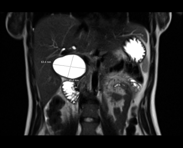
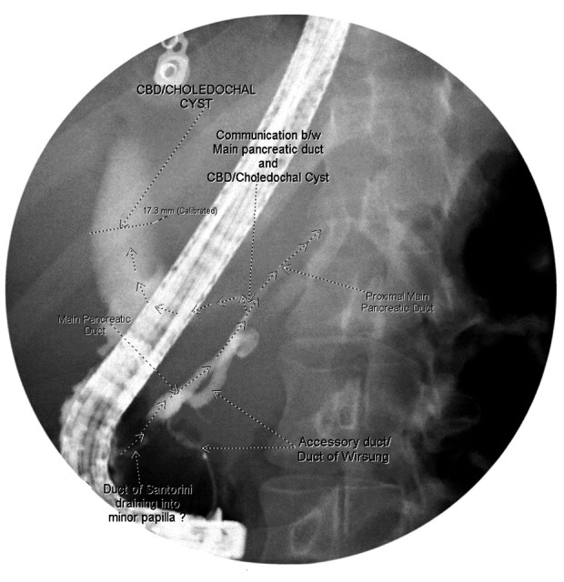
A CT scan of the abdomen was performed, showing evidence of acute pancreatitis, multiple biliary filling defects, pneumobilia, and a fluid collection extending into the right retroperitoneum thought to be a pancreatic pseudocyst versus abscess. An MRI of the abdomen revealed this fluid collection to measure 63.4×45.4 mm (Figure 1), but a diagnosis could not be confirmed at this time. Following fluid resuscitation, pain management, and resolution of acute pancreatitis, a diagnostic ERCP was performed 2 weeks later. ERCP findings confirmed this fluid collection to be a Type I CC, and a new diagnosis of incomplete pancreatic divisum was made (Figure 2). It was determined that a pancreaticoduodenectomy could potentially be a definitive treatment, and she was scheduled for surgery 1 month later. Pancreaticoduodenectomy was recommended given the failure of treatment with sphincterotomy.
Figure 1.

Magnetic resonance imaging of the abdomen revealing a 63.4×45.4 mm choledochal cyst.
Figure 2.

Endoscopic retrograde cholangiopancreatography (ERCP) revealing pancreatic divisum.
The patient underwent a planned laparoscopic-assisted pancreaticoduodenectomy with reconstruction, including a double hepaticojejunostomy, pancreaticojejunostomy, and gastrojejunostomy, without complications. The Whipple specimen that was resected included the head of the pancreas, duodenum, distal stomach, and choledochal cyst, and the bile duct was transected above the bifurcation of the right and left hepatic ducts. Peripancreatic, portacaval, and hepatic artery lymph-adenectomies were also performed. Histological examination determined that the resected lymph nodes and the hepatic duct were negative for malignancy, and chronic inflammation was identified within the CC without dysplasia or carcinoma.
The postoperative course was complicated by a pseudoaneurysm of the right hepatic artery identified by CT scan 2 weeks into the postoperative period, associated with diarrhea and emesis, and she was admitted to the surgical intensive care unit following coiling of the pseudoaneurysm. Cultures of biliary fluid taken during the coiling procedure grew S. aureus, Enterococcus, Streptococcus viridans, Klebsiella, and Enterobacter. The patient was started on ciprofloxin and metronidazole and discharged at 22 days. She was evaluated in the ED 2 weeks after discharge for abdominal pain radiating to the back, with lipase over 3000 u/L and mildly elevated ASL/ALT, and was diagnosed with acute pancreatitis. This presentation was attributed as a complication of to the pancreaticoduodenectomy. She was discharged after 1 week. Over the following 9 years, she was hospitalized over 2 dozen times for recurrent pancreatitis.
Discussion
CC and PD are rare concomitantly, with this case report being the 15th in the literature [1,2,9,14–21]. This case demonstrates that pancreaticoduodenectomy is likely not a suitable treatment option for CC with PD, and the procedure carries more associated risks than other surgical interventions that have been shown to result in good patient outcomes.
According to Todani et al, the most common clinical presentation of CC is intermittent recurrent episodes of upper abdominal pain, intermittent jaundice, and a palpable right upper quadrant (RUQ) mass [22]. However, our patient presented with recurrent pancreatitis without a palpable mass, despite the CC measuring 63.4×45.4 mm, making it the largest CC with PD identified in the literature [1,2,16,20]. This presentation is common, as 5 of the 14 documented cases presented with recurrent pancreatitis and/or cholangitis, and none reported a palpable mass [1,2,14,15,16]. Pancreatitis appears to be the most common presentation of CC with PD besides abdominal pain, which was experienced by all patients in the literature.
Additionally, 9 of the 14 patients in the literature with these concomitant abnormalities were female, with a mean age at time of diagnosis of 32.0 years old and a range of 3–63 years. For the 5 male patients, the mean age was 39.2 years old with a range of 28–77 years. Therefore, it appears that CC with PD is more common in females. This is not particularly surprising, as CCs have been shown to have a female predominance, while PD does not appear to show a sex bias. If females are more predisposed to develop CCs compared to males, and PD is considered to be roughly equivalent in males and females, it follows that females are more likely to have both abnormalities concomitantly.
Eleven cases in the literature reported lab values, with 4 patients having elevated amylase levels, 4 patients with elevated GGT, 4 patients with elevated total bilirubin, 3 patients with elevated lipase, 3 patients with elevated alkaline phosphatase, and 2 with elevated LDH. Thus, while it has been observed that patients with CC and PD often present with acute pancreatitis and hyperamylasemia [1], most patients in the literature did not have elevated amylase levels, and there does not appear to be a useful, consistent laboratory marker to identify patients with CC and PD. Thus, this diagnosis can easily be missed unless there is a high index of suspicion and a thorough diagnostic investigation is performed.
There is some contention regarding the best method of diagnosing these concomitant anomalies. The current criterion standard for diagnosing CC is ERCP, but MRCP has been shown to have a higher sensitivity and is noninvasive [9], with 40% of patients with high-risk characteristics, such as female sex and prior pancreatitis, developing post-ERCP pancreatitis [23]. Despite both of these characteristics appearing frequently in patients with CC and PD, in nearly all cases in the literature, confirmatory diagnosis was achieved via ERCP, often following attempted MRCP. Thus, despite the advantages of MRCP, its utility may be overestimated for patients with concomitant CC and PD, but a more thorough investigation is required before conclusions can be made. Nevertheless, it is recommended that the diagnostic workup begin with abdominal ultrasonography, followed by MRCP in patients with suspected CC, with ERCP reserved for patients who cannot be diagnosed by MRCP [24]. However, despite this general consensus, which was formulated to diagnose CC alone, the evidence supporting the utility of MRCP for patients with concomitant CC and PD in the literature is questionable. The diagnosis of CC with PD in our patient shows that MRCP may lack utility in the diagnosis of patients with both abnormalities, as the confirmed diagnosis came from ERCP findings.
The most widely used classification system of CCs was developed by Todani et al and categorizes CCs based on their morphology. Type I CCs comprise 50–80% of all CCs and are divided into 3 subtypes: IA is saccular, IB is segmental, and IC is fusiform. Type II accounts for represent 2–5% of all CC’s and are characterized by a supraduodenal diverticulum. Type III are characterized by a choledochocele or intraduodenal cyst and make up 4% of CCs. Type IV-A are multiple intra- and extrahepatic cysts, while IV-B are multiple cysts in only the extrahepatic bile duct; together they represent 10–20% of CCs. Finally, Type V, or Caroli disease, is characterized by multiple dilations of the intrahepatic bile ducts, making up 1% of CCs [22].
The most common type of CC observed in patients with concomitant PD was Type III, with 5 of the 11 cases that included classifications diagnosing a choledochocele [14–17,20]. Of the remaining CCs with PD in the literature, 4 (including our patient) were Type I [2,16,18] and 2 were Type IV [2]. According to Todani et al, Type I and IV CCs are the first and second most common types, respectively, and together they represent the overwhelming majority of CCs [22]. Thus, despite the predominance of Type I and IV CCs overall, there appears to be an over-representation of Type III CCs in patients with concomitant PD.
The classification of CC by type is an important factor in determining treatment. Treatment of Type I, II, and, IV-A CC consists of complete extrahepatic bile duct cyst excision to the level of communication with the pancreatic duct, cholecystectomy, and restoration of bilioenteric continuity. Both hepaticoduodenostomy and Roux-en-Y hepaticojejunostomy reconstruction after choledochal cyst resection are reported in the literature, but Roux-en-Y hepaticojejunostomy is preferred [25]. Two patients in the literature with a Type I CC and PD underwent CC excision; one was reported to be asymptomatic at 5 months [2], and the other was complicated by recurrent pancreatitis after 8 years postoperatively, followed by acute cholangitis 7 an additional years later, treated by Roux-en-Y hepaticojejunostomy [1]. The 2 patients with Type IV CC and PD in the literature both underwent successful CC excision and were reported to be asymptomatic at 5 years [2].
Our patient is the only patient with a Type I CC and PD to have undergone a pancreaticoduodenectomy, or for that matter, the first patient in the literature with any type of CC and PD to have undergone an operation directly involving the pancreas. The rationale for the pancreaticoduodenectomy was that by removing the head of the pancreas, the inciting stimulus for that patient’s recurrent pancreatitis would be removed. Furthermore, given the association of PD with pancreatic tumors, particularly in the dorsal pancreas [26], it was thought that pancreaticoduodenectomy would be beneficial to treat both the CC and PD. However, patient outcomes in this instance were dubious given the many hospitalizations for recurrent pancreatitis postoperatively. Moreover, the association of pancreatitis in patients with PD is debatable, as some evidence suggests that PD alone does not in and of itself predispose to pancreatitis, but rather its associated mutations, especially CFTR mutations, are responsible for pancreatitis [12,27]. Thus, pancreaticoduodenectomy performed to prevent pancreatitis in the setting of PD may not address the underlying cause. Additionally, this patient’s pancreatitis very likely could have been caused by the pancreaticoduodenectomy, as a meta-analysis showed a 33% rate of postoperative acute pancreatitis following this procedure [28]. Thus, like the other cases treated with more extensive surgery like hepaticoduodenostomy [1], treatment with pancreaticoduodenectomy may have more postoperative complications than those treated with CC excision alone [2].
Type III CCs without PD can be treated if symptomatic, or in asymptomatic young patients, by sphincterotomy, often with biopsy of the cyst epithelium to assess dysplasia and identify the type of epithelium lining the cyst, as the biliary mucosal lining is associated with a heightened risk of malignancy [3]. Three of 4 patients with Type III CC and PD in the literature underwent sphincterotomy/sphincteroplasty, with 2 patients being asymptomatic at 1 year [15,16], and 1 asymptomatic at 6 months [15]. The remaining patient with a Type III CC and PD underwent a more extensive surgery involving bile duct resection, excision of the papilla, end-to-side hepaticojejunostomy, and a pancreatic duct jejunostomy end-to-side into the same jejunal loop, as well as a Roux-en-Y jejunal reconstruction, although patient follow-up was not provided in that case report [14].
In contrast to other publications on this topic in the literature, this paper evaluates all documented cases of concomitant CC with PD and reports on the similarities and differences in their presentation, treatment, and outcomes. Much of the literature is focused on the pathophysiology of CC and PD and treats them as separate entities, but they should be thought of as interlinked. Moreover, previous publications have underestimated the total number of CC with PD cases in the literature. This paper does, however, have limitations. A minority of publications in the literature provide follow-up data beyond 1 year, and several do not specify the classification of CC. Without classification and long-term data on patient outcomes, the analysis of surgical outcomes is limited.
Conclusions
This report presents a complex case of a combined choledochal cyst and pancreas divisum, which was surgically managed by pancreaticoduodenectomy. The association of CC with PD is a rare condition that should be suspected in patients with recurrent acute pancreatitis and/or cholangitis with no identifiable cause. Surgical treatment of CC with PD depends on the classification of the CC, and complications can include recurrent pancreatitis, although the prognosis is often favorable. The goal of this paper is to emphasize that a pancreaticoduodenectomy is not the proper treatment for CC with PD, and argue that other, less extensive, surgical interventions such as CC excision can produce better patient outcomes.
Footnotes
Publisher’s note: All claims expressed in this article are solely those of the authors and do not necessarily represent those of their affiliated organizations, or those of the publisher, the editors and the reviewers. Any product that may be evaluated in this article, or claim that may be made by its manufacturer, is not guaranteed or endorsed by the publisher
Declaration of Figures’ Authenticity
All figures submitted have been created by the authors who confirm that the images are original with no duplication and have not been previously published in whole or in part.
References:
- 1.Ransom-Rodríguez A, Blachman-Braun R, Sánchez-García Ramos E, et al. A rare case of choledochal cyst with pancreas divisum: Case presentation and literature review. Ann Hepatobiliary Pancreat Surg. 2017;21(1):52–56. doi: 10.14701/ahbps.2017.21.1.52. [DOI] [PMC free article] [PubMed] [Google Scholar]
- 2.Pakkala A, Nagari B, Nekarakanti PK, Bansal AK. A case series of choledochal cyst with pancreatic divisum: A rare association. Turk J Surg. 2022;38(3):294–97. doi: 10.47717/turkjsurg.2022.5609. [DOI] [PMC free article] [PubMed] [Google Scholar]
- 3.Hoilat GJ, Savio J. StatPearls Publishing; Choledochal Cyst. [serial online]. Updated 2023 Aug 28 [cited 2024 Aug 21]. Available from: https://www.ncbi.nlm.nih.gov/books/NBK557762/ [Google Scholar]
- 4.Diao M, Li L, Cheng W. Timing of surgery for prenatally diagnosed asymptomatic choledochal cysts: A prospective randomized study. J Pediatr Surg. 2012;47(3):506–12. doi: 10.1016/j.jpedsurg.2011.09.056. [DOI] [PubMed] [Google Scholar]
- 5.Tashiro S, Imaizumi T, Ohkawa H, et al. Pancreaticobiliary maljunction: Retrospective and nationwide survey in Japan. J Hepatobiliary Pancreat Surg. 2003;10(5):345–51. doi: 10.1007/s00534-002-0741-7. [DOI] [PubMed] [Google Scholar]
- 6.Bhavsar MS, Vora HB, Giriyappa VH. Choledochal cysts: A review of literature. Saudi J Gastroenterol. 2012;18(4):230–36. doi: 10.4103/1319-3767.98425. [DOI] [PMC free article] [PubMed] [Google Scholar]
- 7.Liu CL, Fan ST, Lo CM, Lam CM, et al. Choledochal cysts in adults. Arch Surg. 2002;137(4):465–68. doi: 10.1001/archsurg.137.4.465. [DOI] [PubMed] [Google Scholar]
- 8.Ye Y, Lui VCH, Tam PKH. Pathogenesis of choledochal cyst: Insights from genomics and transcriptomics. Genes (Basel) 2022;13(6):1030. doi: 10.3390/genes13061030. [DOI] [PMC free article] [PubMed] [Google Scholar]
- 9.Li L, Yamataka A, Segawa O, Miyano T. Coexistence of pancreas divisum and septate common channel in a child with choledochal cyst. J Pediatr Gastroenterol Nutr. 2001;32(5):602–4. doi: 10.1097/00005176-200105000-00022. [DOI] [PubMed] [Google Scholar]
- 10.Bülow R, Simon P, Thiel R, et al. Anatomic variants of the pancreatic duct and their clinical relevance: An MR-guided study in the general population. Eur Radiol. 2014;24(12):3142–49. doi: 10.1007/s00330-014-3359-7. [DOI] [PubMed] [Google Scholar]
- 11.Gutta A, Fogel E, Sherman S. Identification and management of pancreas divisum. Expert Rev Gastroenterol Hepatol. 2019;13(11):1089–105. doi: 10.1080/17474124.2019.1685871. [DOI] [PMC free article] [PubMed] [Google Scholar]
- 12.Bertin C, Pelletier AL, Vullierme MP, et al. Pancreas divisum is not a cause of pancreatitis by itself but acts as a partner of genetic mutations. Am J Gastroenterol. 2012;107(2):311–17. doi: 10.1038/ajg.2011.424. [DOI] [PubMed] [Google Scholar]
- 13.Ferri V, Vicente E, Quijano Y, et al. Diagnosis and treatment of pancreas divisum: A literature review. Hepatobiliary Pancreat Dis Int. 2019;18(4):332–36. doi: 10.1016/j.hbpd.2019.05.004. [DOI] [PubMed] [Google Scholar]
- 14.Hackert T, Hartwig W, Werner J. Symptoms and surgical management of a distal choledochal cyst in a patient with pancreas divisum: Case report and review of the literature. Case Rep Gastroenterol. 2007;1(1):90–95. doi: 10.1159/000108636. [DOI] [PMC free article] [PubMed] [Google Scholar]
- 15.Garrido A, León R, López J, Márquez JL. [An exceptional case of choledochocele and pancreas divisum in an elderly man.] Gastroenterol Hepatol. 2012;35(1):8–11. doi: 10.1016/j.gastrohep.2011.09.002. [DOI] [PubMed] [Google Scholar]
- 16.Arulprakash S, Balamurali R, Pugazhendhi T, Kumar SJ. Pancreas divisum and choledochal cyst. Indian J Med Sci. 2009;63(5):198–201. [PubMed] [Google Scholar]
- 17.Patidar Y, Agarwal N, Gupta S, et al. Choledochocele with pancreas divisum: A rare co-occurrence diagnosed on magnetic resonance cholangiopancreatography. World J Radiol. 2013;5(7):264–66. doi: 10.4329/wjr.v5.i7.264. [DOI] [PMC free article] [PubMed] [Google Scholar]
- 18.Bechtler M, Eickhoff A, Willis S, Riemann JF. Choledochal cyst type IA with drainage through the ventral duct in pancreas divisum. Endoscopy. 2009;41(Suppl. 2):E71–72. doi: 10.1055/s-0028-1119485. [DOI] [PubMed] [Google Scholar]
- 19.Dalvi AN, Pramesh CS, Prasanna GS, et al. Incomplete pancreas divisum with anomalous choledochopancreatic duct junction with choledochal cyst. Arch Surg. 1999;134(10):1150–52. doi: 10.1001/archsurg.134.10.1150. [DOI] [PubMed] [Google Scholar]
- 20.Sonoda M, Sato M, Miyauchi Y, et al. A rare case of choledochocele associated with pancreas divisum. Pediatr Surg Int. 2009;25(11):991–94. doi: 10.1007/s00383-009-2460-5. [DOI] [PubMed] [Google Scholar]
- 21.Takikawa T, Kanno A, Masamune A, et al. Ectopic opening of the common bile duct accompanied by choledochocele and pancreas divisum. Intern Med. 2016;55(9):1097–102. doi: 10.2169/internalmedicine.55.6240. [DOI] [PubMed] [Google Scholar]
- 22.Todani T, Watanabe Y, Narusue M, et al. Congenital bile duct cysts: classification, operative procedures, and review of thirty-seven cases including cancer arising from choledochal cyst. Am J Surg. 1977;134:263–69. doi: 10.1016/0002-9610(77)90359-2. [DOI] [PubMed] [Google Scholar]
- 23.Thaker AM, Mosko JD, Berzin TM. Post-endoscopic retrograde cholangiopancreatography pancreatitis. Gastroenterol Rep (Oxf) 2015;3(1):32–40. doi: 10.1093/gastro/gou083. [DOI] [PMC free article] [PubMed] [Google Scholar]
- 24.Khandelwal C, Anand U, Kumar B, Priyadarshi RN. Diagnosis and management of choledochal cysts. Indian J Surg. 2012;74:401–6. doi: 10.1007/s12262-012-0426-7. [DOI] [PMC free article] [PubMed] [Google Scholar]
- 25.Soares KC, Arnaoutakis DJ, Kamel I, et al. Choledochal cysts: Presentation, clinical differentiation, and management. J Am Coll Surg. 2014;219(6):1167–80. doi: 10.1016/j.jamcollsurg.2014.04.023. [DOI] [PMC free article] [PubMed] [Google Scholar]
- 26.Takuma K, Kamisawa T, Tabata T, et al. Pancreatic diseases associated with pancreas divisum. Dig Surg. 2010;27(2):144–48. doi: 10.1159/000286975. [DOI] [PubMed] [Google Scholar]
- 27.Spicak J, Poulova P, Plucnarova J, et al. Pancreas divisum does not modify the natural course of chronic pancreatitis. J Gastroenterol. 2007;42:135–39. doi: 10.1007/s00535-006-1976-x. [DOI] [PubMed] [Google Scholar]
- 28.Wu Z, Zong K, Zhou B, et al. Incidence and risk factors of postoperative acute pancreatitis after pancreaticoduodenectomy: A systematic review and meta-analysis. Front Surg. 2023;10:1150053. doi: 10.3389/fsurg.2023.1150053. [DOI] [PMC free article] [PubMed] [Google Scholar]


