Figure 2.
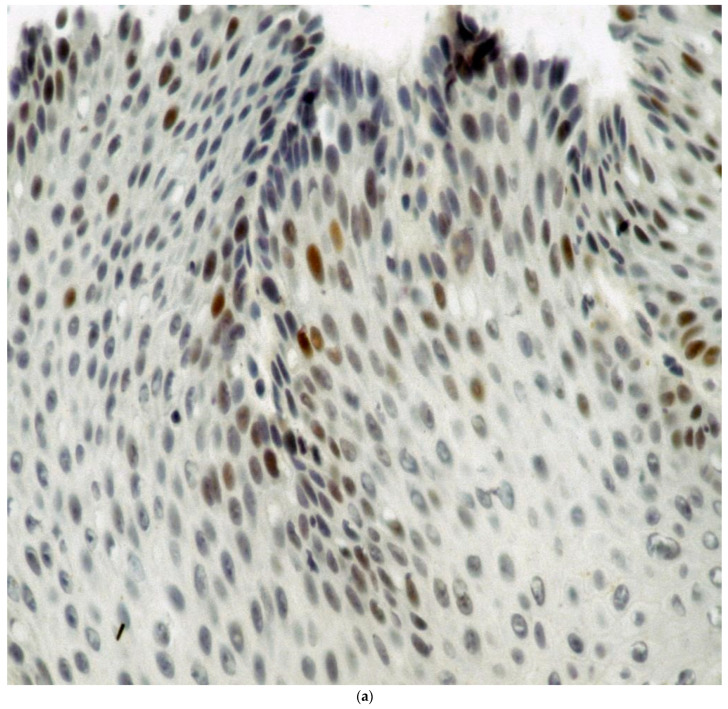
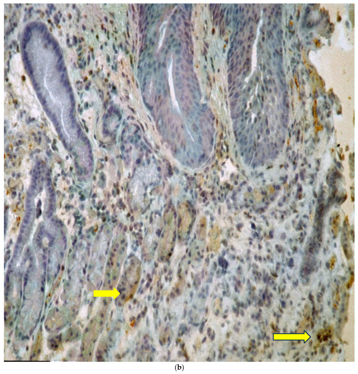
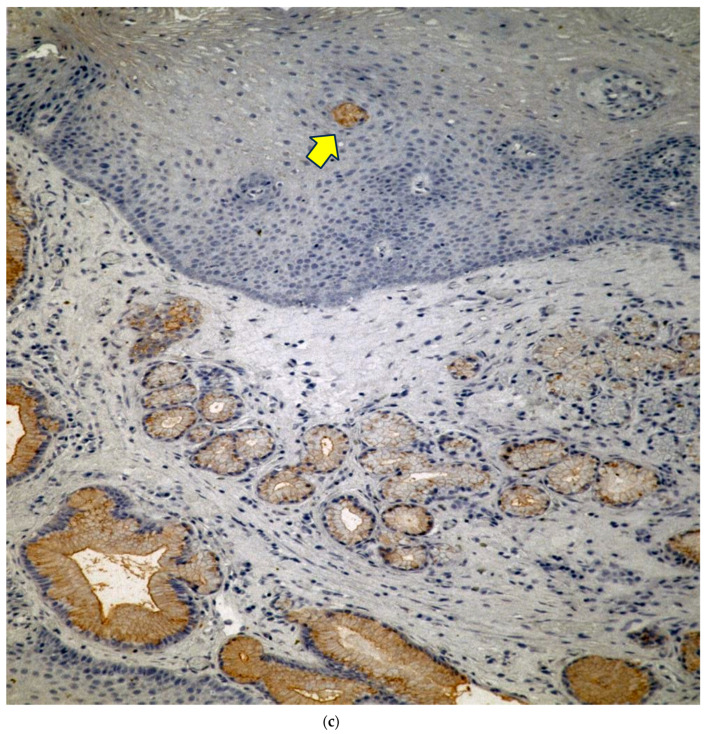
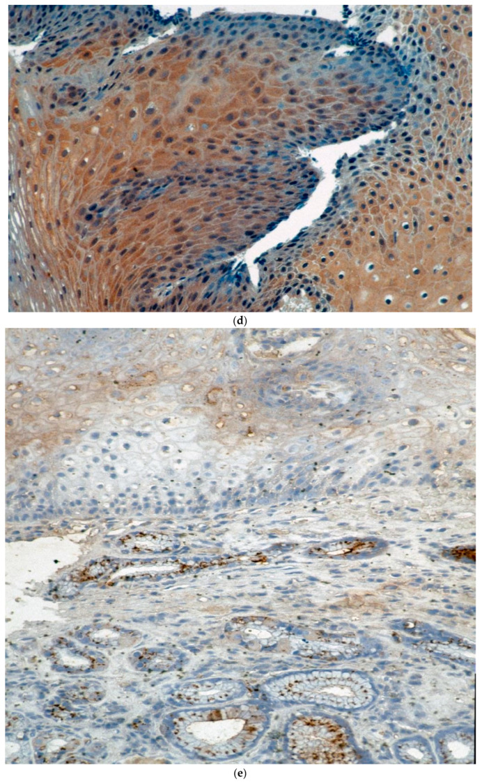
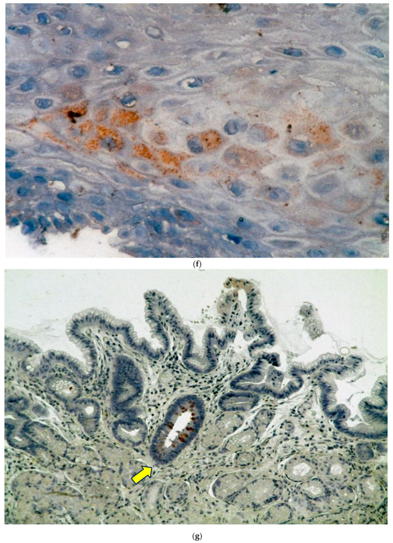
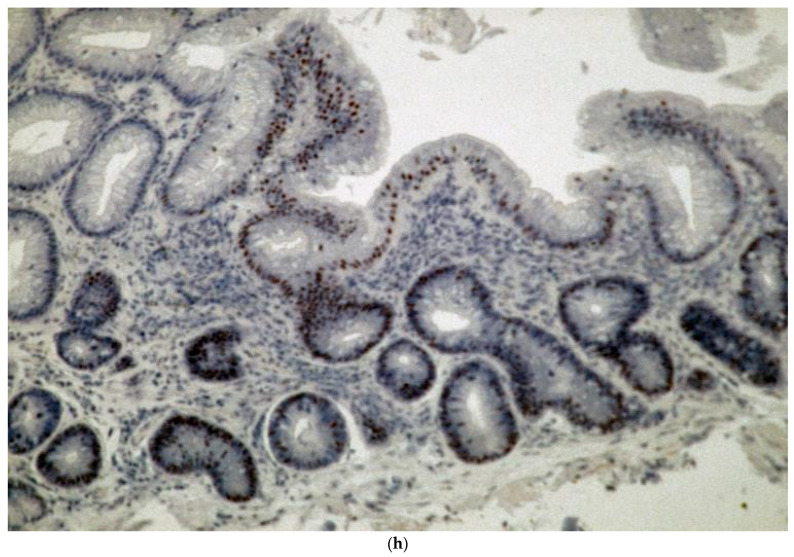
(a) Percentage staining of squamous nuclei in a Barrett’s esophagus patient with p53 nuclear staining as evidenced by the brown nuclei. Magnification 360×. (b) VEGF staining in a glandular section of a specimen taken from a patient with Barrett’s esophagus. (b) shows moderate brown VEGF staining in the glandular epithelium more localized in the cytoplasm (arrows indicate glands and staining). Magnification 50×. (c) Cox2 staining is seen from a section taken through the GEJ in a section taken from a patient with Barrett’s esophagus. Magnification 50×. (d) COX-2 staining in the squamous epithelium is moderate diffuse and cytoplasmic. Magnification 130×. The intensity of stain was 3+ in the areas on the left and 1–2+ on the right. (e) Adnab-9 staining of a section taken from the GEJ of a patient with confirmed Barrett’s esophagus. Esophagus. Magnification 50×. (f) shows Adnab-9 labeling of squamous cells in a patient with Barrett’s esophagus focally with reticulated cytoplasmic staining at a higher power. Magnification 360×. (g) Tn staining of glandular mucosa in a patient with Barrett’s esophagus. Magnification 50×. (h) Intense nuclear CDX2 staining is seen in this section of Barrett’s epithelium and involves most of the glandular epithelium in this section. Magnification 125×.

