Abstract
Gene expression among the nonsegmented negative-strand RNA viruses is controlled by distance from the single transcriptional promoter, so the phenotypes of these viruses can be systematically manipulated by gene rearrangement. We examined the potential of gene rearrangement as a means to develop live attenuated vaccine candidates against Vesicular stomatitis virus (VSV) in domestic swine, a natural host for this virus. The results showed that moving the nucleocapsid protein gene away from the single transcriptional promoter attenuated and ultimately eliminated the potential of the virus to cause disease. Combining this change with relocation of the surface glycoprotein gene yielded a vaccine that protected against challenge with wild-type VSV. By incremental manipulation of viral properties, gene rearrangement provides a new approach to generating live attenuated vaccines against this class of virus.
Gene expression of the nonsegmented negative-strand RNA viruses is controlled by the highly conserved order of genes relative to the single transcriptional promoter (19). We utilized this regulatory mechanism to alter gene expression levels of the prototypic rhabdovirus, Vesicular stomatitis virus (VSV), a significant livestock pathogen, by engineering changes into a cDNA clone and recovering viruses having the order of genes rearranged and as a result, the expression levels of the translocated genes altered (3, 9, 21). This allowed us to test whether gene rearrangement as a means to predictably alter gene expression levels could affect disease potential in a natural host.
The negative-strand RNA genome of VSV has five genes which encode the five viral structural proteins: the nucleocapsid protein, N, required in stoichiometric amounts for encapsidation of genomic RNA during replication; the phosphoprotein, P; the RNA-dependent RNA polymerase, L; the matrix protein, M; and the attachment glycoprotein, G. The genes are in the order 3′-N-P-M-G-L-5′, and their transcription is sequential from a single 3′ polymerase entry site (1, 4). As a result of attenuation of transcription at each gene junction, the genes located closest to the 3′ promoter are transcribed most abundantly, and those located at more promoter distal sites are transcribed in successively lower abundance (12).
In previous work we demonstrated that the translocation of a single gene essential for replication, the nucleocapsid gene, to positions successively farther away from the single transcriptional promoter reduced expression of that gene progressively and lowered growth potential in cell culture and lethality for mice in a stepwise manner (21). The reduction in replication potential did not compromise the ability of the altered viruses to elicit protective immunity against subsequent lethal challenge in mice (21). In subsequent work, we showed additionally that movement of the glycoprotein gene, which encodes the major VSV neutralizing epitopes, closer to the 3′ promoter increased G protein expression in infected cells (9). Viruses engineered to have G in the 3′ position elicited an earlier and enhanced immune response in inoculated mice in comparison to that observed with viruses having the wild-type gene order (9).
Cattle, horses, and swine are naturally infected by VSV, which causes a disease involving vesiculation and ulceration of the tongue and oral epithelia and sometimes the appearance of lesions on the feet and teats (14). These symptoms are indistinguishable from foot-and-mouth disease, one of the most devastating exotic diseases of livestock. Therefore, VSV causes severe economic losses due to quarantine and trade barriers as well as losses due to the lowered productivity caused by the disease itself. Two VSV serotypes, New Jersey (VSV-NJ) and Indiana (VSV-IN), are enzootic from southern Mexico to northern South America (14). In the United States, where VSV occurs sporadically, the most recent outbreaks caused by VSV-IN occurred in 1997 and 1998 and affected mainly horses (16). Prior to that, there was a large outbreak in cattle in 1995 caused by VSV-NJ that had a significant impact on the Colorado beef industry (5). Domestic swine are readily infected by both serotypes of VSV.
Efforts to develop subunit- or DNA-mediated vaccines for VSV have met with limited success (7, 23). Immunization with live field strains has been attempted only under emergency conditions (13). Live attenuated vaccines, however, have not been explored for VSV, despite the success and use of a live attenuated vaccine against another rhabdovirus, rabies virus (2). At present there is no satisfactory vaccine against VSV infection.
In the present study we assessed the effects of rearrangement of the genes of VSV on the ability of the virus to replicate, to cause disease, and to elicit protective immune responses in one of the three natural hosts of VSV, swine. We compared the disease-producing and immunogenic potential of viruses in which the nucleocapsid gene, which is essential for viral replication, had been moved to promoter-distal positions to downregulate its expression and in which the glycoprotein gene had been moved to promoter-proximal positions to increase its expression (9). The data presented below show that manipulation of the position of individual genes allows alteration of the disease potential of VSV. Since monopartite negative-strand RNA viruses have not been reported to undergo homologous recombination (18), gene rearrangement should be irreversible. These studies allowed us to test whether gene rearrangement provides a rational strategy for developing stably attenuated viruses that have the characteristics required for live virus vaccines against this type of virus.
MATERIALS AND METHODS
Animals.
Twenty female Yorkshire pigs (8 to 10 weeks old and 25 to 30 kg) obtained from a local breeder were randomly divided into five groups of four animals, and each group was housed in a separate room under biosafety level 3 (BL-3) isolation conditions at the Plum Island Animal Disease Center. All animals were negative for VSV based on a serum neutralizing antibody assay. The temperature and demeanor of the animals was monitored daily, and samples for virus isolation and antibody titration were collected at intervals as described below by individuals wearing protective clothing and portable HEPA respirators which were changed for entry into each isolation room.
Cells and viruses.
The baby hamster kidney (BHK-21) cell line was used for the growth of virus pools, for virus isolation, and for neutralization assays. Virus pools were titrated by plaque assay on Vero 76 cells. The wild-type virus, having the gene order 3′-N-P-M-G-L-5′ and referred to as N1G4, according to the positions of the N and G genes, and the viruses having rearranged genomes, 3′-G-N-P-M-L-5′ (G1N2), 3′-P-M-G-N-L-5′ (G3N4), and 3′-G-P-M-N-L-5′ (G1N4) were derived from an infectious cDNA clone of VSV-IN. The methods of construction of the cDNA clones and recovery of viruses from these clones have been described previously (9, 21). The cDNA constructions to generate the rearranged genomes utilized methods that did not introduce any other changes into the genome (3). Viruses having rearranged genomes were recovered by transfection as described previously (9, 22). Viruses N1G4(WT) and G1N2 were recovered from cells transfected at 37°C; viruses G3N4 and G1N4 were recovered only from cells transfected at 31°C. As reported previously, the replication of viruses with the N gene moved promoter distally is temperature sensitive to various degrees depending on the order of the genes (21). All subsequent work with these viruses in cell culture was performed at the respective temperatures unless otherwise stated. Viral stocks were amplified by passage at low multiplicity of infection in BHK-21 cells as described previously (9). The recombinant, engineered VSV viruses were banded on 15 to 45% sucrose velocity gradients to separate them from any remaining vTF7-3. Stocks for the infection of animals were prepared by one further passage in BHK-21 cells. The gene order was confirmed by amplifying the rearranged regions of the genomes by using reverse transcription-PCR (RT-PCR) followed by restriction enzyme analysis.
Inoculations and sampling.
The epidermis of the snout of animals that had been sedated with xylazine-ketamine-telazol was pricked 20 times using a dual-tip skin test applicator (Duotip-Test; Lincoln Diagnostics, Decatur, Ill.), and 2.5 × 10⁷ PFU of virus was placed on the scarified area in 100 μl of Dulbecco modified Eagle medium (DMEM). The area under the inoculum was then resensitized by repeating the sacrification procedure, and the animal restrained in a stationary position until the inoculum entered the site. Control animals were treated in an identical manner and given 100 μl of DMEM alone. Virus preparations were all within fourfold of the original titer following the inoculation procedure. Oesophageal-pharyngeal fluid (OPF) was obtained at various times before and following virus inoculation by using a modified pharyngeal or probang scraper (6; J. Lubroth, personal communication). Nasal swabs and serum samples were taken at various times before and following virus inoculation. Swabs of any lesions that appeared were also taken and tested by virus isolation. Swabs and OPF samples were collected into antibiotic-containing DMEM.
At 36 days after the primary inoculations, the pigs were challenged with 4.2 × 10⁷ PFU per animal of N1G4 by scarification of the snout epidermis and inoculation as described above. The temperature and demeanor of the animals was monitored on a daily basis and OPF, nasal, and serum samples were collected as described above.
Assessment of clinical disease.
The extent of disease resulting from inoculation with the different viruses was evaluated by assessing lesion formation using a clinical scoring system based on the size and location of the resulting lesions. A score of 0 indicated no visible lesions; a score of 1 was given for a lesion that occurred at the site of inoculation and was <2 cm in diameter; a score of 2 indicated a lesion that was >2 cm in diameter or multiple lesions at the site of inoculation; a score of 3 indicated a lesion of <2 cm at a noninoculation site; a score of 4 indicated a lesion of >2 cm or multiple lesions at noninoculation sites. Scores of >4 could accumulate from more than one sort of lesion or lesions occurring in a single animal.
Virus isolation.
Nasal swabs, OPF, and lesion swabs were analyzed for the presence of virus by inoculation onto BHK-21 cell monolayers in 96-well plates after the samples had been clarified by centrifugation. The supernatants were serially diluted and added to cells in quadruplicate wells. Samples from animals given N1G4 and G1N2 were incubated at 37°C for 3 days, while samples from G1N4 and G3N4 were cultured at 31°C for 4 days.
Confirmation of virus identity and gene order.
Culture wells that were positive for cytopathic effect (CPE) were collected, and the identity of the virus isolate confirmed by RT-PCR using VSV-specific primers. The VSV primers were designed such that they allowed distinction among the rearranged viral genomes. Primers, named after their gene target (N, P, M, G, or L), nucleotide position, and sense, i.e., forward (f) or reverse (r), were N-1314f/P-1956r and M-2844f/G-3198r for the wild type order, G-4284f/N-121r and M-2844f/L-4925r for G1N2, G-4284f/N-121r and N-1314f/L-4925r for G3N4, and G-4284f/P-1956r for G1N4.
Neutralization assay.
Sera were collected and heat inactivated at 56°C for 30 min. Twofold serial dilutions of the serum were incubated with 150 50% tissue culture infectious doses of N1G4 per ml for 1 h at 37°C. Samples were added to BHK-21 cells in quadruplicate assays in 96-well plates, incubated at 37°C for 3 days, after which they were fixed with 10% formaldehyde and stained with crystal violet. The reciprocal of the dilution giving a 100% inhibition of CPE of the wild-type N1G4 VSV was recorded.
RESULTS
Pathogenesis of the engineered viruses in swine.
The pathogenicity of viruses having the N gene moved from first to second or fourth in the gene order and the G gene moved from fourth to first or third in the gene order, as diagrammed in Fig. 1, was compared to that of virus having the wild-type gene order in swine, a natural host of VSV. Groups of four young female Yorkshire swine were inoculated intradermally by scarification of the snout with 2.5 × 10⁷ PFU per animal of one of the following viruses: N1G4(WT), G1N2, G3N4, or G1N4. The fifth group was inoculated with culture medium alone and maintained as mock-infected controls. Each group was kept in a separate isolation room under BL-3 conditions and monitored daily for the formation of vesicular lesions characteristic of VSV infection and for rectal temperature (11).
FIG. 1.
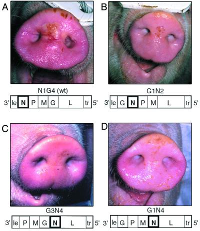
Snouts of pigs inoculated 3 days previously with engineered VSVs having the gene orders rearranged as shown below each panel (“le” and “tr” indicate the 3′ leader and 5′ trailer sequences respectively). Swine were anesthetized, and 2.5 × 10⁷ PFU of the indicated virus was applied as described in Materials and Methods. Animals were infected with N1G4 (WT), G1N2, G3N4, G1N4 (panels A to D, respectively).
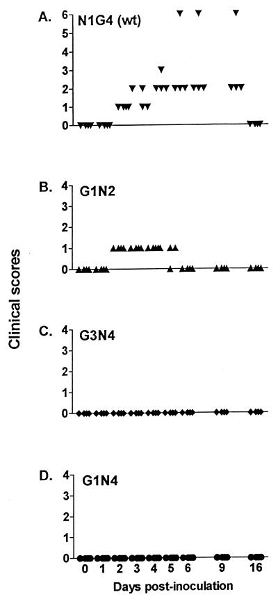
By the second day following inoculation, vesicles of <2 cm in diameter developed at the area of inoculation of all animals that received the wild-type N1G4 virus. The lesions in the N1G4-inoculated animals then increased in size to >2 cm in diameter by days 3 and 4 postinoculation, and multiple lesions developed. An example of these lesions as they appeared on the snout at 3 days postinoculation is shown in Fig. 1A. The clinical scores over time for each pig inoculated with one of the four different viruses are summarized in Fig. 2. The clinical score was determined by the size, the number, and the distribution of resulting lesions as described in Materials and Methods. In three of the four N1G4-inoculated pigs the lesions remained localized to the snout (Fig. 1A and 2A). However, one of the N1G4-inoculated pigs developed a vesicle on its hoof and another one developed a vesicle in the tongue, increasing its clinical score to 6 (Fig. 2A). In general, the size, distribution, and severity of these lesions were indistinguishable from those caused by a field isolate of VSV-IN (L. L. Rodriguez, unpublished results). It is notable that the N1G4 wild-type VSV-IN recovered from our cDNA clone and used in these studies caused clinical disease equivalent to a field isolate of VSV-IN, despite the fact that the recombinant virus had been propagated only a limited number of times in cell culture since recovery and had never been passaged through animals.
FIG. 2.
Clinical scores for each of the four animals per group after inoculation with the rearranged viruses. Swine were inoculated with 2.5 × 10⁷ PFU of virus as described in Materials and Methods, and the formation of lesions was assessed daily. Clinical scores were based on the size and location of the resulting lesions. Each symbol represents an individual animal.
Inoculation with the G1N2 virus resulted in the formation of small vesicles (<2 cm) at the site of inoculation by day 2 in all pigs (Fig. 1B and 2B). However, in contrast to the pathogenesis caused by the wild-type N1G4 virus, the vesicles present on the G1N2 inoculated pigs did not increase in size with time, nor did multiple lesions develop. In addition, the lesions in G1N2-inoculated animals were confined to the site of inoculation. For both N1G4- and G1N2-inoculated animals, lesions started to develop at day 2. Lesions remained visible until day 9 in animals inoculated with the N1G4 wild-type virus, after which healing of the infected tissue became apparent. In contrast, lesions in G1N2-infected animals were completely healed by day 6. Overall, the clinical manifestations resulting from G1N2 infection were less severe than those caused by wild-type virus, indicating that the G1N2 virus was partly attenuated (Fig. 1 and 2, panels A and B).
In contrast to the results observed with wild-type- or G1N2-inoculated animals, no vesicles were observed in animals that received either G3N4 or G1N4 (Fig. 1 and 2, C and D) indicating that these viruses were highly attenuated. No change in daily rectal temperature was observed in any of the animals (data not shown). This is consistent with previous results of intradermal inoculation of field VSV isolates which rarely cause fever (11, 20). Mock-inoculated animals which had received DMEM alone showed no adverse reactions to the inoculation procedure (data not shown).
In summary, the wild-type virus with N in the first position, N1G4, caused disease similar to that seen in VSV Indiana field isolates, whereas G1N2 with N in the second position caused reduced clinical symptoms and viruses with N in the fourth position, G1N4 or G3N4, caused no detectable disease (Fig. 1 and 2). Taken together, these results indicate that translocation of the N gene to successive positions away from the promoter resulted in a stepwise reduction of clinical disease.
Virus isolation following inoculation.
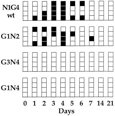
To examine the ability of the viruses to replicate following administration, nasal swabs and OPF, reliable indicators of infection (20). were assayed for virus as described in Materials and Methods. Virus was recovered from day 1 through day 6 postinfection from nasal swabs or OPF samples from all animals given N1G4(WT) (Fig. 3). Virus was recovered also from all animals that received G1N2 starting at day 1 postinfection but, with the exception of a single sample at day 7, G1N2 virus was generally not isolated after day 5. In addition to the nasal and OPF samples, virus was identified from swabs taken from the area of inoculation for up to 6 days in all animals given N1G4. This was in contrast to the group given G1N2 in which no virus was isolated from the snout of one animal, and in the remaining animals virus was not found on the snouts after 3 to 4 days, again suggesting an attenuated phenotype for this virus (data not shown).
FIG. 3.
Virus isolation from swine after inoculation with the rearranged viruses. Swine were inoculated with 2.5 × 10⁷ PFU of virus as described in Materials and Methods. Nasal and probang swabs were taken as indicated. Samples were titrated, and virus was recovered by cultivation on BHK-21 cells at 37°C for 3 days for N1G4(WT) and G1N2 or 31°C for 4 days for G3N4 and G1N4. Each box represents an individual animal, and samples that were positive (■) or negative (□) for viral CPE are shown. The presence of virus and the gene order were confirmed by RT-PCR.
Virus was not recovered from OPF or nasal samples from animals given either G3N4 or G1N4, despite the relative ease with which N1G4 and G1N2 could be detected in the similar samples (Fig. 3). In addition, G3N4 or G1N4 virus was not recovered from swabs of the inoculated areas of the snout (data not shown). These data indicate that these viruses were highly attenuated for replication in swine.
Gene order and intergenic junctions remain stable.
To test whether the viruses that were recovered from N1G4- and G1N2-infected animals were genetically stable after passage in vivo, the viral RNA from a selection of the virus-positive cultures was extracted and, using specific sets of primers, the gene ends and gene junction regions unique to each virus were amplified by RT-PCR. The PCR products were sequenced across the relevant gene junctions. No changes in the gene order or in the sequences of the gene ends or the intergenic junctions were observed (data not shown), indicating that the rearranged gene orders were stable during replication in a natural host.
Humoral immune response.
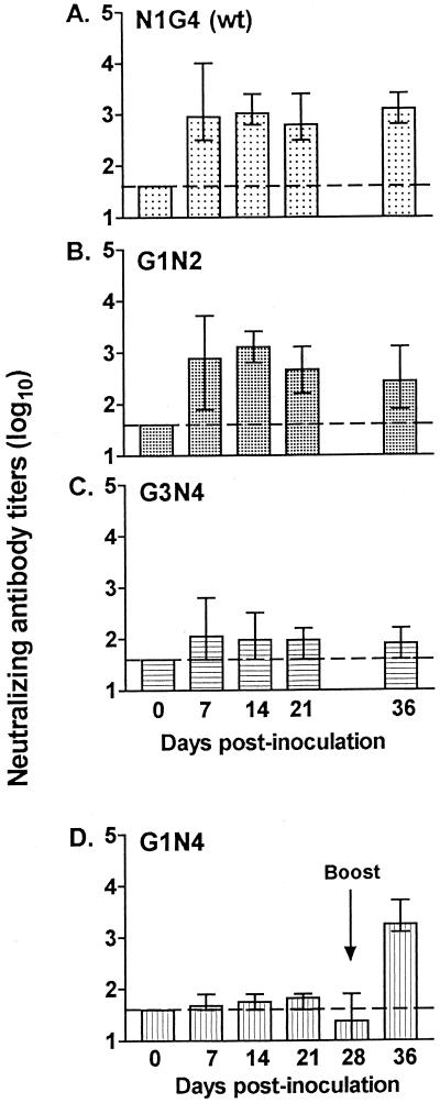
The ability of the wild-type and rearranged viruses to stimulate a humoral immune response was measured by assessing the level of neutralizing antibodies present in serum at various times postinoculation. Neutralizing antibody levels in the serum of N1G4(WT)-inoculated animals ranged from 2.5 to 4.0 log10, with an average titer of 2.9 log10 by 7 days postimmunization (Fig. 4A). These levels of neutralizing antibody were maintained throughout the ensuing 4 weeks of observation. Animals receiving G1N2 had similarly high titers, with an average of 2.9 log10 on day 7, and titers remained high throughout the study period (Fig. 4B). Animals receiving G3N4 showed a rise in neutralizing antibody titer to an average value of 2.0 log10. This value, although 10-fold lower than the titers seen following N1G4 or G1N2 inoculation, showed that G3N4 virus was able to stimulate a humoral response with a single inoculation in the absence of detectable levels of replicating virus in nasal swabs or OPF samples (Fig. 4C).
FIG. 4.
Serum neutralizing antibody titers after inoculation with the rearranged viruses. Swine were inoculated with 2.5 × 10⁷ PFU of virus as described in Materials and Methods. Serum was collected at 7-day intervals, and the level of neutralizing antibody was determined. Animals given G1N4 were boosted with 5.7 × 10⁷ PFU on day 28. Bars show the means and ranges of each group. (The dotted line indicates the limit of the sensitivity of the assay.)
Animals receiving G1N4 virus showed a very low rise in titer that was just detectable above background levels, and these titers did not increase substantially with time (Fig. 4D). As a result of this poor response, the animals in this group were reinoculated on day 28, on the snout, with 5.7 × 10⁷ PFU of the G1N4 virus per pig. No clinical symptoms were detected in these animals. Sera were collected 7 days later, and a sharp rise in titer to 3.3 log10 was observed in all four animals (Fig. 4D). These data showed that, despite our inability to detect replicating virus in these animals, they had been primed such that they had an immune response 7 days following the boost that was equal or greater in magnitude to that seen with the N1G4 wild-type virus 7 days after immunization. The average titers of neutralizing antibody present in each group of immunized animals on the day of challenge (day 36) are shown in Fig. 4. Animals inoculated with N1G4 and G1N4 had average titers of 3.1 and 3.3 log10, respectively. Animals inoculated with G1N2 had titers of 2.4 log10, and those inoculated with G3N4 had titers of 1.9 log10.
Viruses with rearranged gene orders have the ability to protect against wild-type virus challenge.
To assess the vaccine potential of the rearranged viruses, all virus-inoculated animals and the mock-inoculated control group were challenged with 4.2 × 10⁷ PFU of the wild-type N1G4 virus per pig 36 days after the initial administration of the viruses and 7 days after the second inoculation of the G1N4 group. The route of challenge was the same as that used for the original inoculation: via scarification of the epidermis of the snout. Animals were observed for clinical symptoms and scored as described above. Additionally, nasal swabs and OPF samples were taken daily for the first week postchallenge and assayed for the presence of virus.
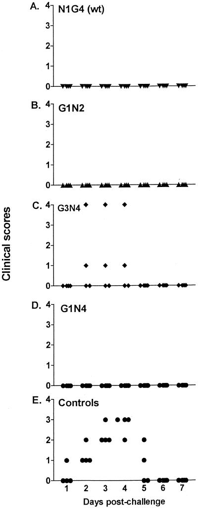
Following challenge, no lesions were observed on any of the animals from groups inoculated with N1G4, G1N2, or G1N4, indicating that these animals were completely protected from clinical disease (Fig. 5A, B, and D). Partial protection was obtained in the animals given G3N4, with lesions visible in two animals beginning on day 2 to day 4 after challenge. The lesions were small (<2 cm) in one case and >2 cm in the second. In both cases the lesions were only present at the site of inoculation and were completely healed by day 5 (Fig. 5C).
FIG. 5.
Clinical scores in swine after challenge with N1G4(WT) virus. Swine were anesthetized and challenged 36 days after the initial administration of viruses with 4.2 × 10⁷ PFU of wild-type virus as described in Materials and Methods. The formation of lesions was monitored daily. Clinical scores were based on the size and location of the resulting lesions as described in Materials and Methods. Each symbol represents an individual animal.
In contrast, all animals in the unvaccinated control group developed lesions following challenge beginning on days 1 and 2 and lasting through day 5 (Fig. 5E). Initially, the lesions were small (<2 cm), but they increased to >2 cm in all cases. In three of four animals, lesions were present at sites other than the site of inoculation; in two cases lesions appeared on the tongue, and in another they appeared on the coronary band of the hoof.
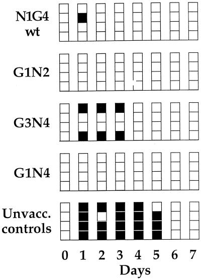
Virus was recovered from nasal swabs or OPF of all of the challenged unvaccinated control animals beginning on day 1 and extending, through day 5, confirming the clinical data (Fig. 6). Virus was recovered from day 1 to 3 from nasal swabs of the two G3N4-inoculated animals that showed lesions on the snout following challenge; no virus was recovered after challenge from the G3N4-inoculated animals which did not have lesions (Fig. 6). To determine whether the virus recovered from the two G3N4-immunized animals after challenge was the N1G4 challenge virus or the immunizing G3N4 genotype, the viral RNA was extracted from the tissue culture samples and the intergenic junctions analyzed by RT-PCR followed by sequence analysis. For both animals, the recovered virus was found to be the N1G4 challenge virus (data not shown).
FIG. 6.
Virus isolation from swine after challenge with N1G4(WT) virus. Following the administration of challenge virus, nasal and probang swabs were taken at the time points indicated. Samples were titrated as described in Materials and Methods. Each box represents an individual animal, and samples that were positive (■) or negative (□) for viral CPE are shown. The presence of virus and the gene order were confirmed by RT-PCR.
Virus was cultured from a nasal swab of one animal without clinical signs from the N1G4-immunized group at a single time point on day 1 after challenge (Fig. 6). It is not possible to know whether this was recovery of the input challenge virus or replication of the challenge virus.
These data taken together show a strong association between protection from clinical symptoms and neutralizing antibody titer. The two animals immunized with G3N4 which exhibited no disease after challenge had higher antibody titers prior to challenge (2.1 and 2.3 log10) than did the two animals that experienced clinical disease (1.6 and 1.9 log10).
Antibody levels following challenge.
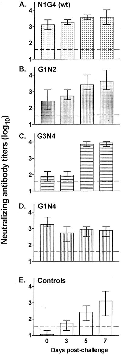
Neutralizing antibody titers were analyzed for all animals on the day of challenge and at days 3, 5, and 7 thereafter. Titers of animals immunized with N1G4, already high at the time of challenge, remained near that level and increased slightly (Fig. 7A). Animals originally immunized with G1N2 experienced an approximate 10-fold rise in titer by 5 to 7 days after challenge to levels near those seen with N1G4 (Fig. 7B). Animals immunized with G3N4, which had the lowest titers at time of challenge (1.9 log10), showed increased titers by nearly 100-fold between days 3 and 5 following challenge to levels that equaled those seen with the wild type (Fig. 7C). The G1N4-immunized animals, which had low titers on day 28 and were given a second immunization 7 days before challenge, maintained the high titer achieved by the boost but did not experience a further rise following challenge (Fig. 7D).
FIG. 7.
Serum neutralizing antibody titers after challenge with N1G4(WT) virus. Serum was collected at the indicated time points following administration of challenge virus as described in the legend to Fig. 5, and the level of neutralizing antibody was determined by an endpoint assay as described in Materials and Methods. Bars show the means and ranges of each group. (The dotted line indicates the limit of the sensitivity of the assay.)
DISCUSSION
We tested whether gene rearrangement as a means to predictably alter gene expression levels could alter the disease potential of VSV in a natural host as we had previously shown it did in a small animal model (9). The data show that moving the nucleocapsid gene of VSV to successively promoter-distal positions, which downregulate its expression, resulted in a systematic and stepwise reduction of clinical disease in a natural host. To our knowledge, this is the first report of an approach that allows the incremental manipulation of clinical disease caused by a negative-strand RNA virus.
Gene rearrangement gives a stepwise reduction in disease.
Remarkably, the VSV-IN recovered from our cDNA clone caused clinical disease in domestic swine equivalent to that observed with infection by a field isolate. This is the first time clinical disease has been induced in a natural host using VSV derived from a cDNA clone. Furthermore, the rearrangement of the genes of this cDNA clone to recover viruses having the N gene moved promoter distally to the second position (G1N2) resulted in reduction of the extent of clinical lesions in infected swine (Fig. 1 and 2). Replication of this virus in pigs also appeared to be slightly attenuated since virus was recovered from snout, nasal, and OPF swabs for a shorter duration of time than with the N1G4 virus (Fig. 3). The G1N2 virus also had the G gene moved closer to the promoter, and previous experiments in mice showed that this resulted in an earlier and increased immune response (9). The immune response in swine infected with G1N2 virus was almost identical to that observed with the wild type. These findings suggest that the attenuation of disease potential of this virus is due to a reduction in N gene expression and reduced replication potential. Movement of the G gene forward may have helped compensate for reduced replication potential to aid in achieving a level of neutralizing antibody similar to that obtained following inoculation with wild-type virus.
Movement of the N gene farther from the promoter, to the fourth position, yielded viruses G3N4 and G1N4, neither of which caused clinical disease in a natural host. They were also reduced in their ability to replicate in pigs since no virus could be detected in snout, nasal, or OPF samples following inoculation. Concomitant with this, the immune responses to a single inoculation of these viruses resulted in lower neutralizing antibody responses than were seen with N1G4 or G1N2 (Fig. 4). However, despite the severely reduced replication potential in swine, these viruses were still capable of inducing an immune response. The G3N4 virus showed a modest increase in antibody levels following administration. Additionally, upon challenge with the wild-type virus on day 36, G3N4 animals responded rapidly 5 days later with a substantial 100-fold increase in antibody titer.
Similarly, the G1N4 virus induced low antibody titers following the initial inoculation; however, when a second, booster inoculation of G1N4 was administered on day 28, there was a rapid response that was equal to or higher than that seen with the wild-type virus at 7 days following its initial administration (Fig. 4). In previous studies in mice, the G1N4 and G3N4 viruses induced an earlier and higher antibody response than the G1N2 or N1G4 viruses (9). In swine this was not the case, and we attribute these differences to differences in the route of inoculation and replication capacity of the viruses in the natural host versus the mouse model. These results emphasize the importance of studies of virus-host interactions in the natural host wherever possible.
Protection against challenge with wild-type virus.
Following challenge, no clinical disease was observed in any animal that had received a single inoculation with N1G4 or G1N2 or had received two inoculations with G1N4. The neutralizing antibody titers present in these three groups were all high on the day of challenge (Fig. 4) and were equal to or greater than the neutralizing antibody titers reported following inoculation of domestic swine with field isolates of VSV via a variety of routes (20; L. Rodriguez, unpublished results). Two of the four G3N4-immunized animals were protected against challenge. These animals proved to be the ones that showed the highest neutralizing antibody titers prior to challenge. These data show a strong association between neutralizing antibody titer and protection from clinical disease. Similar findings were reported using a recombinant vaccinia virus expressing VSV-G to immunize cattle (15).
Further, these data show that despite its reduced disease potential, the G1N2 virus could induce as strong a protective immune response as the wild-type virus with only a single inoculation. The viruses having N in the fourth position which were completely attenuated in the natural host showed a lower immune response following a single inoculation. However, a single further inoculation with G1N4 induced an immune response that was equal to that induced by the wild-type virus. Thus, these data show that it is possible to reduce disease potential by gene rearrangement and still retain a virus that can replicate sufficiently to induce a protective immune response in the absence of clinical disease.
Since vesicular stomatitis is a disease of mandatory report, any live-attenuated vaccine must not only be safe and efficacious but also distinguishable from field strains, in order to gain approval for its use in domestic animals. We showed that viruses with rearranged genomes can readily be distinguished from field strains by RT-PCR of intergenic regions. Furthermore, by using a cDNA derived virus, it is possible to introduce specific changes into the genome such as deleting specific antigenic determinants that could be used for serological testing in order to distinguish vaccinated from naturally infected animals.
Advantages of gene rearrangement for vaccine development.
Since the Mononegavirales have not been observed to undergo homologous recombination (18), gene rearrangement is predicted to be irreversible. Our work to date with these viruses and others having rearranged gene orders has provided no reason to doubt this expectation. For several reasons, gene rearrangement may provide a rational, alternative method for developing stably attenuated live vaccines against the nonsegmented negative-strand RNA viruses. Live-attenuated viruses have proven effective vaccines against many diseases both in humans and animals, for example, smallpox, yellow fever, canine rabies, measles, mumps, and poliomyelitis. However, the strategies for attenuation have been largely empirical. RNA virus genomes are notable for their high mutation rate, which is attributable to their polymerase error rate and lack of proofreading and error correction mechanisms. Most RNA viruses exist as complex quasispecies populations (8, 10), which has significant implications for their biology. The most obvious is that the viral population constitutes a huge reservoir of variants with potentially useful phenotypes in the face of environmental change. Advances in understanding the complex relationships that exist between the error rate of a viral polymerase, the viral population size, its passage history, and changes in the fitness landscape have revealed that empirical methods of attenuation may be intrinsically less reliable than previously thought (17; reference 10 and references therein). Rearrangement of genes essential for replication utilizes the basic principle for control of gene expression, gene position, to effect changes in expression of genes requisite for replication and thus capitalizes on the lack of a natural means to reverse the rearrangement, i.e., the lack of homologous recombination. This means of attenuation avoids the inherent potential of viruses attenuated by a limited number of single base mutations to revert to virulence through polymerase error and subsequent selection. The gene orders of the rearranged viruses used in these studies were stable following their introduction into and replication in one of their natural hosts, swine.
Studies to evaluate the long-term evolution and competitive fitness of these viruses are under way. Selection for compensatory mutations is to be expected; however, mutations that might compensate for the highly altered expression of genes as dictated by gene rearrangement are difficult to imagine, since any mutation affecting the expression level of one gene will also affect the expression of all downstream genes and thus have limited ability to restore virulence.
ACKNOWLEDGMENTS
We are grateful to our colleagues for constructive review, to the animal handlers at the Plum Island Animal Disease Center for their assistance, and to Thomas Bunch for technical help.
This work was supported by Public Health Service grants R37 AI 12464 to G.W.W. and AI 18270 to L.A.B. from the NIAID.
REFERENCES
- 1.Abraham G, Banerjee A K. Sequential transcription of the genes of vesicular stomatitis virus. Proc Natl Acad Sci USA. 1976;73:1504–1508. doi: 10.1073/pnas.73.5.1504. [DOI] [PMC free article] [PubMed] [Google Scholar]
- 2.Artois M, Cliquet F, Barrat J, Schumacher C L. Effectiveness of SAG1 oral vaccine for the long-term protection of red foxes (Vulpes vulpes) against rabies. Vet Rec. 1997;140:57–59. doi: 10.1136/vr.140.3.57. [DOI] [PubMed] [Google Scholar]
- 3.Ball L A, Pringle C R, Flanagan B, Perepelitsa V P, Wertz G W. Phenotypic consequences of rearranging the P, M, and G genes of vesicular stomatitis virus. J Virol. 1999;73:4705–4712. doi: 10.1128/jvi.73.6.4705-4712.1999. [DOI] [PMC free article] [PubMed] [Google Scholar]
- 4.Ball L A, White C N. Order of transcription of genes of vesicular stomatitis virus. Proc Natl Acad Sci USA. 1976;73:442–446. doi: 10.1073/pnas.73.2.442. [DOI] [PMC free article] [PubMed] [Google Scholar]
- 5.Bridges V E, McCluskey B J, Salman M D, Hurd H S, Dick J. Review of the 1995 vesicular stomatitis outbreak in the western United States. J Am Vet Med Assoc. 1997;211:556–560. [PubMed] [Google Scholar]
- 6.Calvarin R, Gayot G. L'epreuve de la curette pharyngienne (Probang-test). Dix annees au service des exportations francaises et europeennes. Rec Med Vet. 1978;154:649–655. [Google Scholar]
- 7.Cantlon J D, Gordy P W, Bowen R A. Immune responses in mice, cattle and horses to a DNA vaccine for vesicular stomatitis. Vaccine. 2000;18:2368–2374. doi: 10.1016/s0264-410x(00)00007-4. [DOI] [PubMed] [Google Scholar]
- 8.Domingo E, Escarmis C, Sevilla N, Moya A, Elena S, Quer J, Novella I, Holland J. Basic concepts in RNA virus evolution. FASEB J. 1996;10:859–864. doi: 10.1096/fasebj.10.8.8666162. [DOI] [PubMed] [Google Scholar]
- 9.Flanagan E B, Ball L A, Wertz G W. Moving the glycoprotein gene of vesicular stomatitis virus to promoter-proximal positions accelerates and enhances the protective immune response. J Virol. 2000;74:7895–7902. doi: 10.1128/jvi.74.17.7895-7902.2000. [DOI] [PMC free article] [PubMed] [Google Scholar]
- 10.Holland J J, Torre J, Steinhauer D. RNA virus populations as a quasispecies. Curr Top Microbiol Immunol. 1992;176:1–20. doi: 10.1007/978-3-642-77011-1_1. [DOI] [PubMed] [Google Scholar]
- 11.Howerth E W, Stallknecht D E, Dorminy M, Pisell T, Clarke G R. Experimental vesicular stomatitis in swine: effects of route of inoculation and steroid treatment. J Vet Diagn Investig. 1997;9:136–142. doi: 10.1177/104063879700900205. [DOI] [PubMed] [Google Scholar]
- 12.Iverson L E, Rose J K. Localized attenuation and discontinuous synthesis during vesicular stomatitis virus transcription. Cell. 1981;23:477–484. doi: 10.1016/0092-8674(81)90143-4. [DOI] [PubMed] [Google Scholar]
- 13.Lauerman L H, Hanson R P. Field trial of live virus vaccination procedure for the prevention of vesicular stomatitis in dairy cattle. III. Evaluation of emergency vaccination in Georgia. US Livestock Sanitary Assoc Proc. 1963;67:473–482. [Google Scholar]
- 14.Letchworth G J, Rodriguez L L, Barrera J D C. Vesicular stomatitis. Vet J. 1999;157:239–260. doi: 10.1053/tvjl.1998.0303. [DOI] [PubMed] [Google Scholar]
- 15.Mackett M, Yilma T, Rose J K, Moss B. Vaccinia virus recombinants: expression of VSV genes and protective immunization of mice and cattle. Science. 1985;227:433–435. doi: 10.1126/science.2981435. [DOI] [PubMed] [Google Scholar]
- 16.McCluskey B J, Hurd H S, Mumford E L. Review of the 1997 outbreak of vesicular stomatitis in the western United States. J Am Vet Med Assoc. 1999;215:1259–1262. [PubMed] [Google Scholar]
- 17.Novella I, Clarke D, Quer J, Duarte E, Lee C, Weaver S, Elena S, Moya A, Domingo E, Holland J. Extreme fitness differences in mammalian and insect hosts after continuous replication of vesicular stomatitis virus in sandfly cells. J Virol. 1995;69:6805–6809. doi: 10.1128/jvi.69.11.6805-6809.1995. [DOI] [PMC free article] [PubMed] [Google Scholar]
- 18.Pringle C R. Enhanced mutability associated with a temperature-sensitive mutant of vesicular stomatitis virus. J Virol. 1981;39:377–389. doi: 10.1128/jvi.39.2.377-389.1981. [DOI] [PMC free article] [PubMed] [Google Scholar]
- 19.Pringle C R, Easton A J. Monopartite negative strand RNA genomes. Semin Virol. 1997;8:49–57. [Google Scholar]
- 20.Stallknecht D E, Howerth E W, Reeves C L, Seal B S. Potential for contact and mechanical vector transmission of vesicular stomatitis virus New Jersey in pigs. Am J Vet Res. 1999;60:43–48. [PubMed] [Google Scholar]
- 21.Wertz G W, Perepelitsa V P, Ball L A. Gene rearrangement attenuates expression and lethality of a nonsegmented negative strand RNA virus. Proc Natl Acad Sci USA. 1998;95:3501–3506. doi: 10.1073/pnas.95.7.3501. [DOI] [PMC free article] [PubMed] [Google Scholar]
- 22.Whelan S P J, Ball L A, Barr J N, Wertz G W. Efficient recovery of infectious vesicular stomatitis virus entirely from cDNA clones. Proc Natl Acad Sci USA. 1995;92:8388–8392. doi: 10.1073/pnas.92.18.8388. [DOI] [PMC free article] [PubMed] [Google Scholar]
- 23.Yilma T, Breeze R G, Ristow S, Gorham J R, Leib S R. Immune responses of cattle and mice to the G glycoprotein of vesicular stomatitis virus. Adv Exp Med Biol. 1985;185:101–115. doi: 10.1007/978-1-4684-7974-4_6. [DOI] [PubMed] [Google Scholar]


