Abstract
Passage of Ross River virus strain NB5092 in avian cells has been previously shown to select for virus variants that have enhanced replication in these cells. Sequencing of these variants identified two independent sites that might be responsible for the phenotype. We now demonstrate, using a molecular cDNA clone of the wild-type T48 strain, that an amino acid substitution at residue 218 in the E2 glycoprotein can account for the phenotype. Substitutions that replaced the wild-type asparagine with basic residues had enhanced replication in avian cells while acidic or neutral residues had little or no observable effect. Ross River virus mutants that had increased replication in avian cells also grew better in BHK cells than the wild-type virus, whereas the remaining mutants were unaffected in growth. Replication in both BHK and avian cells of Ross River virus mutants N218K and N218R was inhibited by the presence of heparin or by the pretreatment of the cells with heparinase. Binding of the mutants, but not of the wild type, to a heparin-Sepharose column produced binding comparable to that of Sindbis virus, which has previously been shown to bind heparin. Replication of these mutants was also adversely affected when they were grown in a CHO cell line that was deficient in heparan sulfate production. These results demonstrate that amino acid 218 of the E2 glycoprotein can be modified to create an heparan sulfate binding site and this modification expands the host range of Ross River virus in cultured cells to cells of avian origin.
Alphaviruses are blood-borne pathogens transmitted by arthropod vectors. The major vertebrate reservoir for many of these viruses is birds, but many alphaviruses utilize small mammals and some use only mammals as vertebrate hosts. The host range of these viruses is extremely broad, and they infect many types of animals and most cells in culture (27). Even within an infected animal, the virus replicates in many tissues of the host (10). This extended host and tissue specificity is governed primarily by the ability of the viruses to attach to and enter target cells. It has been suggested that alphaviruses utilize multiple receptors to gain access to a wide variety of hosts or, alternatively, that ubiquitous proteins among diverse species serve as cellular receptors for these viruses (26).
Ross River virus (RRV) is the causative agent of human epidemic polyarthritis in Australia (7). The prototype T48 strain of RRV, originally isolated in northern Queensland, Australia, is responsible for yearly outbreaks of disease in Australia, and its recent spread to Fiji, American Samoa, and the Cook Islands has had significant consequences for its human hosts (1, 19). Unlike many of its alphavirus relatives, there is no evidence for the association of the virus with birds, and it appears to utilize small mammals, particularly marsupials, as a reservoir (19). In cell culture, RRV replicates poorly in cells of avian origin, and thus there may be a restriction to the growth of the virus in birds (13).
The replication efficiencies of two genetically distinct strains of RRV, the prototype strain T48 and an attenuated mouse strain, NB5092, were examined in chicken cells in a study by Kerr et al. (13). Neither virus grew in chicken embryo fibroblasts (CEF) at 37°C, but both could be propagated at 30°C. NB5092 reached titers that were 100-fold higher than that of the T48 strain. Repeated passage of NB5092 in chicken cells resulted in decreasing virus titers until the fifth passage, at which point the titer increased. Three independently derived adapted variants were selected for further analysis. Sequencing of the subgenomic RNA, which contains the coding region for the alphavirus structural proteins, revealed that all three variants had a single amino acid change in the E2 glycoprotein. Two variants had a lysine for asparagine at position 218, and the third variant had a lysine for glutamate at position 4. When virulence was examined in 1-day-old mice, the N218K viruses were found to be attenuated and to produce less virus, whereas the E4K virus showed little alteration in replication efficiency. In addition, the N218K viruses were resistant to neutralization by monoclonal antibody T10C9, whereas the E4K virus was resistant to neutralization by monoclonal antibody NB3C4. Taken together, these results suggested that these residues may be involved in receptor binding or entry of RRV into the cell. However, the possibility that changes in the nonstructural proteins contributed to, or were responsible for, the phenotype could not be ruled out.
Structural studies using cryoelectron microscopy and image reconstruction techniques to map features of the virus spike place E2 residues 4 and 218 close to surface-exposed regions of the virus. In a study by Paredes et al. (23), a Sindbis virus (SINV) mutant that was unable to cleave the E2 precursor, PE2, was examined. Since these mutant virus particles retained the E3 peptide, which is N-terminal to E2 in the PE2 precursor, a difference map comparing the wild type and the mutant viruses revealed the position of E3. This placed the E3 peptide and, by inference, the N terminus of E2 in a distal portion of the glycoprotein spike. In a separate study, Smith and colleagues (25) also used cryoelectron microscopy to map the binding site of a neutralizing monoclonal antibody on the surface of RRV. The antibody used was T10C9, which bound in the vicinity of E2 residue 216, based on sequencing of an escape variant (29). This antibody bound to an outer portion of the trimeric spike that did not overlap the E3 site and suggested that residue 218 was also on the exposed exterior surface of the spike.
We have used a full-length cDNA clone of RRV RNA to generate a panel of mutants with substitutions at residue 218 of glycoprotein E2. Mutants with basic amino acid substitutions at position 218 replicated better in chicken cells than wild-type RRV or mutants with other changes at position 218. Two mutants, N218R and N218K, infected 40 and 15% of cultured chicken cells, respectively, whereas the parental RRV infected less than 2%. Further characterization showed that mutants that had arginine or lysine at position 218 interacted with the glycosaminoglycan (GAG) heparan sulfate (HS). This demonstrates that changes in this region of the E2 glycoprotein expand the host range of RRV in cell culture by allowing virus to interact with cell surface HS moieties.
MATERIALS AND METHODS
Viruses and cells.
Baby hamster kidney (BHK) and Vero cells were maintained in minimum essential media (MEM; Life Technologies, Rockville, Md.) supplemented with 7.5% fetal bovine serum (FBS; Life Technologies), 1× PSN (0.05 mg of penicillin per ml, 0.05 mg of streptomycin per ml, and 0.1 mg of neomycin per ml), 2 mM l-glutamine, and 0.1 mM nonessential amino acids (Life Technologies). CEF were prepared from 10-day-old White Leghorn embryos, and secondary CEF were used in all experiments. The Chinese hamster ovary (CHO-K1) cells (ATCC CCL-61) and the derivative cell line CHO18.4m were generously provided by Andrew Byrnes and Diane Griffin (12). Both CHO cell lines were maintained in Ham's F-12 medium supplemented with 10% FBS and 1× PSN (Life Technologies).
A laboratory-adapted strain of SINV, Toto 1101, and the wild-type T48 strain of RRV, RR64, were generated from in vitro-transcribed RNA as previously described (17, 24). Mutant viruses, derived from pRR64, were isolated in an identical manner and were plaque purified twice in BHK cells. The resulting virus stocks were sequenced to confirm the inserted mutation and were used in all subsequent experiments.
Site-directed mutagenesis.
Single amino acid substitutions at residue 218 of the RRV E2 glycoprotein were generated by two methods. Substitutions of the wild-type asparagine to threonine, lysine, and arginine were carried out by M13 site-directed mutagenesis as described by Kunkel (18). For all other substitutions at residue 218, a degenerate oligonucleotide was used for DNA amplification by using PCR. The 5′ degenerate primer annealed to nucleotides (nt) 9194 to 9229 [5′CACTACCAGTACTGACAAGACCATC(G/C/T)N(G/T)ACATGCAA3′] of the pRR64 cDNA clone. The 3′ oligonucleotide primer annealed to nt 9640 to 9665 (5′CGGGATC CGGGTTATAGTCCATAATA3′) of the pRR64 cDNA clone. Amplification with Taq DNA polymerase (Promega, Madison, Wis.) produced a 471-bp product that was subsequently digested with the restriction endonucleases ScaI and CspI. The resulting 367-bp fragment was inserted into a similarly digested pRR64 plasmid. Full-length clones were screened by DNA sequencing using the Thermo Sequenase Cycle Sequencing kit (Amersham Pharmacia Biotech, Piscataway, N.J.). Clones containing substitutions were sequenced across the entire amplified region from the ScaI (nt 9201) to the CspI (nt 9568) restriction sites.
Growth analysis.
For one-step growth analyses, 24-well plates containing approximately 105 cells per well were used. Virus, at a multiplicity of infection (MOI) of 5, was adsorbed to cells for 1 h at room temperature. The cells were then overlaid with media and placed at 37°C. Cells were washed twice with fresh media every 0.5 h for the first 2 h of infection, after which media were harvested every 1 h. For cumulative growth analysis, 100-μl aliquots of media were harvested at various times posttransfection. Virus titers were determined by plaque assay in BHK cells.
Virus inhibition assays.
To examine the inhibitory effects of polyanions on the size of plaques formed, between 100 to 200 PFU of virus was adsorbed to cells for 1 h at room temperature. The inoculum was removed, and the cells were rinsed twice with phosphate-buffered saline (PBS) containing 1% FBS and were overlaid with either 1.0% agarose (Life Technologies), 0.8% Bacto agar (Difco Laboratories, Detroit, Mich.), or 0.8% Bacto agar + 150 μg of DEAE-dextran (Sigma, St. Louis, Mo.) in 1× MEM containing 1% FBS.
To examine the effects of heparin on plaque formation, 100 to 200 PFU of virus was incubated with heparin (sodium salt derived from porcine intestinal mucosa, catalogue no. H9399; Sigma) in a final volume of 250 μl for 30 min at 4°C. Control viruses were incubated with 0.5 mg of bovine serum albumin (BSA)/ml in place of heparin. Viruses were adsorbed to cells for 1 h at 4°C, at which time the inoculum was removed and the cells were washed twice with PBS containing 1% FBS, followed by the addition of an agarose overlay. Treatment with heparinase I (Sigma) was performed essentially as previously described (4). Briefly, cells were treated with heparinase I at 37°C for 1 h, followed by adsorption of 100 to 200 PFU of virus to cells for 1 h at 4°C, and then treated as described above.
Virus purification.
To prepare high-titer virus stocks, BHK cells were infected with virus at an MOI of 0.1 PFU/cell for 1 h at 37°C. Cells were overlaid with MEM supplemented with 1% FBS, PSN, 2 mM l-glutamine, and 0.1 mM nonessential amino acids. Virus was harvested 16 to 18 h postinfection, when cells were infected with SINV, or 20 h postinfection, when cells were infected with RRV or the RRV mutants. Supernatants were clarified, followed by polyethylene glycol precipitation (final concentration, 10% polyethylene glycol 8000 and 1 M NaCl) for 6 h at 4°C. Precipitated virus was collected by centrifugation at 1,800 × g for 10 min at 4°C. The pellet was resuspended in 2 ml of TNE buffer (0.05 M Tris-HCl [pH 7.2], 0.15 M NaCl, and 0.001 M EDTA) and layered on top of a linear 20 to 60% (wt/vol) sucrose gradient. The gradient was centrifuged for 2 h at 160,000 × g at 4°C. The virus band was collected with a syringe and concentrated by centrifugation through a 30% sucrose cushion for 1.5 h at 190,000 × g. Virus was resuspended in TNE buffer and assayed for infectivity by plaque assay and for concentration by measurement of absorbance at 280 nm. The titer was 1.0 × 10¹⁰ PFU/ml (approximately 2 mg/ml) for SINV and 3.3 × 10⁹ PFU/ml for RRV.
Binding to heparin.
These experiments were performed essentially as described by Byrnes and Griffin (4). A 1-ml heparin-Sepharose column (Amersham Pharmacia Biotech) was equilibrated with buffer A (0.1 M NaCl, 5 mM phosphate [pH 7.5]). Virus (109 PFU; approximately 200 ng) was applied to the column, followed by washing with 10 ml of buffer A. Virus was eluted with a 50% linear gradient of buffer B (1 M NaCl, 5 mM phosphate [pH 7.5]) at a rate of 1 ml/min, followed by washing with 100% buffer B for 10 min. Absorbance at 280 nm and conductivity were measured, and 500-μl fractions were collected. Virus titers were determined by plaque assay of peak fractions on BHK cells, and Western blot analysis was carried out using a polyclonal anti-E2 for RRV and a polyclonal anti-E1 for SINV.
RESULTS
RRV is restricted in entry into chicken cells.
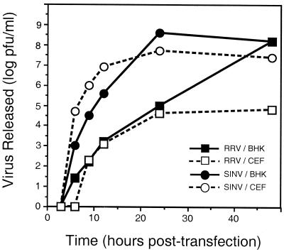
Most cells in culture are susceptible to infection by RRV, but in avian cells, replication is severely reduced (13, 26). Attempts to infect 100% of primary or secondary CEF with RRV by using MOIs of up to 50 failed to produce infection in greater than 5% of the cells, as determined by immunofluorescence microscopy (data not shown). This restriction could be due to a defect in entry or to a postentry event such as RNA synthesis. To discriminate between these two possibilities, we circumvented the attachment and entry steps by transfecting RRV RNA transcribed in vitro from the cDNA clone pRR64 into secondary CEF and compared the amount of virus released against that from a permissive cell line, BHK (Fig. 1). During the initial round of replication, in which approximately 1% of the cells were transfected with RNA, the amounts of RRV released from CEF and BHK cells were comparable (≤24 h posttransfection) (Fig. 1). This similarity in replication kinetics suggested that postentry processes occurred at roughly equivalent rates. However, as secondary infections occurred, the amount of virus released from infected BHK cells continued to increase, whereas the amount of virus released from CEF remained constant. This suggested the restriction in CEF was due to a block in the entry of RRV. In contrast, the Toto 1101 SINV replicated to equivalent levels in both CEF and BHK cells over the course of the experiment (the production of RRV was delayed relative to SINV in these cells) (Fig. 1).
FIG. 1.
Differential growth analysis of RRV and SINV following transfection with RNA. CEF or BHK cells were transfected with 100 ng of either RRV (pRR64) or SINV (pToto1101) RNA that had been transcribed in vitro. At various times posttransfection, 100 μl of supernatant was harvested and virus titers were determined by plaque assay in BHK cell monolayers.
Mutants at E2–218 have altered replication kinetics.
Kerr et al. (13) found that serial passage of the NB5092 strain of RRV in chicken cells selected for variants that had changes in E2, one of which was N218K. Furthermore, a strain of RRV that caused an epidemic of polyarthritis in the South Pacific some years ago in which humans were the primary, or only, vertebrate hosts had the change E2-T219A (3). Thus, it is possible that residue 218 (and 219) might be involved in interaction of the virus with the surface of susceptible cells. To examine whether E2–218 was responsible for the increased growth in chicken cells, we generated a panel of RRV mutants that had amino acid substitutions at this residue (Table 1).
TABLE 1.
RRV and RRV mutants with substitutions at amino acid 218 of E2
| Virus | Codon | Plaque size (mm)a |
|---|---|---|
| RRV | ACC | 2.0 |
| N218R | AGG | 3.5 |
| N218K | ACA | 3.0 |
| N218Y | UAU | 2.0 |
| N218E | GAG | 2.0 |
| N218G | GGG | 2.0 |
| N218Q | CAG | 2.0 |
| N218T | ACC | 2.0 |
| N218V | GUG | 2.0 |
As determined in BHK cells at 48 h postinfection with an agarose overlay.
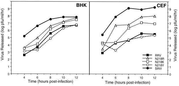
Wild-type RRV and the N218 mutants were assayed for growth in BHK cells by one-step growth analyses (Fig. 2A and data not shown). BHK cells infected with N218K and N218R released 5- and 10-fold more virus at 12 h postinfection, respectively, than the wild type. All other RRV mutants had growth kinetics similar to that of the parental virus (Fig. 2A and data not shown). This group of N218 mutants (N218Y, N218E, N218G, N218Q, N218T, and N218V) behaved similarly to the wild-type virus in this and the following assays, and for the sake of clarity, only the N218Y data are shown.
FIG. 2.
One-step growth analyses in BHK cells and CEF. BHK cells or CEF were infected with the indicated virus at an MOI of 5. Media were replaced every 30 min for the first 2 h and then every hour until 12 h postinfection. Supernatants were collected at the indicated times, and released virus was assayed by titration in BHK cell monolayers at 37°C. The results represent data obtained from two independent experiments. For ease of viewing, results from only a selected set of mutants are shown. The results with N218Y are representative of the data obtained with N218E, N218G, N218Q, N218T, and N218V.
Similar one-step growth analyses were carried out with secondary CEF (Fig. 2B and data not shown). The N218K and N218R mutants produced approximately 32- and 320-fold more virus, respectively, than did wild-type RRV at 12 h postinfection. Again, the wild-type virus and the other mutants tested had similar rates of virus released (Fig. 2B and data not shown). While N218R and N218K produced more virus than wild-type RRV in CEF, they were still unable to infect 100% of the cells. N218R and N218K infected approximately 40% and 10 to 20% of CEF, respectively, as determined by immunofluorescence microscopy and were unable to form plaques on CEF monolayers (data not shown). In contrast, wild-type RRV infected less than 2% of CEF under similar infection conditions (26). Consistent with these observations, the adapted mutants as well as the RRV parent produced much less virus than did SINV, included as a control, which infects 100% of CEF.
The effect of polyanions upon plaque size.
Unpassaged isolates of SINV bind poorly to cultured cells and replicate to low levels in comparison to cell culture-adapted strains of the virus (4, 15). It was shown that passaged strains of SINV had acquired the ability to attach to cell surface HS moieties, whereas the unpassaged strain could not. Analysis of the T48 strain of RRV indicated that it did not attach to HS moieties or several other GAGs that were tested (4). However, it was possible that the enhanced replication observed in the RRV N218R and N218K mutants is the result of interaction with a GAG such as HS.
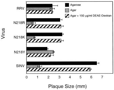
It had been previously shown that polyanions present in agar affect the plaque size of the SINV Toto 1101 strain (4, 5). Presumably, the positively charged polyanions inhibit the growth of virus in agar by acting as a competitor in the interaction of the virus with the cell surface. This results in a slower-growing plaque in agar than in agarose, which has a higher purity and lower GAG concentrations. This assay has previously been used as a preliminary screen to determine possible viral interactions with GAGs (5). Wild-type RRV, isolated from the pRR64 full-length cDNA, was examined by this assay, and the plaque size was not affected by the composition of the overlay medium, in agreement with previous observations (Fig. 3). However, the E2 mutants N218R and N218K formed smaller plaques in the presence of agar than in the presence of agarose (Fig. 3). The inhibitory effect seen with the agar overlay could be reversed by the addition of positively charged DEAE-dextran into the overlay medium. Mutants containing substitutions other than arginine or lysine at E2 residue 218 were not affected by the type of overlay present (Fig. 3 and data not shown). These results suggested that the N218R and N218K mutants interact with cell surface GAGs.
FIG. 3.
The effect of polyanions on virus plaque size. BHK cells were infected with virus (100 to 200 PFU) for 1 h at room temperature. The inoculum was removed, and cells were rinsed twice with PBS containing 1% FBS. Cells were overlaid with MEM supplemented with 1% FBS and agarose (solid bars), agar (gray bars), or agar containing 150 μg of DEAE-dextran/ml (striped bars). Infection was allowed to proceed for 48 h, at which time the cells were stained with 5% neutral red in PBS for 6 h. At 55 h postinfection, the stain was removed and the size of the plaques was determined. The data are the averages of 20 independent plaque measurements, and error analysis was carried out using the Student t test.
Heparin interferes with the binding of RRV mutants to cultured cells.
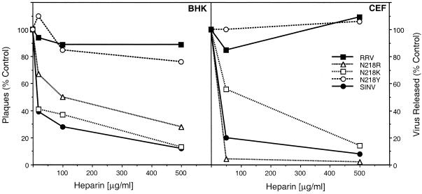
To examine the possibility that the N218R and N218K viruses interacted with GAGs, we tested the ability of heparin, a highly sulfated form of HS, to interfere with the binding of RRV and the RRV mutants to cultured cells. The first approach used plaque assays in BHK cells in the presence or absence of heparin. A virus-heparin mixture was inoculated onto the BHK monolayers to test whether heparin competed with the putative cell surface HS for interaction with virus. In these experiments, as well as in the previous experiments carried out with SINV, a vast molar excess of heparin (5.2 × 10⁸ to 2.6 × 10¹⁰ molecules of heparin/E2 molecule) was used (4, 15). As shown in Fig. 4A, the presence of heparin inhibited plaque formation by N218R, N218K, and SINV in a dose-dependent manner but had little effect on wild-type RRV or other E2–218 mutants.
FIG. 4.
The effect of heparin on infection by viruses. Various concentrations of heparin were added to solutions of viruses and incubated for 30 min at 4°C. Media were removed from monolayers of cells and replaced with the virus-heparin inoculum. Following adsorption of virus for 1 h at 4°C, the inoculum was removed and an agarose overlay (BHK cells) or MEM supplemented with 2% FBS (CEF) was added. In the case of BHK cells, plaques were counted at 50 h postinfection. For CEF, the infection was allowed to proceed for 18 h, at which time the supernatants were collected and virus was assayed by plaque titration of BHK cells. Results are presented as a percentage of the control, where the control is the same virus in the absence of heparin but containing 0.5 mg of BSA/ml.
Heparin and other free GAGs are known to interact with cell surface proteins such as fibroblast growth factor receptor and could possibly interfere with virus attachment by interacting with the cellular receptor (21, 22, 32). However, it seems unlikely that heparin nonspecifically blocked the attachment of virus to the cell surface or inhibited virus replication, as the inhibitory effect appeared to be specific for the E2–218 mutants that had basic amino acid substitutions. The amount of virus released from heparin-incubated BHK cells that were infected with wild-type RRV or the other E2–218 mutants was similar to the amount of virus released from the BSA-incubated BHK control (Fig. 4A).
A similar experiment, examining the ability of heparin to inhibit virus replication, was performed with CEF cells (Fig. 4B). A virus-heparin mixture was inoculated onto CEF monolayers for 1 h at 4°C. Since RRV is unable to form plaques in CEF, the inhibitory effect of heparin was measured by assaying the release of virus into a liquid overlay following a 12-h infection. As with BHK cells, incubation with heparin reduced the yield of SINV, N218R, and N218K but had little or no effect on the yield of wild-type RRV or the other RRV E2–218 mutants. The reduction in virus released for N218R and N218K suggests an interaction between these viruses and heparin, as has been previously established for SINV.
Binding of RRV E2–218 mutants to heparin.
To examine the relative affinities of the RRV E2–218 mutants for heparin, 200 ng of virus was bound to heparin-immobilized Sepharose beads (4, 15). Once bound, virus was eluted with a linear NaCl gradient. Fractions were assayed for the presence of virus by plaque titration in BHK cells and by Western blot analysis using a polyclonal anti-E2 antibody for RRV or a polyclonal anti-E1 antibody for SINV. The N218R and N218K RRV mutants eluted at 343 and 388 mM NaCl, respectively, compared to 305 mM for SINV (Table 2). Thus, the N218R and N218K mutants have an affinity for heparin similar to that of SINV. This contrasts with the wild-type RRV, which eluted at 134 mM NaCl. Interestingly, the profile of the wild-type RRV produced a single elution peak, unlike the two peaks of virus seen by Byrnes and Griffin (4). In our experiments, we used virus derived from a full-length cDNA clone of RRV RNA, pRR64, whereas Byrnes and Griffin used an uncloned stock of the T48 strain (17). They concluded that their preparation of virus contained two genetically distinct populations that differed in their affinity for heparin, eluting from the column at 118 and 189 mM NaCl, respectively. Our preparation of RRV eluted at an intermediate concentration. It is clear that the RRV mutants E2-N218R and E2-N218K have a much higher affinity for heparin than does the parental RRV or any of the other E2–218 mutants.
TABLE 2.
Properties of RRV and RRV E2-218 mutants
| Virus | Heparin- Sepharose elution (mM NaCl)a | Plaque diam (mm)b
|
Titer log (PFU/ml)b
|
||
|---|---|---|---|---|---|
| CHO | CHO18.4m | CHO | CHO18.4m | ||
| RRV | 134 | 1.8 ± 0.1 | 1.5 ± 0.1 | 7.7 ± 0.3 | 7.6 ± 0.1 |
| N218R | 343 | 2.0 ± 0.1 | 0.3 ± 0.1 | 7.6 ± 0.3 | 6.5 ± 0.5 |
| N218K | 388 | 2.0 ± 0.1 | 0.3 ± 0.1 | 7.8 ± 0.3 | 5.5 ± 0.2 |
| N218Y | ND | 1.8 ± 0.1 | 1.5 ± 0.2 | 7.8 ± 0.1 | 6.3 ± 0.1 |
| SINV | 305 | 3.0 ± 0.2 | 0.3 ± 0.2 | 8.3 ± 0.1 | 6.9 ± 0.2 |
Salt concentration of peak virus elution. ND, not determined.
Measured 55 h postinfection. The experiment was performed twice in duplicate.
Attachment to cell surface GAGs.
A CHO cell line, CHO18.4m, that has a reduced susceptibility to infection by SINV was generated by the retrovirus-directed insertion of the neomycin resistance gene into a transcriptionally active gene (12). This cell line has been shown to be deficient in binding laboratory-adapted strains of SINV due to the loss of expression of HS at the cell surface. These cells were used to examine the replication of N218R and N218K as well as wild-type RRV and the Toto1101 SINV. Plaque assays carried out in parallel with the parental CHO cells and the CHO18.4m cells demonstrated that the plaque size was smaller in the CHO18.4m cells than in the parental cells in the case of N218R, N218K, and SINV (Table 2). Furthermore, the yield of virus from infected CHO18.4m cells was also less than that from the parental cells for these viruses (Table 2). In contrast, the wild-type RRV showed little difference in either plaque size or virus release in the two cell lines. The N218Y virus was also relatively insensitive to the cell line. These data strongly suggest that the N218K and N218R mutants utilize HS for entry.
Heparinase I reduces plaque formation of RRV E2-218 mutants in BHK cells.
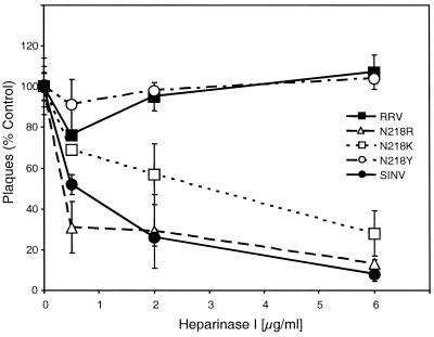
Heparinase I degrades the α1–4 glycosidic bond of HS. BHK cells were pretreated with various amounts of heparinase I and used in plaque assays with a constant amount of virus (Fig. 5 and data not shown). The number of N218R, N218K, and SINV plaques formed in cells treated with heparinase I was reduced, whereas plaques produced by RRV and the other RRV 218 mutants were not affected by such treatment of cells. This inhibition is unlikely to be the result of nonspecific proteolysis because it is specific for the N218R and N218K mutants and because incubation of CHO18.4m with heparinase I did not reduce the number of plaques formed on these cells (4). These data demonstrate that the N218K and N218R mutants utilize the cell surface GAG, HS, for entry into cells.
FIG. 5.
Treatment of BHK cells with heparinase I. BHK cells were treated with heparinase I at 37°C for 1 h. The treated cells were overlaid with inoculum containing 100 to 200 PFU of virus (as assayed on untreated BHK cells), and virus was allowed to adsorb for 1 h at 4°C. An agarose overlay containing MEM supplemented with 2% FBS was added, and plaques were counted at 50 h postinfection. Data are presented as a percentage of control virus without heparinase I treatment of cells. This experiment was carried out twice.
DISCUSSION
The region around amino acid 216 of RRV has been previously implicated as a site of interaction with the host. Molecular genetic studies described here demonstrate that a single amino acid change in this region of the protein adapts the virus to cell surface HS moieties. This adaptation for HS attachment appears to require a positively charged amino acid, as arginine and lysine confer binding, whereas acidic or neutral residues have little or no effect. Mutations at the neighboring E2 threonine-216 have a similar affect on HS binding, suggesting that HS binding occurs in this region of the glycoprotein (data not shown). In addition, cryoelectron microscopy and image reconstruction studies of the E2 N218R virus complexed with heparin have shown that the binding site for heparin overlaps a significant portion of the footprint of monoclonal antibody T10C9 (W. Zhang, M. L. Heil, T. S. Baker, and R. J. Kuhn, unpublished data).
Although substitution of a basic amino acid at either the 216 or 218 residue confers HS binding, the data do not establish whether this binding site is conformational or linear. Studies of HS binding have defined the sequences XBXBBBX or XBBXBX (where X is any residue and B is a basic residue) as linear HS-binding motifs that allow proteins to attach to HS. This motif is not present in the E2–216 to -218 region even after the basic amino acid substitutions. An HS-binding motif is present in E2 at residue 249 but does not appear to confer HS binding in the wild-type virus. In studies of SINV, one region that binds HS has been found to overlap a predicted HS interaction domain (E2 arginine-1), but other binding sites do not, suggesting that they may be conformational (14, 15). This is not a unique observation, as foot-and-mouth disease virus type O is known to interact with HS in a conformation-dependent manner. Structural studies revealed that heparin bound near the south wall of the picornavirus canyon, in contact with the three capsid proteins VP1, VP2, and VP3. The most critical contact, as shown by the crystal structure of the complex, is an ionic interaction between arginine-56 of VP3 and the 2-N-sulfate of GlcN-2 from heparin (8).
Numerous viruses have been shown to utilize HS for infection of cells, including the alphaherpesviruses (31), the betaherpesviruses (20), foot-and-mouth disease virus type O strains (8), papillomavirus (9), respiratory syncytial virus (16), dengue virus (6), and SINV (4, 15). It has been proposed that at least in some cases, HS does not directly serve as a receptor. Instead, virus is concentrated on the two-dimensional cell surface upon attachment to HS, which enhances the probability of virus-receptor interaction. For some viruses, the affinity for HS appears to be a consequence of passage in cell culture. It was shown that newly isolated, unpassaged strains of SINV do not bind to heparin and attach poorly to cells in culture relative to laboratory-adapted strains of the virus (4, 15). After as few as three passages in cell culture, these strains can acquire changes that allow them to attach to HS. We show here that the RRV T48 strain does not bind to heparin but can be adapted to bind by passage in CEF or by site-directed mutagenesis of specific amino acid residues in the E2 glycoprotein.
The N218R and N218K mutants replicated to 32- and 320-fold-higher titers, respectively, than wild-type RRV in CEF but did not approach the levels of SINV replication. The binding of N218R and N218K to immobilized heparin, however, was similar to that of SINV. This suggests that the N218R and N218K viruses have an affinity for cell surface HS similar to that of SINV, but this similar affinity does not lead to an equivalent level of entry for the viruses, since fewer than half the cells are infected by the RRV mutants. Since primary and secondary CEF are heterogeneous in their expression of certain genes, it is possible that only some of the cells express HS. However, repeated passage of the CEF up to 10 times did not alter the infection pattern of RRV or the mutants (data not shown). Thus, by itself, binding to HS is probably insufficient to account for virus entry into cells, and the functional receptor is likely to be a discrete molecular entity.
Based on the results with SINV, the amino acid changes that allow the virus to adapt to HS can arise in different regions of the primary amino acid sequence of the E2 glycoprotein. It is likely that multiple sites in the RRV E2 sequence also confer HS binding to the virus. Passaging of NB5092 through CEF resulted in two genetically distinct viruses (13). One virus had a single amino acid change in E2 at residue 4 (E4R) and the other had a change at residue 218 (N218K). Both of these substitutions were located at or near residues that are changed in mutants that were selected to escape neutralization by certain monoclonal antibodies, suggesting that they were exposed on the surface of the virus. Most of the mutants with changes at residue 218, including N218R and N218K, are no longer neutralized by monoclonal antibody T10C9 (data not shown), indicating that residue 218 is a determinant of reactivity with T10C9. Residue 4 is part of an epitope for monoclonal antibody NB3C4, as a mutant in E2 (E4K) is poorly neutralized by the antibody (13, 30). This epitope is most likely conformational, as escape mutants of NB3C4 map to residues 232 and 234. Since the N terminus of E2 is likely to be found at the top of the glycoprotein spike based on cryoelectron microscopy of a PE2-containing mutant virus, the substitution of a lysine residue at position 4 in the CEF-adapted RRV presumably converts it into an HS-interaction site. Interestingly, a RRV mutant dE2 (a mutant with a deletion of residues 55 to 61 of the E2 glycoprotein), is also less reactive with NB3C4 (11, 28), and preliminary results suggest that this virus has acquired the ability to bind HS (M. L. Heil, J. H. Strauss, and R. J. Kuhn, unpublished data).
It has been shown that both lab strains of SINV and Venezuelan equine encephalitis virus, which attach to HS, are less virulent than wild-type isolates (2, 5). It has been suggested that these mutants participate in a greater number of nonproductive cellular interactions, such as with the basement membrane, and may be cleared more rapidly from the blood than the parental virus. A similar effect on pathogenesis may also be true in the case of HS-binding RRV strains. Infection of mice with the N218R mutant resulted in the doubling of the 50% lethal dose compared with the wild-type T48 strain of virus (R. Weir, R. J. Kuhn, and J. H. Strauss, data not shown). These results are consistent with previous suggestions that changes that adapt an alphavirus to HS decrease its virulence in vivo (2, 5, 15).
ACKNOWLEDGMENTS
We thank Andrew P. Byrnes and Diane E. Griffin for the generous gift of the CHO-K1 cells and the mutant cell line CHO18.4m.
This research was supported by Public Health Service grants AI 20612 to J.H.S. and GM 56279 to R.J.K.
REFERENCES
- 1.Aaskov J G, Mataika J U, Lawrence G W, Rabukawaga V, Tacker M M, Miles J A R, Daglish D A. An epidemic of Ross River virus infection in Fiji, 1979. Am J Trop Med Hyg. 1981;30:1053–1059. doi: 10.4269/ajtmh.1981.30.1053. [DOI] [PubMed] [Google Scholar]
- 2.Bernard K A, Klimstra W B, Johnston R E. Mutations in the E2 glycoprotein of Venezuelan equine encephalitis virus confer heparan sulfate interaction, low morbidity, and rapid clearance from blood of mice. Virology. 2000;276:93–103. doi: 10.1006/viro.2000.0546. [DOI] [PubMed] [Google Scholar]
- 3.Burness A T, Pardoe I, Faragher S G, Vrati S, Dalgarno L. Genetic stability of Ross River virus during epidemic spread in nonimmune humans. Virology. 1988;167:639–643. [PubMed] [Google Scholar]
- 4.Byrnes A P, Griffin D E. Binding of Sindbis virus to cell surface heparan sulfate. J Virol. 1998;72:7349–7356. doi: 10.1128/jvi.72.9.7349-7356.1998. [DOI] [PMC free article] [PubMed] [Google Scholar]
- 5.Byrnes A P, Griffin D E. Large-plaque mutants of Sindbis virus show reduced binding to heparan sulfate, heightened viremia, and slower clearance from the circulation. J Virol. 2000;74:644–651. doi: 10.1128/jvi.74.2.644-651.2000. [DOI] [PMC free article] [PubMed] [Google Scholar]
- 6.Chen Y, Maguire T, Hileman R E, Fromm J R, Esko J D, Linhardt R J, Marks R M. Dengue virus infectivity depends on envelope protein binding to target cell heparan sulfate. Nat Med. 1997;3:866–868. doi: 10.1038/nm0897-866. [DOI] [PubMed] [Google Scholar]
- 7.Doherty R L, Whitehead R H, Gorman B M, O'Gower A K. The isolation of a third group A arbovirus in Australia, with preliminary observations on its relationship to epidemic polyarthritis. Aust J Sci. 1963;26:183–184. [Google Scholar]
- 8.Fry E E, Lea S M, Jackson T, Newman J W, Ellard F M, Blakemore W E, Abu-Ghazaleh R, Samuel A, King A M, Stuart D I. The structure and function of a foot-and-mouth disease virus-oligosaccharide receptor complex. EMBO J. 1999;18:543–554. doi: 10.1093/emboj/18.3.543. [DOI] [PMC free article] [PubMed] [Google Scholar]
- 9.Giroglou T, Florin L, Schäfer F, Streeck R E, Sapp M. Human papillomavirus infection requires cell surface heparan sulfate. J Virol. 2001;75:1565–1570. doi: 10.1128/JVI.75.3.1565-1570.2001. [DOI] [PMC free article] [PubMed] [Google Scholar]
- 10.Griffin D E. Alphavirus pathogenesis and immunity. In: Schlesinger S, Schlesinger M J, editors. The Togaviridae and Flaviviridae. New York, N.Y: Plenum Publishing Corp.; 1986. pp. 209–250. [Google Scholar]
- 11.Hislop A D, Weir R C, Kuhn R J, Dalgarno L. Characterisation of a genetically engineered E2 deletion mutant of Ross River virus. In: Uren M F, Kay B H, editors. Arbovirus research in Australia—proceedings of the 6th symposium. Vol. 6. Brisbane, Australia: CSIRO; 1993. [Google Scholar]
- 12.Jan J T, Byrnes A P, Griffin D E. Characterization of a Chinese hamster ovary cell line developed by retroviral insertional mutagenesis that is resistant to Sindbis virus infection. J Virol. 1999;73:4919–4924. doi: 10.1128/jvi.73.6.4919-4924.1999. [DOI] [PMC free article] [PubMed] [Google Scholar]
- 13.Kerr P J, Weir R C, Dalgarno L. Ross River virus variants selected during passage in chick embryo fibroblasts: serological, genetic, and biological changes. Virology. 1993;193:446–449. doi: 10.1006/viro.1993.1143. [DOI] [PubMed] [Google Scholar]
- 14.Klimstra W B, Heidner H W, Johnston R E. The furin protease cleavage recognition sequence of Sindbis virus PE2 can mediate virion attachment to cell surface heparan sulfate. J Virol. 1999;73:6299–6306. doi: 10.1128/jvi.73.8.6299-6306.1999. [DOI] [PMC free article] [PubMed] [Google Scholar]
- 15.Klimstra W B, Ryman K D, Johnston R E. Adaptation of Sindbis virus to BHK cells selects for use of heparan sulfate as an attachment receptor. J Virol. 1998;72:7357–7366. doi: 10.1128/jvi.72.9.7357-7366.1998. [DOI] [PMC free article] [PubMed] [Google Scholar]
- 16.Krusat T, Streckert H J. Heparin-dependent attachment of respiratory syncytial virus (RSV) to host cells. Arch Virol. 1997;142:1247–1254. doi: 10.1007/s007050050156. [DOI] [PubMed] [Google Scholar]
- 17.Kuhn R J, Niesters H G M, Hong Z, Strauss J H. Infectious RNA transcripts from Ross River virus cDNA clones and the construction and characterization of defined chimeras with Sindbis virus. Virology. 1991;182:430–441. doi: 10.1016/0042-6822(91)90584-x. [DOI] [PubMed] [Google Scholar]
- 18.Kunkel T A. Rapid and efficient site-specific mutagenesis without phenotypic selection. Proc Natl Acad Sci USA. 1985;82:488–492. doi: 10.1073/pnas.82.2.488. [DOI] [PMC free article] [PubMed] [Google Scholar]
- 19.Marshall I D, Miles J A R. Ross River virus and epidemic polyarthritis. In: Harris K F, editor. Current topics in vector research. Vol. 2. New York, N.Y: Praeger Publications; 1984. pp. 31–56. [Google Scholar]
- 20.Neyts J, Snoeck R, Schols D, Balzarini J, Esko J D, Van Schepdael A, De Clercq E. Sulfated polymers inhibit the interaction of human cytomegalovirus with cell surface heparan sulfate. Virology. 1992;189:48–58. doi: 10.1016/0042-6822(92)90680-n. [DOI] [PubMed] [Google Scholar]
- 21.Ornitz D M, Herr A B, Nilsson M, Westman J, Svahn C M, Waksman G. FGF binding and FGF receptor activation by synthetic heparan-derived di- and trisaccharides. Science. 1995;268:432–436. doi: 10.1126/science.7536345. [DOI] [PubMed] [Google Scholar]
- 22.Ornitz D M, Yayon A, Flanagan J G, Svahn C M, Levi E, Leder P. Heparin is required for cell-free binding of basic fibroblast growth factor to a soluble receptor and for mitogenesis in whole cells. Mol Cell Biol. 1992;12:240–247. doi: 10.1128/mcb.12.1.240. [DOI] [PMC free article] [PubMed] [Google Scholar]
- 23.Paredes A M, Heidner H, Thuman-Commike P, Prasad B V, Johnston R E, Chiu W. Structural localization of the E3 glycoprotein in attenuated Sindbis virus mutants. J Virol. 1998;72:1534–1541. doi: 10.1128/jvi.72.2.1534-1541.1998. [DOI] [PMC free article] [PubMed] [Google Scholar]
- 24.Rice C M, Levis R, Strauss J H, Huang H V. Production of infectious RNA transcripts from Sindbis virus cDNA clones: mapping of lethal mutations, rescue of a temperature sensitive marker, and in vitro mutagenesis to generate defined mutants. J Virol. 1987;61:3809–3819. doi: 10.1128/jvi.61.12.3809-3819.1987. [DOI] [PMC free article] [PubMed] [Google Scholar]
- 25.Smith T J, Cheng R H, Olson N H, Peterson P, Chase E, Kuhn R J, Baker T S. Putative receptor binding sites on alphaviruses as visualized by cryoelectron microscopy. Proc Natl Acad Sci USA. 1995;92:10648–10652. doi: 10.1073/pnas.92.23.10648. [DOI] [PMC free article] [PubMed] [Google Scholar]
- 26.Strauss J H, Rümenapf T, Weir R C, Kuhn R J, Wang K, Strauss E G. Cellular receptors for alphaviruses. In: Wimmer E, editor. Cellular receptors for animal viruses. Cold Spring Harbor, N.Y: Cold Spring Harbor Press; 1994. pp. 141–163. [Google Scholar]
- 27.Strauss J H, Strauss E G. The alphaviruses: gene expression, replication, and evolution. Microbiol Rev. 1994;58:491–562. doi: 10.1128/mr.58.3.491-562.1994. [DOI] [PMC free article] [PubMed] [Google Scholar]
- 28.Vrati S, Faragher S G, Weir R C, Dalgarno L. Ross River virus mutant with a deletion in the E2 gene: properties of the virion, virus-specific macromolecule synthesis, and attenuation of virulence for mice. Virology. 1986;151:222–232. doi: 10.1016/0042-6822(86)90044-9. [DOI] [PubMed] [Google Scholar]
- 29.Vrati S, Fernon C A, Dalgarno L, Weir R C. Location of a major antigenic site involved in Ross River virus neutralization. Virology. 1988;162:346–353. doi: 10.1016/0042-6822(88)90474-6. [DOI] [PubMed] [Google Scholar]
- 30.Vrati S, Kerr P J, Weir R C, Dalgarno L. Entry kinetics and mouse virulence of Ross River virus mutants altered in neutralization epitopes. J Virol. 1996;70:1745–1750. doi: 10.1128/jvi.70.3.1745-1750.1996. [DOI] [PMC free article] [PubMed] [Google Scholar]
- 31.WuDunn D, Spear P G. Initial interaction of herpes simplex virus with cells is binding to heparan sulfate. J Virol. 1989;63:52–58. doi: 10.1128/jvi.63.1.52-58.1989. [DOI] [PMC free article] [PubMed] [Google Scholar]
- 32.Yayon A, Klagsbrun M, Esko J D, Leder P, Ornitz D M. Cell surface, heparin-like molecules are required for binding of basic fibroblast growth factor to its high affinity receptor. Cell. 1991;64:841–848. doi: 10.1016/0092-8674(91)90512-w. [DOI] [PubMed] [Google Scholar]


