Abstract
Brucellosis is a zoonosis caused by bacteria of the genus Brucella, which results in economic losses relating to livestock and threatens public health. A cross-sectional study was conducted to determine the molecular prevalence of Brucella species in smallholder dairy cattle in six regions of Tanzania from July 2019 to October 2020. Dairy cattle (n = 2048) were sampled from 1371 farms. DNA extracted from blood and vaginal swabs was tested for Brucella using qPCR targeting the IS711 gene and positives were tested for the alkB marker for B. abortus and BMEI1172 marker for B. melitensis. The molecular prevalence was 3.5% (95% CI: 2.8–4.4) with the highest prevalence 8.1% (95% CI: 4.6–13.0) in Njombe region. B. melitensis was the predominant species detected (66.2%). Further studies are recommended to understand the source of B. melitensis and its implications for veterinary public health. Livestock keepers should be informed of the risks and biosecurity practices to reduce the introduction and control of Brucella. Cattle and small ruminant vaccination programs could be implemented to control brucellosis in high-risk populations in the country.
Keywords: dairy cattle, brucellosis, qPCR, molecular prevalence, Brucella, Tanzania
1. Introduction
Brucellosis is a zoonotic bacterial disease that causes economic loss in dairy cattle production systems. Brucellosis is considered to be one of the most widespread zoonotic diseases globally [1]. The disease is caused by a bacterium of the genus Brucella. Of the twelve Brucella species that are known to affect mammals, the common species that affect domestic animals are B. abortus in cattle, B. melitensis in goats, B. ovis in sheep, B. suis in pigs and B. canis in dogs [2,3]. Brucella spp. are somewhat host-specific; however, recent studies have highlighted the importance of cross-species infection [4,5,6]. Studies have found that brucellosis in cattle can also be caused by B. melitensis or B. suis [2,7,8]. This renders eradication through vaccination with B. abortus-derived vaccines ineffective, since the efficacy of the S.19 vaccine, which is widely used in endemic areas, has not been fully validated against B. melitensis, and those vaccines which confer cross-protection may not be available, especially in low- and middle-income countries (LMICs) [9,10,11].
In Tanzania, the first isolates of B. abortus and B. melitensis from cattle and goats, respectively, were obtained in 1967 [12]; however, no typing of the isolates was performed [12]. In 2015, B. abortus was isolated from aborted materials of dairy cattle in Njombe region and the first typing identified B. abortus biovar 3 [13]. Around the same time, B. abortus biovar 1 was detected and typed from cow’s milk [14]. In recent years, mixed farming practices have been reported to be associated with brucellosis reemergence in Tanzania [15,16]. Research in neighboring countries has identified cattle infected with other Brucella species. Studies in Kenya, Uganda and Rwanda have identified B. melitensis and B. abortus in dairy cattle [6,8,17,18]. Furthermore, B. melitensis, the most pathogenic of the classical Brucella species, has been frequently isolated from febrile human patients in northern Tanzania [19,20]; however, the possible sources of infection in humans have yet to be established. The authors, concluded that to control human brucellosis, vaccination should also target small ruminants by using the B. melitensis REV1 vaccine [19]. In Tanzania, there have not been any reports of isolation or molecular detection of B. melitensis in cattle. Therefore, the objective of the current study was to identify Brucella species circulating in smallholder dairy cattle populations in Tanzania by using molecular techniques.
2. Materials and Methods
2.1. Study Area and Design
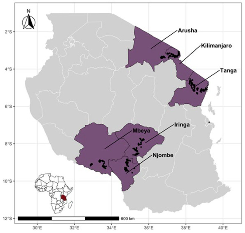
A cross-sectional study was conducted from July 2019 to October 2020 to identify the Brucella species circulating in smallholder dairy farming systems in two agroecological zones comprising six administrative regions of Tanzania. The study was conducted in three regions in the northern zone (Kilimanjaro, Arusha and Tanga), involving 252,554 dairy cattle, and three regions in the southern highland zone (Iringa, Njombe and Mbeya), involving 103,306 of dairy cattle (Figure 1). These regions have the highest density of smallholder dairy cattle in Tanzania [21,22]. The number of dairy cattle in each study region and the sample size estimation are elaborated upon in our previously published article [16]. According to the household budget survey of 2018, all six regions were above the food poverty line of TSH 33,748 (USD 13) per person per month [23]. All the study regions practice mixed farming, in which dairy cattle interact with other domestic animals [16].
Figure 1.
Map of Tanzania showing study regions (in purple) with large populations of smallholder dairy cattle and unstudied regions in gray (right). Black dots indicate the locations of the sampled cattle. The inset shows the location of Tanzania in Africa.
2.2. Study Population
Dairy cattle kept under smallholder farming systems were the target of our study. The dairy cattle in these regions are mainly Friesian, Ayrshire and Jersey crossbreeds, with Tanzanian Short Horn Zebu (TSHZ) and Friesian crosses comprising the largest proportion (80%) of breeds. The feeding management systems of these dairy cattle were twofold, involving (1) an intensive management system in which pastures were cut and carried to the farm for them to feed on and (2) an extensive system in which cattle were left to graze on private or communal land. The dairy cattle in this study were selected from a subset of the dairy cattle registry of the Africa Dairy Genetics Gains (ADGGs) program (https://data.ilri.org/portal/dataset/adgg-tanzania, accessed on 1 June 2019). The ADGGs project randomly registered over 52,500 cattle across the study regions in the database. Furthermore, 4000 dairy cattle were randomly selected and genotyped [24]. The sample for this study was selected from the genotyped animals, although at the time of the study, not all the genotyped animals were available because of the high rate of animal removal from the farms due to the sale, natural death or slaughter and the time that elapsed between genotyping and sampling. The sample size estimation for a concurrent seroprevalence study was calculated with a seroprevalence estimate of 5% (with 3% precision) and 95% confidence interval for the smallest region, assuming simple random sampling [16].
2.3. Blood and Swab Sampling from Dairy Cattle and Samples Storage
A cross-sectional survey was conducted at 1371 farms. A total of 2049 dairy cattle were sampled, and 5 mL blood was collected aseptically by venipuncture into EDTA, as explained in a previous study [25]. The animal’s identification number, the date of collection and the field barcode were labeled on each tube. In the field, all samples were kept in a cool box containing ice packs and transported to the field laboratory for storage on a daily basis. Similarly, vaginal swabs were collected aseptically after the vulva was cleaned using a chlorhexidine-soaked paper towel. The vulva lips were opened with fingers and a long shaft swab was inserted per vaginum, and the mucosa was gently swabbed by rotating the swab shaft left and right while removing it. After its removal, the swab was then inserted in a cryovial tube containing 1 mL sterile phosphate-buffered saline (PBS) and squeezed on the tube wall while the solution was mixed. The shaft was then cut off and thrown in a waste disposal bin, leaving the swab tip in the PBS cryovial. The PBS cryovial was then labeled with the collection date, barcoded, and scanned into the ODK form. Both EDTA blood tubes and PBS swab tubes were stored at −20 °C in an upright position until they were transported to the Nelson Mandela African Institution of Science and Technology laboratory in Arusha for longer-term storage.
2.4. DNA Extraction from EDTA Blood and PBS Swabs
EDTA tubes containing blood and PBS tubes containing a swab were allowed to thaw at room temperature on a table. After thawing, each tube was briefly mixed by vortexing. Three hundred microliters (300 µL) of blood/PBS was aliquoted and placed in a sample autoplate of TANBeads®. Total genomic DNA extraction was performed by using a TANBeads® Nucleic Acid Extraction Validation Kit (OptiPure Blood DNA Auto Plate) designed for use with the Maelstrom 9600 (Taiwan Advanced Nanotech Inc, Taoyuan City, Taiwan), a robotic system in ILRI laboratories in Nairobi, Kenya. The extraction kit was suitable for isolating DNA from whole blood, including deep-frozen blood, and it used the silicone dioxide layer coated on the magnetic beads. A 100 µL genomic DNA extract was provided. After the extraction, different random DNA samples were tested for quality and degradation by using a Nanodrop spectrophotometer and 1% agar rose gel electrophoresis, respectively (Supplementary Material S1).
2.5. Real-Time PCR for Brucella Genus Detection and Species Characterization
The QuantiStudio 5 qPCR machine (Applied Biosystems, Woodlands, Singapore) with the 96-well plate format and 0.2 mL block installed with QuantiStudio TM Design and Analysis software v1.5 was used for the analysis.
For Brucella genera detection, the DNA samples were tested for the presence of insertion sequence IS711 by using the IS711 primer pair and probe Table 1 in a uniplex assay. The qPCR conditions and reaction volumes used were adopted from Akoko et al. [5]. The following reaction volumes were prepared: 5 µL of ready-to-use Master Mix (Luna Universal Probe qPCR Master Mix, New England BioLabs, MA, USA), 1.25 µL of IS711 Primer and Probe mix (Macrogen, The Netherlands), 1.75 µL molecular grade water and a 3 µL DNA template. The total reaction-mix volume (cocktail) of 11 µL was then thoroughly mixed via gentle vortexing for 1 minute before the 96 wells PCR plate was inserted into the qPCR machine.
Table 1.
Oligonucleotide primers and probes used to perform qPCR assays.
| Target | Targeted Gene | Sequences of Primers and Probes (5′–3′) | Fluorophore/Quencher | Reference |
|---|---|---|---|---|
| Genus Brucella | IS711 | Probe: AAG CCA ACA CCC GGC Forward: GGC CTA CCG CTG CGA AT Reverse: TTG CGG ACA GTC ACC ATA ATG |
FAM/-MGBNFQ | Matero et al. (2011) [26] |
| B. melitensis | IS711 downstream of BMEI1162 | Probe: CAGGAGTGTTTCGGCTCAGAATAATCCACA Forward: AACAAGCGGCACCCCTAAAA Reverse: CATGCGCTATGATCTGGTTACG |
Texas Red/BHQ2 | Probert et al. (2004) [27] |
| B. abortus | IS711 downstream of alkB | Probe: CGCTCATGCTCGCCAGACTTCAATG Forward: GCGGCTTTTCTATCACGGTATTC Reverse: CATGCGCTATGATCTGGTTACG |
JOE/BHQ1 |
The thermo cycler conditions were set as they would be for pretreatment with Uracil-DNA Glycosylases (UDGs) at 50 °C for 2 min, followed by polymerase activation and DNA denaturation at 95 °C for 10 min, an amplification step at 95 °C for 15 s and 1 min of annealing at 60 °C for 42 cycles.
Positive DNA samples based on the IS711 gene marker (positive for the Brucella genus) were further characterized for B. abortus and B. melitensis using alkB and BMEI1172 primers and a probe, respectively.
The reaction volumes used for B. abortus- and B. melitensis-specific assays were 7.5 µL Master mix (Perfecta qPCR ToughMix UNG Low ROX), 0.75 µL primer and probe (species-specific), 1.75 µL molecular-grade water, and 5 µL of DNA template. The final reaction mix (cocktail) of 15 µL was thoroughly mixed by vortexing for 1 min before the 96-well PCR plate was inserted into the qPCR machine.
The PCR conditions for Brucella species-specific assays were as follows: the pretreatment stage with Uracil-N-Glycosylase (UNG-step) was set at 45 °C for 5 min to cleave all contaminated templates containing U bases, followed by DNA denaturation at 95 °C for 5 min, amplification at 95 °C for 15 s, and annealing at 60 °C for 30 s for a total of 42 cycles.
The positive controls for the Brucella strains, B. melitensis 16 M and B. abortus 544, were both sourced from the Friedrich-Loeffler Institute, Germany. For the negative controls, a mixture of RNAase-free molecular-grade water and Master mix was used. The assay efficiency statistics and limit of detection of the tenfold serial dilution of the reference materials for genus and species detection are provided in Supplementary Material S2.
2.6. Spatial Analysis
A spatial scan statistic was used to detect statistically significant spatial clusters of PCR-positive animals in the Njombe, Kilimanjaro and Arusha Regions only. Cluster analyses were performed using the SaTScan™ v10.1 software [26] with a Bernoulli model for binary events (i.e., PCR-positive/PCR-negative). SaTScan uses Monte Carlo hypothesis testing to obtain the p-values and SaTScan adjusts for the underlying spatial homogeneity of a background population. For each location and scanning-window size, the alternative hypothesis was that there was an elevated risk within the window compared with the risk outside the window, and a likelihood ratio test was performed. Multiple different window sizes were used, and the selected locations were the latitude/longitude for each animal with slight jittering to avoid more than one animal being at the same location. The window with the maximum likelihood was the most likely cluster, i.e., the cluster that was least likely to be due to chance. A p-value was assigned to this cluster. For this analysis, we used 9999 Monte Carlo replications, and a cluster was considered to be statistically significant if the p-value was <0.05. There was no adjustment for within-herd clusters.
2.7. Data Analysis for Calculation of Molecular Prevalence
Data analysis was carried out using R software (version 4.2.3) [28,29] with individual animals as primary sampling units and the district as the clustering unit rather than the herd. This was because so many herds were small and had only one animal sampled. The prevalence was estimated as the ratio of PCR positives (numerator) and the total number of animals tested (denominator) for overall and regional prevalences and the binom.test function was used to generate the 95% binomial confidence interval. The design-adjusted overall prevalence was estimated after the different sampling weights were incorporated into the estimation using the svydesign, confint and svyby functions in the survey package [30]. This allowed the stratified study design to be accounted for in the prevalence estimates.
3. Results
3.1. Description of Sampled Dairy Cattle
A total of 2049 dairy cattle were sampled from 1371 farms across the six study regions. The median herd size was two cattle. The majority of sampled cattle were female (97.2%). The predominant breed was SHZ–Friesian crosses (68.7%), with other breeds being SHZ–Ayrshire (20.8%), SHZ–Jersey (6.9%) and indigenous breeds (3.6%).
3.2. Brucellosis Molecular Prevalence of Dairy Cattle in Selected Regions of Tanzania
There were 35/2046 blood and 37/1893 swab samples that were Brucella genus-positive (Table 2). There was no agreement between sample types.
Table 2.
Regional distribution of Brucella genus positive blood and swab DNA samples from individual cattle in Tanzania.
| Region | Total Animals Sampled | Number of Positive Blood Samples (%) | Number of Positive Swab Samples (%) |
|---|---|---|---|
| Arusha | 318 | 5/318 (1.6%) | 10/294 (3.4%) |
| Tanga | 524 | 6/524 (1.0%) | 1/412 (0.2%) |
| Kilimanjaro | 521 | 11/519 (2.1%) | 8/513 (1.6%) |
| Iringa | 281 | 7/281 (2.5%) | 1/273 (0.4%) |
| Njombe | 187 | 1/186 (1.1%) | 14/186 (7.5%) |
| Mbeya | 218 | 5/218 (2.3%) | 3/215 (1.4%) |
| Total | 2049 | 35/2046 (1.7%) | 37/1893 (2.0%) |
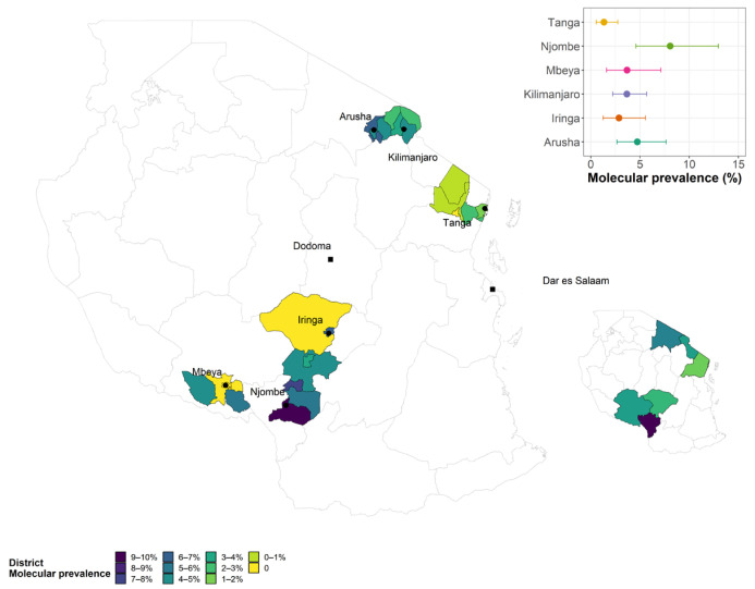
The overall unadjusted animal molecular (PCR) prevalence (with positivity determined either via a blood sample or via a vaginal swab) was 3.5% (95% CI: 2.8–4.4) and the overall design-adjusted prevalence was 3.7% (96% CI: 2.7–3.7). Among the study regions, Njombe region had the highest molecular (PCR) prevalence of 8.1%, followed by Arusha 4.7%, Mbeya 3.8% and Kilimanjaro region 3.7% (Table 3 and Figure 2).
Table 3.
The combined molecular (PCR) prevalence based on Brucella genus-positive results.
| Region | Negative | Positive | Total | PCR Prevalence % |
95% CI | Dairy Cattle Population |
|---|---|---|---|---|---|---|
| Arusha | 303 | 15 | 318 | 4.7 | 2.7–7.7 | 78,637 |
| Tanga | 517 | 7 | 524 | 1.3 | 0.5–2.7 | 41,639 |
| Kilimanjaro | 500 | 19 | 519 | 3.7 | 2.2–5.7 | 161,984 |
| Iringa | 273 | 8 | 281 | 2.8 | 1.2–5.5 | 7081 |
| Njombe | 171 | 15 | 186 | 8.1 | 4.6–13.0 | 7177 |
| Mbeya | 210 | 8 | 218 | 3.8 | 1.7–7.4 | 72,724 |
| Total | 1974 | 72 | 2046 | 3.5 | 2.8–4.4 | 369,242 |
Figure 2.
Choropleth map showing the regional molecular prevalence (insets) and the detailed molecular prevalence by local authority sampled in each region.
3.3. Brucella Species Circulating in Dairy Cattle Population Identified from Brucella Genus-Positive Swab and Blood Samples
The majority of blood samples (19/35 (54.3%)) and vaginal swabs (29/37 (78.4%)) were PCR-positive for B. melitensis only, with a further 10/35 (28.6%) blood samples and 7/37 (18.9%) swabs found to be PCR-positive for both (B. melitensis and B. abortus), meaning that the vast majority of infections involved B. melitensis (Table 4). B. abortus occurred on its own in 2/35 (5.7%) blood samples. There were four blood samples (11.4%) and one swab sample (2.7%) in which no species was determined (Table 4).
Table 4.
Real-time polymerase chain reaction (qPCR) results for Brucella species identified from genus-positive swab and blood samples.
| Region | Sample | B. abortus | B. melitensis | Mixed | Undetermined |
|---|---|---|---|---|---|
| Arusha | Blood n = 5 | 0 | 3 | 2 | 0 |
| Swabs n = 10 | 0 | 9 | 1 | 0 | |
| Kilimanjaro | Blood n = 11 | 1 | 9 | 0 | 1 |
| Swabs n = 8 | 0 | 6 | 2 | 0 | |
| Tanga | Blood n = 6 | 0 | 2 | 3 | 1 |
| Swabs n = 1 | 0 | 1 | 0 | 0 | |
| Njombe | Blood n = 1 | 0 | 1 | 0 | 0 |
| Swabs n = 14 | 0 | 11 | 2 | 1 | |
| Iringa | Blood n = 7 | 0 | 3 | 2 | 2 |
| Swabs n = 1 | 0 | 1 | 0 | 0 | |
| Mbeya | Blood n = 5 | 1 | 1 | 3 | 0 |
| Swabs n = 3 | 0 | 1 | 2 | 0 | |
| Total | Blood n = 35 | 2 | 19 | 10 | 4 |
| Swabs n = 37 | 0 | 29 | 7 | 1 |
Mixed = PCR-positive for both B. abortus and B. melitensis.
3.4. Brucellosis Hotspot Areas
Figure 2 is a spatial choropleth map of Tanzania (main) with the right-bottom inset showing the study regions and their molecular prevalence, with the highest prevalence in Njombe region (dark blue), while the main map shows the molecular prevalence of the study districts (local authorities) for each study region. The main map shows that PCR-positive animals were clustered within a small number of local authorities in the Kilimanjaro, Mbeya, Arusha and Njombe regions.
3.5. Spatial Clustering of Brucella PCR-Positive Animals
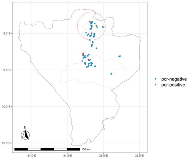
To explore the spatial clustering pattern of animals, the Kilimanjaro, Arusha and Njombe regions were mapped to identify the cluster of PCR-positive animals. SatScan analysis identified five clusters in the three regions, but only one significant cluster was found in Njombe region, and therefore Njombe region was further mapped to have a closer view (Figure 3). In a cluster of five animals, four of them were PCR-positive with a relative risk of 16.98 in a radius of 1.36 km in northern Njombe region.
Figure 3.
Map of Njombe region showing district boundaries, the location of PCR-positive and PCR-negative animals (jittered) and the radius (red circle) of the significant cluster identified by the SaTScan analysis.
4. Discussion
Brucellosis is a globally neglected bacterial zoonosis. It was characterized for the first time in Tanzania, where it was identified in domestic animals in 1967 and again in 2015 [12,13,14]. In Tanzania, brucellosis in dairy cattle is endemic and has continued to affect dairy production and public health, apart from during a short period in the late 1990s when it was successfully controlled [31]. In Tanzania, most brucellosis studies have depended on the use of serological tests to provide recommendations and conclusions on the best way to control the disease in animals and have assumed Brucella host-specificity due to the lack of serological tools with which to differentiate them [32]. However, recent studies have shown that the host-specificity of Brucella species no longer applies, as cross-infections have recently been reported globally [6,33]. Therefore, molecular characterization of Brucella species is becoming increasingly important, as it will allow us to understand the differences in their epidemiology and make appropriate recommendations for control and eradication [34].
The current study reports the overall animal level-adjusted PCR prevalence (molecular prevalence) of 3.5% across the study regions, which represent the major dairy cattle-keeping areas of Tanzania, where roughly 50% of improved dairy cattle are located [21,22]. The molecular prevalence reported in this study is lower than the molecular prevalence of 18.9% reported in Kenya [5] and similar to the 5.6% prevalence reported in Rwanda [6]. The discrepancy in molecular prevalence is likely due to differences in the sample types used, sample size, study population and study locations, as the prevalence of brucellosis has been reported to be lower in highland areas in Kenya [35] which have a similar agroecology to the areas in this study. Clustering analysis revealed one significant cluster of molecular results analysis in northern part of Njombe region; a similar cluster was also revealed in serosurvey results in our previous analysis [16], suggesting that Njombe region is the brucellosis hotspot region that requires urgent interventions. To control the disease throughout the country, high-risk regions such as Njombe, Kilimanjaro, Mbeya and Arusha need to be prioritized for disease interventions [16].
The current study has identified that B. abortus, B. melitensis and undetermined Brucella species are circulating in this dairy cattle population. The study revealed that dairy cattle are predominantly PCR-positive for B. melitensis, which is generally considered to be a pathogen that affects sheep and goats. Our previous work has demonstrated an association between seropositivity and the presence of goats, with the odds of cattle being seropositive on a farm that keeps goats being 3.02 times greater than those odds for cattle on a farm that does not keep goats [16]. The role of goats in the epidemiology of cattle brucellosis in SSA has also been reported in previous studies [15]. It was not possible to revisit farms during this study to sample small ruminants to gain a better understanding of the epidemiology of B. melitensis in this setting. However, a focus on small ruminants is recommended for future studies.
The current study also reports dairy cattle PCR-positive for two Brucella species, B. abortus and B. melitensis, and undetermined Brucella species. Co-infections with more than one Brucella species have also been reported in Rwanda and in other African countries [6,33,36,37]. Furthermore, PCR-positivity for two Brucella species was attributed to the mingling of cattle and small ruminants [38,39]. The detection of an undetermined Brucella species highlights the potential for infection with other Brucella species such as B. canis, and B. suis, which have been identified in dairy cattle following natural infection in different countries [40,41,42] and may be related to the presence of other domestic animals such as sheep, dogs and pigs in dairy farms.
The presence of Brucella in vaginal swabs suggests that bacterial transmission may occur among dairy cattle in a herd and between herds as a result of contaminated drinking water and pasture [43]. The presence of Brucella in vaginal swabs of cattle has also been revealed by other studies [44,45]. Furthermore, the presence of Brucella in the vagina signifies the necessity of veterinarians, veterinary assistants and farmers using personal protective equipment when managing difficult calvings and retained placenta in cows.
The presence of B. melitensis and mixed B. abortus and B. melitensis PCR-positives in dairy cattle pose a challenge when it comes to controlling the disease by vaccination [46,47]. The monovalent B. abortus S19 vaccine which is produced in Tanzania may not be effective in controlling the disease under these scenarios (B. melitensis and co-infections), as the vaccine has not been fully validated for conferring cross-protection and alternative vaccines which may confer protection may not be available for use in LMICs [9,10,11]. Further validation of the currently available vaccines is required; furthermore, production of a bivalent vaccine (containing B. abortus and B. melitensis) might assist with the control and eradication of the disease in cattle [48,49].
This study had limitations, as there was no agreement regarding PCR positivity between vaginal swabs and blood from the same animal, which could be attributed to the short and transient bacteremia in cattle and the fact that shedding of bacteria in vaginal samples tends to occur post-calving.
Furthermore, the poor agreement could have been attributed to the long-term storage of samples, which were kept in a deep freezer at a very low temperature (−20 °C) for over a year; these conditions are likely to have degraded the samples. Poor agreement was also observed between the PCR and cELISA results published in our previous article [16], which could have been attributed to individual animals’ varying bacteremic and immunologic phases and abortion status. This finding is similar to other studies in cattle which found poor agreement between serological and PCR results in different samples from the same animal [50]; however, there was a higher chance of PCR positivity among animals with a history of abortion [51]. Therefore, to provide the highest disease detection rate, it is important to run the serological and molecular detection methods using different samples and therefore reduce the number of false-negative results.
The exclusion of serologic results previously reported [16] in this study may have led to a misclassification of animals with chronic B. melitensis infections and is likely to have resulted in an underestimation of the number of cattle affected. Finally, there was insufficient DNA to allow sequencing for further characterization of the pathogens.
5. Conclusions
The current study confirms that bacteria of the genus Brucella are circulating in smallholder dairy cattle, suggesting that brucellosis is present and is likely to be causing clinical disease in dairy cattle. The importance of B. melitensis infections in smallholder dairy cattle is not clear, and further understanding of the clinical significance and veterinary public health implications of these infections is needed.
This study recommends that further studies be conducted on Brucella species circulating in dairy cattle, and further studies on the roles of small ruminants and other domestic animals in the epidemiology of brucellosis in dairy cattle should also be carried out. Training famers in good biosecurity and control methods is recommended, as is vaccination of dairy cattle and small ruminants in high-risk populations.
Acknowledgments
The Tanzania Livestock Research Institute (TALIRI) and the Local Government Authorities offices are recognized for their support. Thank you to the participants for their time. For the purpose of open access, the author has applied a Creative Commons Attribution (CC BY) license to any Author-Accepted Manuscript version arising from this submission.
Supplementary Materials
The following supporting information can be downloaded at https://www.mdpi.com/article/10.3390/pathogens13090815/s1, Supplementary Material S1: Genomic DNA quality checks; Supplementary Material S2: Statistics for the efficiency of PCR assays.
Author Contributions
Conceptualization, I.J.M., G.M.S., E.A.J.C. and B.M.d.C.B.; methodology, I.J.M., G.M.S., E.A.J.C., B.M.d.C.B., S.K.M., S.F.B. and L.E.H.-C.; formal analysis, I.J.M., B.M.d.C.B. and L.E.H.-C.; resources, E.A.J.C. and B.M.d.C.B.; data curation, I.J.M., S.K.M., S.F.B., B.M.d.C.B. and L.E.H.-C.; writing—original draft preparation, I.J.M.; writing—review and editing, I.J.M., G.M.S., E.A.J.C., B.M.d.C.B., L.E.H.-C., E.L., J.M.A. and D.M.K.; supervision, G.M.S., B.M.d.C.B. and E.A.J.C.; project administration, G.M.S., B.M.d.C.B. and E.A.J.C.; funding acquisition, B.M.d.C.B. and E.A.J.C. All authors have read and agreed to the published version of the manuscript.
Institutional Review Board Statement
The ethics of the study for the animal subjects were reviewed and approved by the International Livestock Research Institute Institutional Animal Care and Use Committee (IL-RI-IACUC2018-27) and a research permit was granted by the Tanzania Commission for Science and Technology (COSTECH), Ref. (2019-207-NA-2019-95).
Informed Consent Statement
Consent forms were signed by the cattle owners prior to the interview and sample collection.
Data Availability Statement
All relevant data are presented within the manuscript.
Conflicts of Interest
The authors declare no conflicts of interest.
Funding Statement
This study was conducted as part of the CGIAR Research Program on Livestock. ILRI is supported by contributors to the CGIAR Trust Fund. CGIAR is a global research partnership for a food-secure future. Its science is carried out by 15 research centers in close collaboration with hundreds of partners across the globe (www.cgiar.org, accessed on 10 December 2018). This research was funded in part by the Bill & Melinda Gates Foundation and with UK aid from the UK Foreign, Commonwealth and Development Office (Grant Agreement OPP1127286) under the auspices of the Centre for Tropical Livestock Genetics and Health (CTLGH), established jointly by the University of Edinburgh, SRUC (Scotland’s Rural College), and the International Livestock Research Institute (ILRI). This work was also supported by funding from the BBSRC (BBS/E/D/30002275). The funders had no role in the study design, data collection and analysis, decision to publish, or preparation of the manuscript. B.M.d.C.B., E.A.J.C., I.J.M., S.F.B., S.K.M. and L.H.C were supported by the Bill & Melinda Gates Foundation and by a UK aid from the UK Foreign, Commonwealth, and Development Office (Grant Agreement OPP1127286) under the auspices of the Centre for Tropical Livestock Genetics and Health established jointly by the University of Edinburgh, Scotland’s Rural College, and the ILRI.
Footnotes
Disclaimer/Publisher’s Note: The statements, opinions and data contained in all publications are solely those of the individual author(s) and contributor(s) and not of MDPI and/or the editor(s). MDPI and/or the editor(s) disclaim responsibility for any injury to people or property resulting from any ideas, methods, instructions or products referred to in the content.
References
- 1.Schelling E.E., Diguimbaye C., Daoud S., Nicolet J., Boerlin P., Tanner M., Zinsstag J. Brucellosis and Q-fever seroprevalences of nomadic pastoralists and their livestock in Chad. Prev. Vet. Med. 2003;61:279–293. doi: 10.1016/j.prevetmed.2003.08.004. [DOI] [PubMed] [Google Scholar]
- 2.World Organization for Animal Health (WOAH) Manual of Diagnostic Tests and Vaccines for Terrestrial Animals. 12th ed. World Organization for Animal Health; Paris, France: 2023. Brucellosis; pp. 1–35. Chapter 3.1.4. [Google Scholar]
- 3.Pappas G. The changing Brucella ecology: Novel reservoirs, new threats. Int. J. Antimicrob. Agents. 2010;36:S8–S11. doi: 10.1016/j.ijantimicag.2010.06.013. [DOI] [PubMed] [Google Scholar]
- 4.Corbel M.J. Brucellosis in Humans and Animals. World Health Organization (WHO); Geneva, Switzerland: Food and Agriculture Organization (FAO); Rome, Italy: World Organization for Animal Health (OIE); Paris, France: 2006. [Google Scholar]
- 5.Akoko J.M., Pelle R., Lukambagire A.S., Machuka E.M., Nthiwa D., Mathew C., Fèvre E.M., Bett B., Cook E.A.J., Othero D., et al. Molecular epidemiology of Brucella species in mixed livestock-human ecosystems in Kenya. Sci. Rep. 2021;11:2045–2322. doi: 10.1038/s41598-021-88327-z. [DOI] [PMC free article] [PubMed] [Google Scholar]
- 6.Ntivuguruzwa J.B., Babaman K.F., Mwikarago E.I., van Heerden H. Seroprevalence of brucellosis and molecular characterization of Brucella spp. from slaughtered cattle in Rwanda. PLoS ONE. 2022;17:e0261595. doi: 10.1371/journal.pone.0261595. [DOI] [PMC free article] [PubMed] [Google Scholar]
- 7.ElTahir Y., Al-Farsi A., Al-Marzooqi W., Al-Toobi A., Gaafar O.M., Jay M., Corde Y., Bose S., Al-Hamrashdi A., Al-Kharousi K. Investigation on Brucella infection in farm animals in Saham, Sultanate of Oman with reference to human brucellosis outbreak. BMC Vet. Res. 2019;15:378. doi: 10.1186/s12917-019-2093-4. [DOI] [PMC free article] [PubMed] [Google Scholar]
- 8.Muendo E.N., Mbatha P.M., Macharia J., Abdoel T.H., Janszen P.V., Pastoor R., Smits H.L. Infection of cattle in Kenya with Brucella abortus biovar 3 and Brucella melitensis biovar 1 genotypes. Trop. Anim. Health Prod. 2012;44:17–20. doi: 10.1007/s11250-011-9899-9. [DOI] [PubMed] [Google Scholar]
- 9.Moriyón I., Grilló M., Monreal D., González D., Marín C., López-Goñi I., Mainar-Jaime R., Moreno E., Blasco J. Rough vaccines in animal brucellosis: Structural and genetic basis and present status. Vet. Res. 2004;35:1–38. doi: 10.1051/vetres:2003037. [DOI] [PubMed] [Google Scholar]
- 10.Schurig G.G., Sriranganathan N., Corbel M.J. Brucellosis vaccines: Past, present and future. Vet. Microbiol. 2002;90:479–496. doi: 10.1016/S0378-1135(02)00255-9. [DOI] [PubMed] [Google Scholar]
- 11.Van Straten M., Bardenstein S., Keningswald G., Banai M. Brucella abortus S19 vaccine protects dairy cattle against natural infection with Brucella melitensis. Vaccine. 2016;34:5837–5839. doi: 10.1016/j.vaccine.2016.10.011. [DOI] [PubMed] [Google Scholar]
- 12.Mahlau E. Further brucellosis surveys in Tanzania. Bulletin of Epizootic Diseases of Africa. Bull. Epizoot. Dis. Afr. 1967;15:373–378. [PubMed] [Google Scholar]
- 13.Mathew C., Stokstad M., Johansen T.B., Klevar S., Mdegela R.H., Mwamengele G., Michel P., Escobar L., Fretin D., Godfroid J. First isolation, identification, phenotypic and genotypic characterization of Brucella abortus biovar 3 from dairy cattle in Tanzania. BMC Vet. Res. 2015;11:156. doi: 10.1186/s12917-015-0476-8. [DOI] [PMC free article] [PubMed] [Google Scholar]
- 14.Assenga J.A., Matemba L.E., Muller S.K., Malakalinga J.J., Kazwala R.R. Epidemiology of Brucella infection in the human, livestock and wildlife interface in the Katavi-Rukwa ecosystem, Tanzania. BMC Vet. Res. 2015;11:189. doi: 10.1186/s12917-015-0504-8. [DOI] [PMC free article] [PubMed] [Google Scholar]
- 15.Ducrotoy M., Bertu W.J., Matope G., Cadmus S., Conde-Álvarez R., Gusi A.M., Welburn S., Ocholi R., Blasco J.M., Moriyón I. Brucellosis in Sub-Saharan Africa: Current challenges for management, diagnosis and control. Acta Trop. 2017;165:179–193. doi: 10.1016/j.actatropica.2015.10.023. [DOI] [PubMed] [Google Scholar]
- 16.Mengele I.J., Shirima G.M., Bwatota S.F., Motto S.K., Bronsvoort B.M.C., Komwihangilo D.M., Lyatuu E., Cook E.A.J., Hernandez-Castro L.E. The status and risk factors of brucellosis in smallholder dairy cattle in selected regions of Tanzania. Vet. Sci. 2023;10:155. doi: 10.3390/vetsci10020155. [DOI] [PMC free article] [PubMed] [Google Scholar]
- 17.Makita K., Fèvre E., Waiswa C., Eisler M., Thrusfield M., Welburn S. Herd prevalence of bovine brucellosis and analysis of risk factors in cattle in urban and peri-urban areas of the Kampala economic zone, Uganda. BMC Vet. Res. 2011;7:60. doi: 10.1186/1746-6148-7-60. [DOI] [PMC free article] [PubMed] [Google Scholar]
- 18.Khurana S.K., Sehrawat A., Tiwari R., Prasad M., Gulati B., Shabbir M.Z., Chhabra R., Karthik K., Patel S., Pathak M., et al. Bovine brucellosis—A comprehensive review. Vet. Q. 2021;41:61–88. doi: 10.1080/01652176.2020.1868616. [DOI] [PMC free article] [PubMed] [Google Scholar]
- 19.Bodenham R.F., Lukambagire A.S., Ashford R.T., Buza J.J., Cash-Goldwasser S., Crump J.A., Kazwala R.R., Maro V.P., McGiven J., Mkenda N. Prevalence and speciation of brucellosis in febrile patients from a pastoralist community of Tanzania. Sci. Rep. 2020;10:7081. doi: 10.1038/s41598-020-62849-4. [DOI] [PMC free article] [PubMed] [Google Scholar]
- 20.Nyawale H.A., Simchimba M., Mlekwa J., Mujuni F., Chibwe E., Shayo P., Mngumi E.B., Majid K.S., Majigo M., Mshana S.E. High Seropositivity of Brucella melitensis Antibodies among Pregnant Women Attending Health Care Facilities in Mwanza, Tanzania: A Cross-Sectional Study. J. Pregnancy. 2023;2023:2797441. doi: 10.1155/2023/2797441. [DOI] [PMC free article] [PubMed] [Google Scholar]
- 21.Njombe A., Msanga Y., Mbwambo N., Makembe N. Dairy Industry Status in Tanzania. Ministry of Livestock Development and Fisheries; Proceedings of the 7th African Dairy Conference and Exhibition; Dar Es Salaam, Tanzania. 25–27 May 2011; [(accessed on 10 May 2021)]. Available online: https://dairyafrica.com/ [Google Scholar]
- 22.National Bureau of Statistics (NBS) National Sample Census of Agriculture 2019–2020. Ministry of Finance and Planning, United Republic of Tanzania; Dodoma, Tanzania: 2021. pp. 1–317. (National Report). [Google Scholar]
- 23.Ministry of Finance and Planning (MoFP) Tanzania Mainland Household Budget Survey 2017–2018, Key indicators. Poverty Eradication Division, National Bureau of Statistics, United Republic of Tanzania; Dodoma, Tanzania: 2019. (Report: Poverty Eradication Division, National Bureau of Statistics, United Republic of Tanzania). [Google Scholar]
- 24.Mrode R., Ojango J., Ekine-Dzivenu C., Aliloo H., Gibson J., Okeyo M.A. Genomic prediction of crossbred dairy cattle in Tanzania: A route to productivity gains in smallholder dairy systems. J. Dairy Sci. 2021;104:11779–11789. doi: 10.3168/jds.2020-20052. [DOI] [PubMed] [Google Scholar]
- 25.Shirima G.M. Ph.D. Thesis. University of Glasgow; Glasgow, UK: 2005. The Epidemiology of Brucellosis in Animals and Humans in Arusha and Manyara Regions in Tanzania. [Google Scholar]
- 26.Matero P., Hemmilä H., Tomaso H., Piiparinen H., Rantakokko-Jalava K., Nuotio L., Nikkari S. Rapid field detection assays for Bacillus anthracis, Brucella spp., Francisella tularensis and Yersinia pestis. Clin. Microbiol. Infect. 2011;17:34–43. doi: 10.1111/j.1469-0691.2010.03178.x. [DOI] [PubMed] [Google Scholar]
- 27.Probert W.S., Schrader K.N., Khuong N.Y., Bystrom S.L., Graves M.H. Real-time multiplex PCR assay for detection of Brucella spp., B. abortus, and B. melitensis. J. Clin. Microbiol. 2004;42:1290–1293. doi: 10.1128/JCM.42.3.1290-1293.2004. [DOI] [PMC free article] [PubMed] [Google Scholar]
- 28.Kulldorff M. Information Management Services. Software for the Spatial and Space-Time Scan Statistics. SaTScan v10.1.2 64bit. 2023. [(accessed on 15 February 2020)]. Available online: https://www.satscan.org/
- 29.R Core Team . R: A Language and Environment for Statistical Computing. R Foundation for Statistical Computing; Vienna, Austria: 2021. [(accessed on 17 February 2020)]. Available online: https://www.R-project.org/ [Google Scholar]
- 30.Lumley T. Analysis of complex survey samples. J. Stat. Softw. 2004;9:1–19. doi: 10.18637/jss.v009.i08. [DOI] [Google Scholar]
- 31.Shirima G., Lyimo B., Kanuya N. Re-emergence of Bovine Brucellosis in Smallholder Dairy Farms in Urban Settings of Tanzania. J. Appl. Life Sci. Int. 2018;17:1–7. doi: 10.9734/JALSI/2018/40955. [DOI] [Google Scholar]
- 32.Mengele I.J., Shirima G.M., Bronsvoort B.M., Hernandez-Castro L.E., Cook E.A.J. Diagnostic challenges of brucellosis in humans and livestock in Tanzania: A thematic review. CABI One Health. 2023:ohcs20230001. doi: 10.1079/cabionehealth.2023.0001. [DOI] [Google Scholar]
- 33.Aliyev J., Alakbarova M., Garayusifova A., Omarov A., Aliyeva S., Fretin D., Godfroid J. Identification and molecular characterization of Brucella abortus and Brucella melitensis isolated from milk in cattle in Azerbaijan. BMC Vet. Res. 2022;18:71. doi: 10.1186/s12917-022-03155-1. [DOI] [PMC free article] [PubMed] [Google Scholar]
- 34.Oliveira M.S., Dorneles E.M.S., Soares P.M.F., Junior A.A.F., Orzil L., de Souza P.G., Lage A.P. Molecular epidemiology of Brucella abortus isolated from cattle in Brazil, 2009–2013. Acta Trop. 2017;166:106–113. doi: 10.1016/j.actatropica.2016.10.023. [DOI] [PubMed] [Google Scholar]
- 35.Akoko J.M., Muturi M., Wambua L., Abkallo H.M., Nyamota R., Bosire C., Oloo S., Limbaso K.S., Gakuya F., Nthiwa D., et al. Mapping brucellosis risk in Kenya and its implications for control strategies in sub-Saharan Africa. Sci. Rep. 2023;13:20192. doi: 10.1038/s41598-023-47628-1. [DOI] [PMC free article] [PubMed] [Google Scholar]
- 36.Mitterran K.N.R., Barberine S.A., Oumarou F., Simo G. Detection of Brucella abortus and Brucellla melitensis in cattle and sheep from southern Cameroon. Res. Sq. 2020;2:1–13. doi: 10.21203/rs.3.rs-21575/v2. [DOI] [Google Scholar]
- 37.Abnaroodheleh F., Emadi A., Dashtipour S., Jamil T., Khaneghah A.M., Dadar M. Shedding rate of Brucella spp. in the milk of seropositive and seronegative dairy cattle. Heliyon. 2023;9:1–8. doi: 10.1016/j.heliyon.2023.e15085. [DOI] [PMC free article] [PubMed] [Google Scholar]
- 38.Kolo F.B., Adesiyun A.A., Fasina F.O., Katsande C.T., Dogonyaro B.B., Potts A., Matle I., Gelaw A.K., Van Heerden H. Seroprevalence and characterization of Brucella species in cattle slaughtered at Gauteng abattoirs, South Africa. Vet. Med. Sci. 2019;5:545–555. doi: 10.1002/vms3.190. [DOI] [PMC free article] [PubMed] [Google Scholar]
- 39.Morales-Estrada A.I., Joel C., Ahide L., Maria R.M., Juan G.V., Araceli C. Characterization of Brucella species in Mexico by Bruce-Ladder polymerase chain reaction (PCR) Afr. J. Microbiol. Res. 2012;6:2793–2796. doi: 10.5897/AJMR12.074. [DOI] [Google Scholar]
- 40.Ewalt D.R., Payeur J.B., Rhyan J.C., Geer P.L. Brucella suis biovar 1 in naturally infected cattle: A bacteriological, serological, and histological study. J. Vet. Diagn. Investig. 1997;9:417–420. doi: 10.1177/104063879700900414. [DOI] [PubMed] [Google Scholar]
- 41.Fretin D., Mori M., Czaplicki G., Quinet C., Maquet B., Godfroid J., Saegerman C. Unexpected Brucella suis biovar 2 infection in a dairy cow, Belgium. Emerg. Infect. Dis. 2013;19:2053. doi: 10.3201/eid1912.130506. [DOI] [PMC free article] [PubMed] [Google Scholar]
- 42.Baek B., Park M.Y., Islam M.A., Khatun M.M., Lee S.I., Boyle S.M. The first detection of Brucella canis in cattle in the Republic of Korea. Zoonoses Public Health. 2012;59:77–82. doi: 10.1111/j.1863-2378.2011.01429.x. [DOI] [PubMed] [Google Scholar]
- 43.Yaeger M.J., Holler L.D. Current Therapy in Large Animal Theriogenology. Elsevier; Amsterdam, The Netherlands: 2007. Bacterial causes of bovine infertility and abortion; pp. 389–399. [Google Scholar]
- 44.Varsha T., Bannalikar A. Molecular characterization of Brucella species detected from clinical samples of cattle and buffaloes. Indian J. Anim. Sci. 2022;92:1274–1279. doi: 10.56093/ijans.v92i11.124795. [DOI] [Google Scholar]
- 45.Efrem G.H., Mihreteab B., Ghebremariam M.K., Yitbarek G., Gezahegne M. Isolation and identification of Brucella abortus and B. melitensis in ruminants with a history of abortion: The first report from Eritrea. Ethiopian Vet. J. 2024;28:122–138. doi: 10.4314/evj.v28i1.8. [DOI] [Google Scholar]
- 46.Hegazy Y.M., Oreiby A.F., Algabbary M.H., Hamdy M.E.R., Beleta E.I., Martínez I., Shahein M.A., García N., Eltholth M. Trans-species transmission of Brucellae among ruminants hampering brucellosis control efforts in Egypt. J. Appl. Microb. 2022;132:90–100. doi: 10.1111/jam.15173. [DOI] [PubMed] [Google Scholar]
- 47.Bardenstein S., Grupel D., Blum S.E., Motro Y., Moran-Gilad J. Public and animal health risks associated with spillover of Brucella melitensis into dairy farms. Microb. Genom. 2023;9:1014. doi: 10.1099/mgen.0.001014. [DOI] [PMC free article] [PubMed] [Google Scholar]
- 48.Sanz C., Sáez J.L., Álvarez J., Cortés M., Pereira G., Reyes A., Rubio F., Martín J., García N., Domínguez L. Mass vaccination as a complementary tool in the control of a severe outbreak of bovine brucellosis due to Brucella abortus in Extremadura, Spain. Prev. Vet. Med. 2010;97:119–125. doi: 10.1016/j.prevetmed.2010.08.003. [DOI] [PubMed] [Google Scholar]
- 49.Lord V.R., Schurig G.G., Cherwonogrodzky J.W., Marcano M.J., Melendez G.E. Field study of vaccination of cattle with Brucella abortus strains RB51 and 19 under high and low disease prevalence. Am. J. Vet. Res. 1998;59:1016–1020. doi: 10.2460/ajvr.1998.59.08.1016. [DOI] [PubMed] [Google Scholar]
- 50.El-Diasty M., Wareth G., Melzer F., Mustafa S., Sprague L.D., Neubauer H. Isolation of Brucella abortus and Brucella melitensis from Seronegative Cows is a Serious Impediment in Brucellosis Control. Vet. Sci. 2018;5:28. doi: 10.3390/vetsci5010028. [DOI] [PMC free article] [PubMed] [Google Scholar]
- 51.Zinka M., Amela J., Orjana S., Maid R. Molecular detection of Brucella spp. in clinical samples of seropositive ruminants in Bosnia and Herzegovina. Comp. Immunol. Microbiol. Infect. Dis. 2022;86:101821. doi: 10.1016/j.cimid.2022.101821. [DOI] [PubMed] [Google Scholar]
Associated Data
This section collects any data citations, data availability statements, or supplementary materials included in this article.
Supplementary Materials
Data Availability Statement
All relevant data are presented within the manuscript.


