Abstract
Tumor necrosis factor-related apoptosis-inducing ligand (TRAIL) induces apoptosis of various tumor cells but not normal cells. However, various cytokines and virus infection differentially regulate TRAIL and TRAIL receptor expression. It has been demonstrated that virus infection changes the pattern of human TRAIL-receptor expression on normal cells, which were resistant to TRAIL-mediated apoptosis, and makes them susceptible to TRAIL-mediated apoptosis. Since previous studies on the function of TRAIL have been performed mainly in vitro, its physiological role in the immune response to virus infection remains unknown. In the present study, we investigated the expression of TRAIL in the lungs of influenza virus-infected mice and the function of TRAIL in the immune response to infection. Influenza virus infection increased TRAIL mRNA expression in the lung. TRAIL protein expression was induced on NK cells in the lung 4 days after infection. At 7 days after infection, TRAIL protein expression was also detected on CD4+ and CD8+ T cells. However, NK cells and T cells in the lungs of uninfected mice did not express a detectable level of TRAIL on their cell surfaces. DR5, which is a mouse TRAIL receptor, was also induced to express after virus infection. Expression of both TRAIL and DR5 mRNAs was reduced to normal level at 6 weeks after virus infection. Administration of anti-TRAIL monoclonal antibody, which blocks TRAIL without killing TRAIL-expressing cells, to mice during influenza virus infection significantly delayed virus clearance in the lung. These results suggest that TRAIL plays an important role in the immune response to virus infection.
Tumor necrosis factor (TNF)-related apoptosis-inducing ligand (TRAIL) is a type II transmembrane protein belonging to the TNF family. Among the members of this family, TRAIL exhibits highest homology to Fas ligand (FasL), which is a well-characterized apoptosis-inducing ligand (26, 29, 38). Thus far, at least four human TRAIL receptors (TRAIL-Rs), i.e., TRAIL-R1/death receptor 4 (DR4), TRAIL-R2/DR5/TRICK2, TRAIL-R3/decoy receptor 1 (DcR1)/TRID/LIT, and TRAIL-R4/DcR2/TRUNDD, have been identified and shown to bind to TRAIL with similar affinities (1, 3, 27, 28, 33). In mice, only mouse DR5 has been identified as a human TRAIL-R2/DR5 homologue (39). TRAIL-R1 and TRAIL-R2 contain a cytoplasmic death domain and induce apoptotic signals by binding with trimeric TRAIL. Aggregation of the death domain recruits caspase-8 or -10 via Fas-associated domain or a Fas-associated domain-like adaptor molecule and leads to activation of the caspase cascade, resulting in apoptotic cell death (1, 27, 28, 33). In contrast to these apoptotic receptors, TRAIL-R3 completely lacks a cytoplasmic domain and exists as a glycophospholipid-anchored protein on the cell surface (1, 27, 33). TRAIL-R4 contains a truncated cytoplasmic death domain that cannot transduce apoptotic signals. Furthermore, TRAIL-R4 can activate NF-κB, a known survival factor that inhibits apoptosis (1, 3). TRAIL-R3 and TRAIL-R4 have been reported to act as decoy receptors and suppress the apoptotic cell death induced by TRAIL and TRAIL-R1/R2 interaction.
TRAIL preferentially induces apoptotic cell death of a variety of transformed cells but not normal cells (29, 36, 39). Recent studies have indicated that stimulation with anti-CD3 monoclonal antibody (MAb) and alpha/beta interferon (IFN-α/β) rapidly induces a remarkable TRAIL expression on the cell surface of CD4+ and CD8+ human peripheral blood T cells and that stimulation with interleukin-2 (IL-2) and IL-15 induces TRAIL expression on murine splenic NK cells. TRAIL induced on these cells mediates cytotoxicity against a variety of tumor cell lines (15, 16). On the other hand, recent investigations have shown that various cytokines and virus infection differentially modulate TRAIL and TRAIL-R expression and NF-κB activation (2, 32). It was shown that human cytomegalovirus infection directly up-regulates the expression of TRAIL, TRAIL-R1, and TRAIL-R2 on virus-infected fibroblast cells. These virus-infected cells become susceptible to apoptosis via TRAIL. Furthermore, IFN-γ or TNF-α treatment up-regulates the expression of TRAIL, TRAIL-R1, and TRAIL-R2 on the virus-infected cells, and the cells have increased susceptibility to TRAIL-mediated apoptosis. In contrast, IFN-γ or TNF-α down-regulates the expression of TRAIL-R1 and TRAIL-R2 on the surface of uninfected cells (32). Both IFNs and TNFs are antiviral cytokines, and therefore a role of TRAIL in the immune response to virus infection is strongly implied. It has been also demonstrated that TRAIL can induce apoptosis of normal dendritic cells (DCs), monocytes, and T cells (8, 31, 37). Furthermore, it has been shown that TRAIL mediates activation-induced cell death of human T cells (23). Thus, TRAIL is thought to act as a modulator of immune regulation. These results suggest that TRAIL plays a role in elimination of virus-infected cells and/or in immune modulation after viral infection. Previous studies on the function of TRAIL have been performed mainly in vitro, and thus the role of TRAIL during virus infection in vivo remains to be investigated.
In this study, we first examined the expression of TRAIL and DR5 mRNAs in the lungs of influenza A virus-infected mice by reverse transcription-PCR (RT-PCR). Next, we examined TRAIL expression on mononuclear cells isolated from the lungs of influenza A virus-infected mice by flow cytometry. In addition, to determine the role of TRAIL in the immune response to influenza A virus infection, we investigated the effect of anti-TRAIL MAb treatment during influenza A virus infection on the pulmonary virus titer. The results obtained from this study indicated that TRAIL may play an important role in the immune response to influenza A virus infection.
MATERIALS AND METHODS
Mice.
Six- to 8-week-old male C57BL/6 mice were purchased from Japan SLC. Mice were kept under specific-pathogen-free conditions approved by the Institutional Animal Care and Use Committee of Yokohama City University School of Medicine.
Virus and virus infection.
The mouse-adapted strain of influenza A/PR/8/34 (PR8, H1N1) virus was prepared as previously described (41). We used the diluted virus solution at a titer of 5 × 10² PFU/ml for infection. Mice were inoculated intranasally with 50 μl of diluted virus solution under anesthesia with diethyl ether. In some experiments, mice were injected intraperitoneally with 200 μg of anti-mouse TRAIL MAb (N2B2) 4 h before infection and 2, 5, and 7 days after infection. Normal rat immunoglobulin G (IgG) was administrated as an isotype control on the same schedule. Mice were sacrificed and the lungs were excised on days 4, 5, 7, and 10.
Cell lines.
Madin-Darby canine kidney (MDCK) cells were cultured in RPMI 1640 medium supplemented with 10% heat-inactivated fetal calf serum (Biological Industries), 300 U/ml penicillin (Banyu Pharmaceutical Co., Ltd.), 0.3 mg/ml streptomycin (Meiji Seika Co., Ltd.), and 2 mM l-glutamine (WAKO) at 37°C under 5% CO2 in air.
MAbs.
Cy-chrome-conjugated anti-mouse CD3 and anti-mouse CD4, fluorescein isothiocyanate (FITC)-conjugated anti-mouse NK1.1, phycoerythrin-conjugated streptavidin, and purified anti-mouse CD16/32 (Fc block) were purchased from BD PharMingen. FITC-conjugated anti-mouse CD8 was purchased from Immunotech. Normal rat IgG was purchased from Caltag, and normal mouse IgG was obtained from Chemicon. Anti-mouse TRAIL was a generous gift from N. Kayagaki.
RT-PCR.
Lungs were excised, treated with 1 ml of TRIzol reagent (GIBCO-BRL), snap frozen in liquid nitrogen, and stored at −80°C until used for isolation of total RNA. The lung was thawed and homogenized with a plastic Dounce tissue homogenizer, and total RNA of the lung was extracted. To avoid contamination of genomic DNA, this extract was suspended in diethyl pyrocarbonate-H2O and treated with 1 U of RNase-free DNase (Promega) for 1 h at 37°C. DNase was inactivated by incubation at 70°C for 10 min. After ethanol precipitation, total RNA was suspended in 30 μl of diethyl pyrocarbonate-H2O, and 7.5 μl of this suspension was reverse transcribed into cDNA with 200 U of Moloney murine leukemia virus reverse transcriptase (GIBCO-BRL), using oligo(dT) primer (Promega). The reverse transcription was conducted at 37°C for 1 h, and the reaction was stopped by incubation at 70°C for 15 min. The density of cDNA was calculated with GeneQuant II (Pharmacia Biotech), and 1 μg of cDNA was used as the template for PCR amplification. The cDNA was amplified using specific primers for mouse GAPDH (glyceraldehyde-3-phosphate dehydrogenase) as a control. The primers used were 5′-CAATGCATCCTGCACCACCAA-3′ (sense) and 5′-GTCATTGAGAGCAATGCCAGC-3′ (antisense) for GAPDH, 5′-GAAGACCTCAGAAAGTGGC-3′ (sense) and 5′-GACCAGCTCTCCATTCCTA-3′ (antisense) for TRAIL, and 5′-AAGTGTGTCTCCAAAACGG-3′ (sense) and 5′-AATGCACAGAGTTCGCACT-3′ (antisense) for DR5. The quantitative PCR was performed at Nihon Idenshi Kenkyuujo (Miyagi, Japan).
Cell preparation.
For lung mononuclear cell isolation, mice were sacrificed with diethyl ether and exsanguinated. The lungs were excised, rinsed in RPMI 1640 medium, minced, and digested for 90 min at 37°C with RPMI 1640 containing 0.1% collagenase (Wako), 0.05% DNase I (Boehringer Mannheim), and 5% fetal calf serum. The pieces of lung were ground with slide glasses for single-cell suspension and passed through gauze. Erythrocytes were depleted with buffered ammonium chloride solution. The mononuclear cells in the single-cell suspension were isolated by density gradient centrifugation with 70% and 30% Percoll (Amersham Pharmacia). The mononuclear cells at the interface of the 70% and 30% Percoll were collected, washed, and counted.
Flow cytometry.
Mononuclear cells isolated from the lung were incubated with normal rat IgG, mouse IgG, and anti-mouse CD16/32 to block nonspecific binding of Abs. After blocking, the cells were stained directly with FITC- or Cy-chrome-conjugated reagents or indirectly with biotinylated Abs followed by phycoerythrin-conjugated streptavidin. The cells were analyzed with a flow cytometer (FACScan; BD PharMingen). Ten thousands cells were acquired into the list mode, and data were analyzed with CELLQuest software.
Lung virus titration by MDCK cell plaque formation assay.
Lungs were removed at the bronchi, snap frozen in liquid nitrogen, and stored at −80°C until used for titration. The lung was thawed at 4°C in 2 ml of phosphate-buffered saline and homogenized with a Dounce tissue grinder (Ikemoto R.K.I.), and the homogenate was centrifuged at 3,000 × g for 15 min at 4°C (Tomy CX-200). The supernatant was used for virus titration. The supernatant was diluted with Dulbecco's modified Eagle's medium supplemented with 0.12% NaHCO3. One hundred microliters of each diluted supernatant was added to a confluent monolayer of MDCK cells in six-well cell culture plates (Sumilon) and incubated at room temperature for 90 min. Each well then received 3 ml of agar overlay medium, which is 1% agar (Difco Laboratories, Inc.) in Dulbecco's modified Eagle's medium supplemented with 0.4% bovine serum albumin (Biosciences NZ, Ltd.), 0.12% NaHCO2, 7.5 mM HEPES, 2 mM l-glutamine, 0.1% dextrose, 0.3% 1,000× viotin, 3% 100× vitamin solution, 0.1% Fungizone (amphotericin B), and 0.0005% trypsin. After 3 days of incubation at 37°C in 5% CO2, cells were fixed with 3 ml of 10% formaldehyde for 1 h. The agar overlays were then removed, and fixed monolayers were stained with 0.1% cresyl violet in 20% ethanol. The results are presented as PFU per 1/20 volume of homogenate of lung = number of plaques/(1/dilution factor)
RESULTS
Expression of TRAIL and DR5 mRNAs in lungs of influenza virus-infected mice.
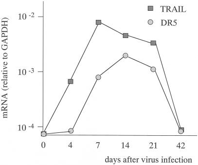
Primary cultured human foreskin fibroblast cells are normally resistant to TRAIL-mediated apoptosis (32). However, the expression of TRAIL, TRAIL-R1, and TRAIL-R2 on the cell surface of the foreskin fibroblast cells is increased by human cytomegalovirus infection in vitro, and virus-infected cells become susceptible to apoptosis via TRAIL (32). It is unclear whether expression of TRAIL and TRAIL-Rs is affected by virus infection in vivo. First, we investigated the effect of virus infection on the expression of TRAIL and DR5 in vivo. Quantitative PCR analysis was performed to study the relative abundances of TRAIL and DR5 mRNAs in the lungs of influenza A virus (25 PFU/mouse)-infected or uninfected mice. Mice were sacrificed 4, 7, 14, 21, and 42 days after influenza A virus infection. In the lungs of uninfected mice, TRAIL mRNA was not detected (Fig. 1). The expression of TRAIL mRNA was increased 4 days after influenza A virus infection, and this increased TRAIL mRNA expression peaked at 7 days after infection. DR5 mRNA was not also expressed in the lungs of uninfected mice and was expressed at very low levels at 4 days after influenza A virus infection (Fig. 1). DR5 mRNA expression peaked at 14 days after virus infection. Expression of both TRAIL and DR5 mRNAs became very low at 42 days after virus infection. mRNA of the GAPDH housekeeping gene was detected at relatively similar levels irrespective of influenza A virus infection. Taken together, the data from PCR analysis indicated that clear differences exist in the relative abundances of TRAIL and DR5 mRNAs in the lungs of mice after influenza A virus infection compared to those in the lungs of uninfected mice.
FIG. 1.
RT-PCR analysis of TRAIL and DR5 mRNA expression in lungs of influenza virus-infected mice and uninfected mice. Mice were inoculated intranasally with 25 PFU of influenza virus. Lungs were excised 0, 4, 7, 14, 21, and 42 days after infection. RNAs were isolated from lungs of uninfected and influenza virus-infected mice. GAPDH-normalized TRAIL and DR5 mRNA abundances are shown.
Expression of TRAIL on surfaces of NK cells and T cells in lungs during influenza virus infection.
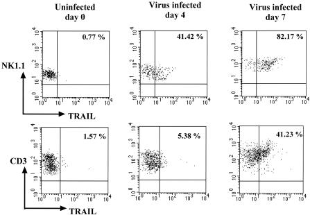
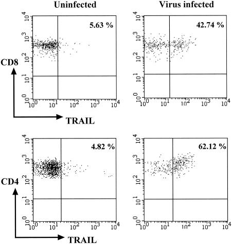
It was determined that influenza virus infection increased the TRAIL mRNA expression in the lung. Next, to determine the TRAIL expression on cell surface, mice were infected with influenza A virus (25 PFU/mouse), and TRAIL expression as the cell surface protein on mononuclear cells isolated from the lungs of virus-infected or uninfected mice was investigated. Mice were sacrificed 4 and 7 days after infection, and TRAIL expression was analyzed by three-color flow cytometry. Neither T cells nor NK cells from the lungs of uninfected mice expressed TRAIL on the cell surface (Fig. 2). However, TRAIL expression was detected on NK cells and T cells isolated from the lung after influenza A virus infection. On day 4 postinfection, surface TRAIL expression was detected on NK cells but not on T cells (Fig. 2). On day 7 postinfection, TRAIL was expressed not only on NK cells but also on T cells (Fig. 2). The abundance of TRAIL expression on the surface of the NK cells and the number of TRAIL-expressing NK cells in the lung on day 7 postinfection were higher than those on day 4 postinfection. Furthermore, it was investigated which T-cell subset expresses TRAIL on the cell surface. Both CD4+ T cells and CD8+ T cells expressed TRAIL on their cell surfaces 7 days after infection (Fig. 3). These data indicated that influenza A virus infection of mice influenced the expression of TRAIL protein on the cell surfaces of NK cells and CD4+ and CD8+ T cells in the lung.
FIG. 2.
Flow cytometric analysis of TRAIL expression on NK cells and T cells in lungs of influenza virus-infected mice and uninfected mice. Mice were inoculated intranasally with 25 PFU of influenza virus. Lungs were excised 4 and 7 days after infection. Mononuclear cells were isolated from the pooled lungs of three uninfected mice or three influenza virus-infected mice, and TRAIL expression was analyzed by flow cytometry. Dot plots were acquired by gating on lymphocytes at FSC-SSC and NK1.1+ or CD3+ cells. Data shown are cell surface expression of TRAIL on NK cells (upper panels) and CD3+ T cells (lower panels). The percentage of TRAIL-expressing cells in each gated population is indicated.
FIG. 3.
Flow cytometric analysis of TRAIL expression on CD8+ and CD4+ T cells in lungs of influenza virus-infected mice and uninfected mice. Mononuclear cells were isolated from the pooled lungs of three influenza virus-infected mice 7 days after infection, and TRAIL expression on T-cell subsets was analyzed by flow cytometry. Dot plots were acquired by gating on lymphocytes at FSC-SSC and CD8+ or CD4+ cells. The data shown are cell surface expression of TRAIL on CD8+ T cells (upper panels) and CD4+ T cells (lower panels). The percentage of TRAIL-expressing cells in each gated population is indicated.
Effect of anti-TRAIL MAb treatment during influenza virus infection on pulmonary virus titer.
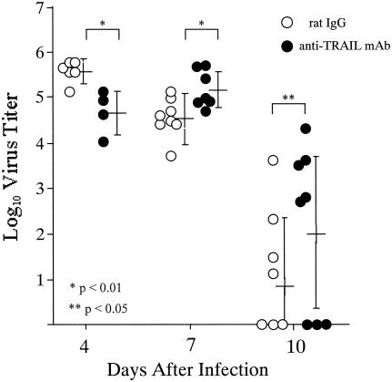
To investigate the function of TRAIL in the immune response to virus infection in vivo, anti-TRAIL MAb was administered to mice to block TRAIL and TRAIL-R interaction during influenza A virus infection. Mice were injected intraperitoneally with 200 μg of anti-TRAIL MAb. Normal rat IgG was used as an isotype control. Antibodies were injected 4 h before influenza A virus infection and 2, 5, and 7 days after infection, and the virus titer in the lung homogenate was assessed by plaque-forming assay with MDCK cells on 4, 7, and 10 days after infection. The virus titers in the lungs from mice treated with anti-TRAIL MAb reached a peak 4 to 7 days after infection, whereas those in lungs from control mice reached a peak 4 days after infection (Fig. 4). On day 4, the mean pulmonary virus titer of mice treated with anti-TRAIL MAb was sixfold lower than that of control mice. However, on days 7 and 10, anti-TRAIL MAb-treated mice showed four- to fivefold-higher mean pulmonary virus titers than control mice. These results showed that the clearance of influenza A virus from lungs was delayed by administration of anti-TRAIL MAb.
FIG. 4.
Pulmonary virus titers in mice administered anti-TRAIL MAb or rat IgG during influenza virus infection. Mice were infected intranasally with 25 PFU of influenza virus and treated with anti-TRAIL MAb or rat IgG 4 h before infection and 2, 5, and 7 days after infection. Lungs were excised 4, 7, and 10 days after infection and homogenized, and the supernatants were diluted for virus titer analysis. Circles indicate virus titers in individual mice. The virus titers in 1/20 volume of lung homogenates of rat IgG-treated mice (open circles) and in those of anti-TRAIL MAb-treated mice (closed circles) are shown. Means ± standard deviations are shown to the right of circles belonging to each group. Statistical analysis was performed using the Mann-Whitney U test.
DISCUSSION
In the present study, we showed that the abundance of TRAIL mRNA in the lung is increased after influenza A virus infection. Furthermore, the analysis of the expression of TRAIL on the cell surface of lung mononuclear cells revealed that influenza A virus infection induces TRAIL expression on NK cells and T cells. These data indicate that the increased level of TRAIL mRNA caused by influenza A virus infection reflects induction of TRAIL expression on the surfaces of NK cells and T cells. The induction of TRAIL expression has been analyzed extensively in both humans and mice. In humans, remarkable TRAIL expression is induced on peripheral blood T cells upon stimulation with anti-CD3 MAb and IFN-α/β (15). In mice, in vitro activation with concanavalin A/IL-2 and restimulation with anti-CD3 MAb result in significant up-regulation of TRAIL expression on splenic T cells (22). In addition, treatment with IL-2 or IL-15 of NK cells isolated from spleens of mice and administration of IFN-γ to liver NK cells isolated from IFN-γ-deficient mice induce remarkable expression of TRAIL on the cell surface (16, 34). Thus, TRAIL expression on T cells and NK cells is regulated by T-cell receptor signaling and/or cytokines. It is well known that IFN-α/β and IFN-γ as well as IL-2 can be inducible by infection with various viruses (5, 24, 30). Influenza A virus infection of mice also induces IFNs from day 2 postinfection (11). Hence, these cytokines induced by influenza A virus infection might trigger TRAIL expression on NK cells and T cells in the lungs of the virus-infected mice. On the other hand, TRAIL mRNA was present in the lungs of uninfected mice at a low level. There might be other cells stably expressing TRAIL. Some investigators have shown that TRAIL is detected on the surface of freshly isolated splenic B cells of mice (22). Furthermore, it was reported that human macrophages and DCs also express TRAIL upon activation with IFN-α/β and IFN-γ (4, 8, 20). Further study is needed to clarify the cell populations expressing TRAIL consistently or upon influenza A virus infection.
DR5 mRNA in the lung was also induced to express after virus infection. TRAIL-mediated apoptosis might be regulated by the expression of ligand, TRAIL, and the expression of receptor DR5 in mice. In humans, resistance of normal cells to TRAIL-induced apoptosis is thought to be mediated by two mechanisms. One is the expression of decoy receptors, such as TRAIL-R3 and TRAIL-R4, and another is the presence of intracellular inhibitory factors of apoptosis, such as the cellular inhibitor of caspase 8/FLICE-inhibitory protein, Bcl-2, and inhibitor of apoptosis (6, 7, 9, 17, 25, 42). Decoy receptors for mouse DR5 have not been found so far, and little is known about intracellular inhibitory molecules for the TRAIL-mediated apoptotic pathway in mice. The molecules involved in the regulation of TRAIL-mediated apoptosis in mice should be investigated. The cells expressing DR5 in the lung after virus infection of mice have not been determined yet, so this should be examined by immunohistochemistry.
NK cells and CD8+ T cells are well characterized as effector cells with cytotoxic activity, and they are able to kill directly transformed cells and virus-infected cells via two major effector pathways, the perforin-mediated and FasL-mediated pathways (12, 13, 18, 21). It was demonstrated that upon influenza A virus infection, CD8+ T cells utilize perforin- and FasL-mediated pathways as primary mechanisms to clear the viruses (35). For an alternative killing mechanism, TRAIL expressed on NK cells and CD8+ T cells during influenza virus infection might contribute to virus clearance as well as FasL. In this study, TRAIL was found to be expressed on more than 40% of NK cells 4 days after infection and to also be induced on more than 40% of T cells 7 days after infection. This result coincides with the role of NK cells in innate immunity at early phases of infection and the role of CD8+ T cells in acquired immunity at late phases of infection. Both CD8+ and CD4+ T cells expressed TRAIL on their surface 7 days after infection. Although CD8+ T cells have been generally considered to act as cytotoxic T lymphocytes (CTLs), some CD4+ T cells have been also demonstrated to express cytotoxic activity (10, 40). It was reported that CD4+ CTLs preferentially lyse target cells via Fas-FasL interaction. We wonder whether CD4+ T cells, expressing TRAIL, could also induce apoptosis of virus-infected cells via TRAIL, as well as NK cells and CD8+ T cells. However, further study is required to clarify the functional relevance of TRAIL to virus clearance. To determine whether influenza A virus-specific T cells induce apoptosis of the virus-infected cells via TRAIL, it is necessary to investigate the cytotoxic activity of virus-specific T cells in the presence or absence of anti-TRAIL MAb by in vitro CTL assay.
Interestingly, 4 days after infection, the pulmonary virus titers of anti-TRAIL MAb-treated mice were lower than those of control mice. That is to say, the time when the virus titer reached a peak was delayed in mice treated with anti-TRAIL MAb. It is difficult to explain why the virus titer is decreased in the early phase of influenza A virus infection in this model. However, this suggests that TRAIL may play a role in immune suppression rather than elimination of virus or virus-infected cells in an early phase of the infection. It is well known that macrophages and DCs play a crucial role in front line of the innate immune defense against infection by producing IFN-α and IL-12 upon virus infection. These cytokines inhibit proliferation of viruses and activate NK cells (19). It was reported that macrophages and DCs are susceptible to TRAIL-mediated apoptosis (14). TRAIL expressed on NK cells after influenza A virus infection might induce apoptosis of macrophages or DCs, which play some role in elimination of the virus-infected cells at the early phase of the infection. Thus, TRAIL suppresses the immune response to the infection. In fact, at 4 days after virus infection anti-TRAIL MAb-treated mice had a slightly higher percentage of CD11c+ major histocompatibility complex class II+ cells in the lung than did mice infected with virus without anti-TRAIL MAb treatment (15.7% ± 2.3% and 12.3% ± 1.6%, respectively). TRAIL might have two conflicting functions in the immune response to influenza A virus infection: immune suppression and clearance of virus-infected cells.
In this study, influenza A virus infection of mice increased the level of TRAIL mRNA in the lung and induced TRAIL expression on NK cells and T cells. Administration of neutralizing MAb for TRAIL to mice during influenza A virus infection delayed virus clearance from the lung. These results imply that TRAIL plays an important role in the immune response to virus infection, and the findings would be a clue to understand the physiological function of TRAIL.
REFERENCES
- 1.Ashkenazi, A., and V. M. Dixit. 1998. Death receptors: signaling and modulation. Science 281:1305-1308. [DOI] [PubMed] [Google Scholar]
- 2.Clarke, P., S. M. Meintzer, S. Gibson, C. Widmann, T. P. Garrington, G. L. Johnson, and K. L. Tyler. 2000. Reovirus-induced apoptosis is mediated by TRAIL. J. Virol. 74:8135-8139. [DOI] [PMC free article] [PubMed] [Google Scholar]
- 3.Degli-Esposti, M. A., W. C. Dougall, P. J. Smolak, J. Y. Waugh, C. A. Smith, and R. G. Goodwin. 1997. The novel receptor TRAIL-R4 induces NF-kappaB and protects against TRAIL-mediated apoptosis, yet retains an incomplete death domain. Immunity 7:813-820. [DOI] [PubMed] [Google Scholar]
- 4.Fanger, N. A., C. R. Maliszewski, K. Schooley, and T. S. Griffith. 1999. Human dendritic cells mediate cellular apoptosis via tumor necrosis factor-related apoptosis-inducing ligand (TRAIL). J. Exp. Med. 190:1155-1164. [DOI] [PMC free article] [PubMed] [Google Scholar]
- 5.Gresser, I. 1997. Wherefore interferon? J. Leukoc. Biol. 61:567-574. [DOI] [PubMed] [Google Scholar]
- 6.Griffith, T. S., W. A. Chin, G. C. Jackson, D. H. Lynch, and M. Z. Kubin. 1998. Intracellular regulation of TRAIL-induced apoptosis in human melanoma cells. J. Immunol. 161:2833-2840. [PubMed] [Google Scholar]
- 7.Griffith, T. S., C. T. Rauch, P. J. Smolak, J. Y. Waugh, N. Boiani, D. H. Lynch, C. A. Smith, R. G. Goodwin, and M. Z. Kubin. 1999. Functional analysis of TRAIL receptors using monoclonal antibodies. J. Immunol. 162:2597-2605. [PubMed] [Google Scholar]
- 8.Griffith, T. S., S. R. Wiley, M. Z. Kubin, L. M. Sedger, C. R. Maliszewski, and N. A. Fanger. 1999. Monocyte-mediated tumoricidal activity via the tumor necrosis factor-related cytokine, TRAIL. J. Exp. Med. 189:1343-1354. [DOI] [PMC free article] [PubMed] [Google Scholar]
- 9.Gura, T. 1997. How TRAIL kills cancer cells, but not normal cells. Science 277:768. [DOI] [PubMed] [Google Scholar]
- 10.Hahn, S., R. Gehri, and P. Erb. 1995. Mechanism and biological significance of CD4-mediated cytotoxicity. Immunol. Rev. 146:57-79. [DOI] [PubMed] [Google Scholar]
- 11.Hennet, T., H. J. Ziltener, K. Frei, and E. Peterhans. 1992. A kinetic study of immune mediators in the lungs of mice infected with influenza A virus. J. Immunol. 149:932-939. [PubMed] [Google Scholar]
- 12.Kagi, D., B. Ledermann, K. Burki, R. M. Zinkernagel, and H. Hengartner. 1996. Molecular mechanisms of lymphocyte-mediated cytotoxicity and their role in immunological protection and pathogenesis in vivo. Annu. Rev. Immunol. 14:207-232. [DOI] [PubMed] [Google Scholar]
- 13.Kagi, D., F. Vignaux, B. Ledermann, K. Burki, V. Depraetere, S. Nagata, H. Hengartner, P. Golstein, H. Kojima, N. Shinohara, S. Hanaoka, Y. Someya-Shirota, Y. Takagaki, H. Ohno, T. Saito, T. Katayama, H. Yagita, K. Okumura, et al. 1994. Fas and perforin pathways as major mechanisms of T cell-mediated cytotoxicity. Two distinct pathways of specific killing revealed by perforin mutant cytotoxic T lymphocytes. Science 265:528-530. [DOI] [PubMed] [Google Scholar]
- 14.Kaplan, M. J., D. Ray, R. R. Mo, R. L. Yung, and B. C. Richardson. 2000. TRAIL (Apo2 ligand) and TWEAK (Apo3 ligand) mediate CD4+ T cell killing of antigen-presenting macrophages. J. Immunol. 164:2897-2904. [DOI] [PubMed] [Google Scholar]
- 15.Kayagaki, N., N. Yamaguchi, M. Nakayama, H. Eto, K. Okumura, and H. Yagita. 1999. Type I interferons (IFNs) regulate tumor necrosis factor-related apoptosis-inducing ligand (TRAIL) expression on human T cells: a novel mechanism for the antitumor effects of type I IFNs. J. Exp. Med. 189:1451-1460. [DOI] [PMC free article] [PubMed] [Google Scholar]
- 16.Kayagaki, N., N. Yamaguchi, M. Nakayama, K. Takeda, H. Akiba, H. Tsutsui, H. Okamura, K. Nakanishi, K. Okumura, and H. Yagita. 1999. Expression and function of TNF-related apoptosis-inducing ligand on murine activated NK cells. J. Immunol. 163:1906-1913. [PubMed] [Google Scholar]
- 17.Kim, K., M. J. Fisher, S. Q. Xu, and W. S. el-Deiry. 2000. Molecular determinants of response to TRAIL in killing of normal and cancer cells. Clin. Cancer Res. 6:335-346. [PubMed] [Google Scholar]
- 18.Kojima, H., N. Shinohara, S. Hanaoka, Y. Someya-Shirota, Y. Takagaki, H. Ohno, T. Saito, T. Katayama, H. Yagita, K. Okumura, et al. 1994. Two distinct pathways of specific killing revealed by perforin mutant cytotoxic T lymphocytes. Immunity 1:357-364. [DOI] [PubMed] [Google Scholar]
- 19.Le Page, C., P. Genin, M. G. Baines, and J. Hiscott. 2000. Interferon activation and innate immunity. Rev. Immunogenet. 2:374-386. [PubMed] [Google Scholar]
- 20.Liu, S., Y. Yu, M. Zhang, W. Wang, and X. Cao. 2001. The involvement of TNF-alpha-related apoptosis-inducing ligand in the enhanced cytotoxicity of IFN-beta-stimulated human dendritic cells to tumor cells. J. Immunol. 166:5407-5415. [DOI] [PubMed] [Google Scholar]
- 21.Lowin, B., M. Hahne, C. Mattmann, and J. Tschopp. 1994. Cytolytic T-cell cytotoxicity is mediated through perforin and Fas lytic pathways. Nature 370:650-652. [DOI] [PubMed] [Google Scholar]
- 22.Mariani, S. M., and P. H. Krammer. 1998. Surface expression of TRAIL/Apo-2 ligand in activated mouse T and B cells. Eur. J. Immunol. 28:1492-1498. [DOI] [PubMed] [Google Scholar]
- 23.Martinez-Lorenzo, M. J., M. A. Alava, S. Gamen, K. J. Kim, A. Chuntharapai, A. Pineiro, J. Naval, and A. Anel. 1998. Involvement of APO2 ligand/TRAIL in activation-induced death of Jurkat and human peripheral blood T cells. Eur. J. Immunol. 28:2714-2725. [DOI] [PubMed] [Google Scholar]
- 24.Morris, A., and I. Zvetkova. 1997. Cytokine research: the interferon paradigm. J. Clin. Pathol. 50:635-639. [DOI] [PMC free article] [PubMed] [Google Scholar]
- 25.Munshi, A., G. Pappas, T. Honda, T. J. McDonnell, A. Younes, Y. Li, and R. E. Meyn. 2001. TRAIL (APO-2L) induces apoptosis in human prostate cancer cells that is inhibitable by Bcl-2. Oncogene 20:3757-3765. [DOI] [PubMed] [Google Scholar]
- 26.Nagata, S. 1997. Apoptosis by death factor. Cell 88:355-365. [DOI] [PubMed] [Google Scholar]
- 27.Pan, G., J. Ni, Y. F. Wei, G. Yu, R. Gentz, and V. M. Dixit. 1997. An antagonist decoy receptor and a death domain-containing receptor for TRAIL. Science 277:815-818. [DOI] [PubMed] [Google Scholar]
- 28.Pan, G., K. O'Rourke, A. M. Chinnaiyan, R. Gentz, R. Ebner, J. Ni, and V. M. Dixit. 1997. The receptor for the cytotoxic ligand TRAIL. Science 276:111-113. [DOI] [PubMed] [Google Scholar]
- 29.Pitti, R. M., S. A. Marsters, S. Ruppert, C. J. Donahue, A. Moore, and A. Ashkenazi. 1996. Induction of apoptosis by Apo-2 ligand, a new member of the tumor necrosis factor cytokine family. J. Biol. Chem. 271:12687-12690. [DOI] [PubMed] [Google Scholar]
- 30.Ramshaw, I. A., A. J. Ramsay, G. Karupiah, M. S. Rolph, S. Mahalingam, and J. C. Ruby. 1997. Cytokines and immunity to viral infections. Immunol. Rev. 159:119-135. [DOI] [PubMed] [Google Scholar]
- 31.Secchiero, P., P. Mirandola, D. Zella, C. Celeghini, A. Gonelli, M. Vitale, S. Capitani, and G. Zauli. 2001. Human herpesvirus 7 induces the functional up-regulation of tumor necrosis factor-related apoptosis-inducing ligand (TRAIL) coupled to TRAIL-R1 down-modulation in CD4(+) T cells. Blood 98:2474-2481. [DOI] [PubMed] [Google Scholar]
- 32.Sedger, L. M., D. M. Shows, R. A. Blanton, J. J. Peschon, R. G. Goodwin, D. Cosman, and S. R. Wiley. 1999. IFN-gamma mediates a novel antiviral activity through dynamic modulation of TRAIL and TRAIL receptor expression. J. Immunol. 163:920-926. [PubMed] [Google Scholar]
- 33.Sheridan, J. P., S. A. Marsters, R. M. Pitti, A. Gurney, M. Skubatch, D. Baldwin, L. Ramakrishnan, C. L. Gray, K. Baker, W. I. Wood, A. D. Goddard, P. Godowski, and A. Ashkenazi. 1997. Control of TRAIL-induced apoptosis by a family of signaling and decoy receptors. Science 277:818-821. [DOI] [PubMed] [Google Scholar]
- 34.Takeda, K., Y. Hayakawa, M. J. Smyth, N. Kayagaki, N. Yamaguchi, S. Kakuta, Y. Iwakura, H. Yagita, and K. Okumura. 2001. Involvement of tumor necrosis factor-related apoptosis-inducing ligand in surveillance of tumor metastasis by liver natural killer cells. Nat. Med. 7:94-100. [DOI] [PubMed] [Google Scholar]
- 35.Topham, D. J., R. A. Tripp, and P. C. Doherty. 1997. CD8+ T cells clear influenza virus by perforin or Fas-dependent processes. J. Immunol. 159:5197-5200. [PubMed] [Google Scholar]
- 36.Walczak, H., R. E. Miller, K. Ariail, B. Gliniak, T. S. Griffith, M. Kubin, W. Chin, J. Jones, A. Woodward, T. Le, C. Smith, P. Smolak, R. G. Goodwin, C. T. Rauch, J. C. Schuh, and D. H. Lynch. 1999. Tumoricidal activity of tumor necrosis factor-related apoptosis-inducing ligand in vivo. Nat. Med. 5:157-163. [DOI] [PubMed] [Google Scholar]
- 37.Wang, J., L. Zheng, A. Lobito, F. K. Chan, J. Dale, M. Sneller, X. Yao, J. M. Puck, S. E. Straus, and M. J. Lenardo. 1999. Inherited human caspase 10 mutations underlie defective lymphocyte and dendritic cell apoptosis in autoimmune lymphoproliferative syndrome type II. Cell 98:47-58. [DOI] [PubMed] [Google Scholar]
- 38.Wiley, S. R., K. Schooley, P. J. Smolak, W. S. Din, C. P. Huang, J. K. Nicholl, G. R. Sutherland, T. D. Smith, C. Rauch, C. A. Smith, and R. G. Goodwin. 1995. Identification and characterization of a new member of the TNF family that induces apoptosis. Immunity 3:673-682. [DOI] [PubMed] [Google Scholar]
- 39.Wu, G. S., T. F. Burns, Y. Zhan, E. S. Alnemri, and W. S. El-Deiry. 1999. Molecular cloning and functional analysis of the mouse homologue of the KILLER/DR5 tumor necrosis factor-related apoptosis-inducing ligand (TRAIL) death receptor. Cancer Res. 59:2770-2775. [PubMed] [Google Scholar]
- 40.Yagita, H., S. Hanabuchi, Y. Asano, T. Tamura, H. Nariuchi, and K. Okumura. 1995. Fas-mediated cytotoxicity—a new immunoregulatory and pathogenic function of Th1 CD4+ T cells. Immunol. Rev. 146:223-239. [DOI] [PubMed] [Google Scholar]
- 41.Yamamoto, N., S. Suzuki, A. Shirai, M. Suzuki, M. Nakazawa, Y. Nagashima, and T. Okubo. 2000. Dendritic cells are associated with augmentation of antigen sensitization by influenza A virus infection in mice. Eur. J. Immunol. 30:316-326. [DOI] [PubMed] [Google Scholar]
- 42.Zhang, X. D., X. Y. Zhang, C. P. Gray, T. Nguyen, and P. Hersey. 2001. Tumor necrosis factor-related apoptosis-inducing ligand-induced apoptosis of human melanoma is regulated by smac/DIABLO release from mitochondria. Cancer Res. 61:7339-7348. [PubMed] [Google Scholar]


