Abstract
Introduction Metacarpal fractures are common and have various treatment options, but understanding their morphometry is crucial for optimizing fixation techniques and reducing complications. Accurate assessment of metacarpal anatomy is challenging in conventional radiographs but feasible with computed tomography (CT) scans, which offer precise views. This study aimed to provide accurate anatomical data on metacarpals within an Indian population using CT scans and to compare the results with existing literature. The findings have implications for surgical procedures, including plating, pinning, and intramedullary screw fixation.
Materials and Methods This retrospective analysis utilized CT scans of 100 hands, including 50 males and 50 females, from two hospitals in India. Inclusion criteria included complete metacarpal visualization with a slice thickness of 0.6 mm, while exclusion criteria involved trauma, deformity, or underlying pathologies. Various parameters of all metacarpals were measured using RadiAnt DICOM Viewer 2021.1, providing accurate anteroposterior and lateral views.
Results Male and female cohorts had mean ages of 38.58 ± 12.02 and 43.60 ± 13.61 years, respectively. The study showed good to excellent reliability in measurements. The 2nd metacarpal was consistently the longest, and the general length pattern was 3rd > 4th > 5th > 1st metacarpal in both genders. Men generally had larger metacarpal dimensions than women, except for intramedullary diameter, which showed minimal sex-related differences. Notably, the medullary cavity's narrowest part was at the 4th metacarpal, and the thumb had the widest intramedullary diameter.
Conclusion This study provides valuable anatomical reference data for metacarpals in an Indian population, aiding in optimizing surgical techniques for metacarpal fractures. The 2nd metacarpal consistently stood out as the longest, and men generally had larger metacarpal dimensions than women. These insights into anatomical variations can inform clinical decisions and stimulate further research in this field. However, a larger and more diverse sample would enhance the study's representativeness.
Keywords: metacarpals, metacarpal fractures, CT scans, Indian population, morphometry, normative data
Introduction
The metacarpals link carpals and phalanges and are essential for hand function. Metacarpal fractures account for 18% of all hand and forearm fractures and 30 to 40% of hand fractures. 1 2 3 Many options of treatment are available, from conservative to surgical. Understanding the morphometry of metacarpals is essential to improve the fixation technique and reduce complications. There is a paucity of data regarding metacarpal measurements and intramedullary diameter.
Very few publications are there on measuring metacarpals based on plain radiographs. There is no consensus regarding a standard radiographic measurement method as it is difficult due to the overlap of bones in lateral view. 4 However, this limitation can be overcome by utilizing computed tomography (CT) scans, which offer a more precise evaluation of the anatomy in both anteroposterior (AP) and lateral views compared to conventional radiographs.
Using CT scans for radiographic anatomy measurement proves reliable, as it allows for image manipulation to obtain accurate AP and lateral views for precise measurements. Various software programs like RadiAnt and Invesalius are employed to obtain linear measurements from CT scans, and they have been demonstrated to be dependable and accurate when compared with physical measurements. 5
Our study used CT scans to conduct detailed morphometric measurements of all metacarpals. The primary objective of this research was to assess the accurate anatomy of metacarpals within a sample Indian population, providing valuable reference data. The outcomes of this study can aid in determining the optimization needed for plating, pinning, and intramedullary screw fixation in metacarpal fractures. Moreover, understanding the normal dimensions of metacarpals is crucial in guiding surgical reconstruction for correcting malunions, addressing traumatic bone loss, or facilitating reconstruction after tumor resection.
Materials and Methods
This study is a retrospective analysis based on CT scans of the hands gathered from the medical databases of two hospitals: Sunshine Hospital, Secunderabad, Hyderabad, India and Kasturba Medical College and Hospital, Manipal, , Karnataka, India encompassing the period from 2010 to 2021.
Inclusion criteria : One hundred CT scans (50 males and 50 females) were selected from the hospital radiographic database. Only CT scans with a slice thickness of 0.6 mm and complete visualization of metacarpals were included in the study.
Exclusion criteria : CT scans indicating any previous evidence of trauma, deformity, or association with etiologies such as infection, tumor, congenital anomalies, or inflammatory pathology were excluded from the study.
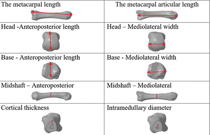
The raw data files obtained from the CT (Philips Incisive 128 slice) scans were reconstructed in three planes (coronal, sagittal, and axial) using RadiAnt DICOM Viewer 2021.1. For each metacarpal, a true AP, lateral, and transverse view was generated based on the geometric axis of the bone, utilizing the RadiAnt software. Image measurement tools were employed to obtain 10 specific parameters for each metacarpal, including total metacarpal length, metacarpal articular length, head AP width, head mediolateral width, midshaft AP diameter, midshaft mediolateral diameter, base AP width, base mediolateral width, midshaft cortical thickness, and midshaft intramedullary diameter ( Fig. 1 ). The accuracy of the imaging software (RadiAnt) used for these measurements had been previously validated in another study and found to be comparable to physical measurements. 6
Fig. 1.

Various measurements of metacarpals.
Metacarpal length was defined as the distance from the midpoint of the head to the most distal part of the base (distal condyle) of the metacarpal.
Metacarpal articular length refers to the distance from the midpoint of the head to the midpoint of the articular surface at the base.
The AP width in the sagittal view and mediolateral width in the coronal view of the head and base were measured at the widest portion from cortex to cortex.
Shaft AP and mediolateral diameters of each metacarpal were measured at the midshaft level (midpoint of articular length) from cortex to cortex.
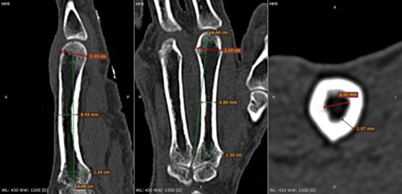
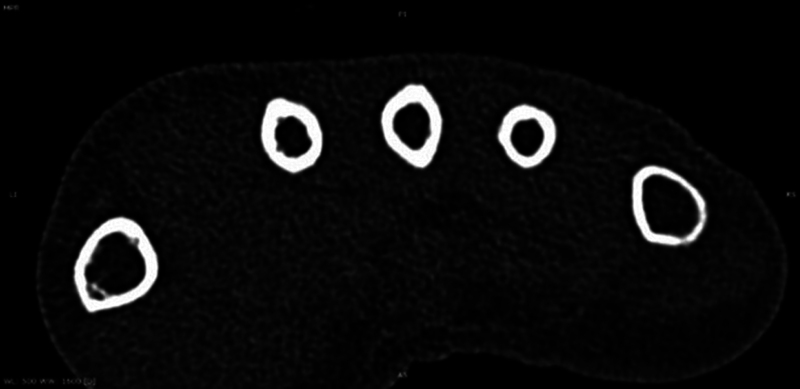
Intramedullary diameter and cortical thickness were measured in axial sections at the midshaft level (midpoint of articular length), and the narrowest diameter was recorded ( Fig. 2 ). It is important to note that the shape of the medullary canal in the transverse section varies among metacarpals; thus, the narrowest diameter was measured ( Fig. 3 ).
Fig. 2.

Computed tomography (CT) scan measurements in sagittal, coronal, and axial views.
Fig. 3.

Shapes of the medullary cavity from 1 to 5 metacarpal at midshaft level.
The study received approval from the Institutional Review Board of both hospitals ensuring adherence to ethical guidelines for research involving human subjects.
Statistical Analysis
The statistical analysis was conducted using SPSS version 23 (IBM Inc., Chicago, Illinois, United States). Descriptive statistics were used to present the study findings, expressing the results as mean and standard deviation with minimum and maximum values. To compare the morphologies of male and female cohorts, the Student's unpaired t -test was applied for parametric distribution, while the Mann–Whitney U test was used for nonparametric distribution. A p -value of < 0.05 was considered statistically significant, with a 95% confidence interval. For assessing the interobserver and intraobserver reliability, the first 20 CT scans were measured separately and twice by two independent investigators at a 1-week interval. The level of agreement between the two observers was determined using the intraclass correlation coefficient (ICC), with values ranging from 0 to 1. ICC values between 0.75 and 0.9 indicated good reliability, while values greater than 0.90 indicated excellent reliability. 5
Results
We assessed CT images from 100 individuals, comprising 50 males and 50 females. The mean age for males was 38.58 ± 12.02 years (min–max: 19–75), while for females, it was 43.60 ± 13.61 years (min–max: 18–77), showing a trend toward statistical significance ( p = 0.054). Table 1 displays the distribution of both male and female right hand (26 images) and left hand (24 images) scans.
Table 1. Descriptive statistics of the studied subjects ( n = 100) by age .
| Sex | N | Minimum | Maximum | Mean | Standard deviation | p -Value |
|---|---|---|---|---|---|---|
| Male | 50 | 19 | 75 | 38.58 | ± 12.02 | 0.054 |
| Female | 50 | 18 | 77 | 43.60 | ± 13.61 |
To evaluate the reliability of our measurements, we calculated the ICC for both intraobserver and interobserver variability. The results ranged from 0.824 to 0.986, indicating good to excellent agreement between the two observers for all 10 parameters measured.
Metacarpal Length
The study involved measuring the articular length from the head of the metacarpal to the articular surface at the base. In men, the lengths were recorded as 4.63, 6.65, 6.49, 5.72, and 5.41 cm for the 1st, 2nd, 3rd, 4th, and 5th metacarpals, respectively, while in women, the lengths were 4.16, 6.04, 5.86, 5.15, and 4.68 cm for the 1st, 2nd, 3rd, 4th, and 5th metacarpals, respectively ( Tables 2 and 3 ). The 2nd metacarpal was found to be the longest in both men and women, and the general pattern followed was 3rd > 4th > 5th > 1st metacarpal. Men generally exhibited greater metacarpal dimensions than women for all measured parameters with statistical significance ( p ≤ 0.05), except for the intramedullary diameter, which showed minimal differences between the sexes ( Table 4 ).
Table 2. Descriptive statistics of various metacarpal measurements in males ( n = 50) .
| Metacarpal measurement | Thumb | 2nd metacarpal | 3rd metacarpal | 4th metacarpal | 5th metacarpal |
|---|---|---|---|---|---|
| Total metacarpal length (cm) | 4.75 ± 0.27 (4.23–5.41) |
6.88 ± 0.37 (6.26–7.83) |
6.66 ± 0.41 (5.35–7.57) |
5.92 ± 0.42 (5.25–7.88) |
5.35 ± 0.36 (5.73–6.65) |
| Metacarpal articular length (cm) | 4.63 ± 0.28 (4.02–5.40) |
6.65 ± 0.37 (5.94–7.47) |
6.49 ± 0.34 (5.82–7.26) |
5.72 ± 0.38 (5.11–7.36) |
5.14 ± 0.36 (4.45–6.43) |
| Head anteroposterior (mm) | 1.28 ± 0.15 (1.06–1.62) |
1.34 ± 0.10 (1.01–1.68) |
1.38 ± 0.11 (1.14–1.65) |
1.28 ± 0.09 (1.07–1.47) |
1.18 ± 0.08 (1.03–1.45) |
| Head mediolateral (mm) |
1.48 ± 0.14 (1.13–1.77) |
1.32 ± 0.12 (1.14–1.76) |
1.28 ± 0.12 (0.99–1.61) |
1.41 ± 0.10 (0.95–1.47) |
1.11 ± 0.14 (0.94–1.90) |
| Base anteroposterior (mm) | 1.45 ± 0.12 (1.02–1.68) |
1.50 ± 0.13 (1.23–1.78) |
1.46 ± 0.09 (1.26–1.65) |
1.20 ± 0.10 (0.98–1.65) |
1.04 ± 0.11 (0.75–1.43) |
| Base mediolateral (mm) |
1.48 ± 0.11 (1.26–1.76) |
1.43 ± 0.14 (1.12–1.71) |
1.22 ± 0.13 (0.90–1.54) |
1.13 ± 0.12 (0.91–1.48) |
1.22 ± 0.13 (1.00–1.53) |
| Midshaft anteroposterior (mm) | 8.60 ± 0.90 (6.92–10.7) |
8.66 ± 0.70 (7.26–9.65) |
8.93 ± 0.86 (7.13–10.4) |
7.38 ± 0.88 (5.91–9.82) |
6.76 ± 0.89 (5.20–9.55) |
| Midshaft mediolateral (mm) | 10.6 ± 1.38 (7.21–14.8) |
8.13 ± 0.77 (6.72–9.60) |
7.60 ± 0.71 (6.02–8.95) |
6.53 ± 0.89 (5.01–8.83) |
7.88 ± 0.85 (6.24–9.77) |
| Cortical thickness (mm) |
1.74 ± 0.39 (1.07–2.90) |
2.57 ± 058 (1.44–3.80) |
2.29 ± 0.52 (1.19–3.53) |
1.86 ± 0.48 (1.01–3.12) |
1.66 ± 0.38 (1.03–2.57) |
| Intramedullary diameter (mm) | 5.92 ± 0.89 (4.55–7.55) |
3.24 ± 0.82 (1.75–5.12) |
3.32 ± 1.02 (1.26–5.54) |
2.90 ± 0.92 (1.21–5.56) |
3.84 ± 1.12 (1.36–6.12) |
Table 3. Descriptive statistics of various metacarpal measurements in females ( n = 50) .
| Metacarpal measurement | Thumb | 2nd metacarpal | 3rd metacarpal | 4th metacarpal | 5th metacarpal |
|---|---|---|---|---|---|
| Total metacarpal length (cm) | 4.28 ± 0.32 (3.69-5.40) |
6.31 ± 0.43 (5.24–7.25) |
6.04 ± 0.42 (5.13–6.96) |
5.29 ± 0.41 (4.27–6.40) |
4.84 ± 0.36 (3.92–5.45) |
| Metacarpal articular length (cm) | 4.16 ± 0.38 (3.63–5.52) |
6.04 ± 0.39 (5.10–6.88) |
5.86 ± 0.39 (5.06–6.66) |
5.15 ± 0.37 (4.27-6.22) |
4.68 ± 0.35 (3.73-5.19) |
| Head anteroposterior (mm) | 1.08 ± 0.09 (0.93–1.46) |
1.21 ± 0.07 (1.07–1.42) |
1.24 ± 0.09 (1.03–1.44) |
1.13 ± 0.08 (1.00-1.34) |
1.05 ± 0.07 (0.87-1.21) |
| Head mediolateral (mm) |
1.37 ± 0.09 (1.15–1.56) |
1.20 ± 0.10 (0.99–1.43) |
1.18 ± 0.11 (0.93–1.45) |
1.05 ± 0.09 (0.85–1.33) |
1.04 ± 0.07 (0.85–1.20) |
| Base anteroposterior (mm) | 1.31 ± 0.11 (1.12–1.83) |
1.30 ± 0.13 (1.02–1.63) |
1.30 ± 0.12 (1.04–1.72) |
1.08 ± 0.10 (0.90–1.39) |
0.94 ± 0.09 (0.78–1.16) |
| Base mediolateral (mm) |
1.35 ± 0.09 (1.15–1.66) |
1.25 ± 0.12 (1.00–1.54) |
1.10 ± 0.08 (0.89–1.32) |
1.02 ± 0.08 (0.85–1.17) |
1.14 ± 0.10 (0.83–1.28) |
| Midshaft anteroposterior (mm) | 7.49 ± 0.81 (5.90–10.2) |
7.59 ± 0.63 (6.37–9-87) |
7.90 ± 0.77 (6.09–9.49) |
6.46 ± 0.81 (4.85–8.72) |
6.17 ± 0.79 (4.36–7.77) |
| Midshaft mediolateral (mm) | 10.1 ± 1.21 (7.49–13.4) |
7.14 ± 0.12 (5.57–8.67) |
6.80 ± 0.65 (5.59–8.36) |
5.81 ± 0.65 (4.76–8.08) |
6.87 ± 0.88 (4.96–9.38) |
| Cortical thickness (mm) |
1.56 ± 0.32 (1.04–2.90) |
2.08 ± 0.49 (1.01–3.73) |
2.01 ± 0.42 (1.27–3.24) |
1.61 ± 0.39 (1.01–2.85) |
1.44 ± 0.26 (1.01–2.26) |
| Intramedullary diameter (mm) | 6.23 ± 0.93 (4.38–8.93) |
3.43 ± 0.77 (2.09–5.22) |
3.12 ± 0.88 (1.24–5.26) |
2.65 ± 0.63 (1.44–3.87) |
3.92 ± 0.74 (2.51–5.76) |
Table 4. Comparing the mean (mm) and standard deviation (SD) of measurements of both men and women of all metacarpals.
| CT scan measurement | N | Sex | 1st metacarpal | 2nd metacarpal | 3rd metacarpal | 4th metacarpal | 5th metacarpal | ||||||||||
|---|---|---|---|---|---|---|---|---|---|---|---|---|---|---|---|---|---|
| Mean | SD ± |
p -Value | Mean | SD ± |
p -Value | Mean | SD ± |
p -Value | Mean | SD ± |
p -Value | Mean | SD ± |
p -Value | |||
| Total MC length | 50 | M | 47.5 | 2.7 | 0.000 | 68.8 | 3.7 | 0.000 | 66.6 | 4.1 | 0.000 | 59.2 | 4.2 | 0.000 | 52.8 | 3.6 | 0.000 |
| 50 | F | 42.8 | 3.2 | 63.1 | 4.3 | 60.4 | 4.2 | 52.9 | 4.1 | 48.4 | 3.6 | ||||||
| MC articular length | 50 | M | 46.3 | 2.8 | 0.000 | 66.5 | 3.7 | 0.000 | 64.9 | 3.4 | 0.000 | 57.2 | 3.8 | 0.000 | 51.4 | 3.6 | 0.000 |
| 50 | F | 48.4 | 3.8 | 60.4 | 3.9 | 58.6 | 3.9 | 51.5 | 3.7 | 46.8 | 3.5 | ||||||
| Head anteroposterior | 50 | M | 12.8 | 1.5 | 0.000 | 13.4 | 1.0 | 0.000 | 13.8 | 1.1 | 0.000 | 12.8 | 0.9 | 0.000 | 11.8 | 0.8 | 0.000 |
| 50 | F | 10.8 | 0.9 | 12.1 | 0.7 | 12.4 | 0.9 | 11.3 | 0.8 | 1.5. | 0.7 | ||||||
| Head mediolateral | 50 | M | 14.8 | 1.4 | 0.000 | 13.2 | 1.2 | 0.000 | 12.8 | 1.2 | 0.000 | 11.4 | 1.0 | 0.000 | 11.1 | 1.4 | 0.003 |
| 50 | F | 13.7 | 0.9 | 12.0 | 1.0 | 11.8 | 1.1 | 10.5 | 0.9 | 10.4 | 0.7 | ||||||
| Base anteroposterior | 50 | M | 14.5 | 1.2 | 0.000 | 15.0 | 1.3 | 0.000 | 14.6 | 0.9 | 0.000 | 12.0 | 1.0 | 0.000 | 10.4 | 1.1 | 0.000 |
| 50 | F | 13.1 | 1.1 | 13.0 | 1.3 | 13.0 | 1.2 | 10.8 | 1.0 | 9.4 | 0.9 | ||||||
| Base mediolateral | 50 | M | 14.8 | 1.1 | 0.000 | 14.3 | 1.4 | 0.000 | 12.2 | 1.3 | 0.000 | 11.3 | 1.2 | 0.000 | 12.2 | 1.3 | 0.000 |
| 50 | F | 13.5 | 0.9 | 12.5 | 1.2 | 11.0 | 0.8 | 10.2 | 0.8 | 11.4 | 1.0 | ||||||
| Midshaft anteroposterior | 50 | M | 8.60 | 0.90 | 0.000 | 8.66 | 0.70 | 0.000 | 8.93 | 0.86 | 0.000 | 7.38 | 0.88 | 0.000 | 6.76 | 0.89 | 0.001 |
| 50 | F | 7.49 | 0.81 | 7.59 | 0.63 | 7.90 | 0.77 | 6.46 | 0.817 | 6.17 | 0.79 | ||||||
| Midshaft mediolateral | 50 | M | 10.6 | 1.38 | 0.118 | 8.13 | 0.77 | 0.000 | 7.60 | 0.71 | 0.000 | 6.53 | 0.89 | 0.000 | 7.88 | 0.85 | 0.000 |
| 50 | F | 10.1 | 1.21 | 7.14 | 0.72 | 6.80 | 0.65 | 5.81 | 0.65 | 6.87 | 0.88 | ||||||
| Intramedullary diameter | 50 | M | 5.92 | 0.89 | 0.102 | 3.24 | 0.82 | 0.240 | 3.32 | 1.02 | 0.291 | 2.90 | 0.92 | 0.124 | 3.84 | 1.12 | 0.672 |
| 50 | F | 6.23 | 0.93 | 3.43 | 0.77 | 3.12 | 0.88 | 2.65 | 0.63 | 3.92 | 0.74 | ||||||
| Cortical thickness |
50 | M | 1.74 | 0.39 | 0.019 | 2.57 | 0.58 | 0.000 | 2.29 | 0.52 | 0.004 | 1.86 | 0.48 | 0.007 | 1.66 | 0.38 | 0.001 |
| 50 | F | 1.56 | 0.32 | 2.05 | 0.49 | 2.01 | 0.42 | 1.61 | 0.39 | 1.44 | 0.26 | ||||||
Note: The bold values signifies the intramedullary diameter measurements showed minimal differences between the sexes without any statistical significance.
Abbreviations: CT, computed tomography; F, female; M, male; MC, metacarpal.
Intramedullary Diameter
The narrowest diameter of the medullary cavity was measured at the midshaft level. For the 1st to 5th metacarpals, the intramedullary diameters in millimeters were recorded as 6.0, 3.3, 3.2, 2.7, and 3.8, respectively. The maximum intramedullary diameter was observed in the thumb, measuring 6.23 ± 0.93 mm, while the 4th metacarpal exhibited the minimum intramedullary diameter at 2.65 ± 0.63 mm. Interestingly, there was no statistically significant difference in intramedullary diameter between men and women.
Tables 5 and 6 compare our study's results with findings from other studies 7 8 9 10 in the literature.
Table 5. Comparison of metacarpal length in various studies (mm).
| Author | n | Study type | 1st MC | 2nd MC | 3rd MC | 4th MC | 5th MC |
|---|---|---|---|---|---|---|---|
| Our study, India |
100 | CT scan | 43.9 | 63.4 | 61.7 | 54.3 | 49.1 |
| Michael Okoli, Philadelphia 8 |
57 | CT scan | 67.6 | 64.2 | 56.7 | 51.4 | |
| Abdullah Örs, Turkey 9 |
105 | CT scan | 66.3 | 63.5 | 54.6 | 50.5 | |
| Andrew Sephien, Florida 10 | 140 | X-ray | 68.7 | 66.6 | 58.9 | 53.9 | |
| Doaa A. El Morsi, Egypt 7 |
100 | X-ray | 46.5 | 68.4 | 65.4 | 57.5 | 54.1 |
| D. Troy Case, North Carolina 13 |
20 | Dry bones Osteometric board |
44.8 | 64.0 | 61.5 | 55.3 | 51.3 |
Abbreviations: CT, computed tomography; MC, metacarpal.
Table 6. Comparison of intramedullary diameter in various studies (mm).
| Author | n | Study type | 1st MC | 2nd MC | 3rd MC | 4th MC | 5th MC |
|---|---|---|---|---|---|---|---|
| Our study, India |
100 | CT scan | 6.0 | 3.3 | 3.2 | 2.7 | 3.8 |
| Michael Okoli, Philadelphia 8 |
57 | CT scan | 2.6 | 2.7 | 2.3 | 3.0 | |
| Abdullah Örs, Turkey 9 |
105 | CT scan | 3.2 | 3.3 | 2.9 | 4.1 | |
| Andrew Sephien, Florida 10 | 140 | X-ray | 3.6 | 3.4 | 3.2 | 4.1 | |
| Don Hoang, Seattle 17 |
100 | CT scan | 5.7 | 3.2 | 3.2 | 2.8 | 3.7 |
| Mark L. Dunleavy, USA 16 |
111 | CT scan | 3.1 | 3.1 | 2.6 | 3.3 |
Abbreviations: CT, computed tomography; MC, metacarpal.
Discussion
In 1914, Martin established a standard system for measuring the human skeleton, 11 and later in 1993, Scheuer and Elkington provided detailed measurements for metacarpals, including interarticular length, base width, base height, head width, head height, and maximum midshaft diameter. 12 Various methods and instruments, such as calipers and mini osteometric boards, have been used by researchers for measuring metacarpals. However, these instruments' measurements often exhibit significant intra- and interobserver variation. 13
Some studies, like El Morsi and Al Hawary, used plain radiographs to measure metacarpal length for sex determination. 7 Meanwhile, Deschênes conducted a geometric morphometric analysis by three-dimensional scanning metacarpals and creating models for measurements, primarily focused on sex determination and biological anthropometry. 14
Previous research on the morphometry of human metacarpals was mainly conducted on dry bones or X-ray radiographs. However, accurate evaluation of true metacarpal anatomy can be challenging in two-dimensional conventional radiograph images due to the overlap of adjacent metacarpals in lateral views and the difficulty in obtaining or standardizing true orthogonal views. CT scans have proven to be a valuable tool for morphometric analysis of bones, as they provide accurate depictions of anatomy and allow manipulation of images to obtain true AP and lateral views without altering size and shape.
Our study used CT scans of 100 hands to measure various parameters of all the metacarpals in Indian individuals and compared the results with other studies. Due to the reconstruction, all metacarpals examined were in true isolated lateral or AP positions, enabling more precise evaluations.
Surgical fixation of metacarpal neck and shaft fractures has gained significance, with various techniques developed, including antegrade and retrograde Kirschner wire fixation, plate, and screw constructs, and intramedullary headless screw (IMHS) fixation. IMHS fixation offers several advantages, such as easy exposure, early mobilization, and reduced hardware prominence, contributing to its growing popularity.
However, when using IMHS fixation, the narrowest part of the medullary cavity is typically found at the midshaft, gradually expanding toward the metaphysis. 15 This variability in medullary cavity size poses challenges in selecting the appropriate screw size. Smaller screws might lead to inadequate intramedullary purchase and instability, while larger diameter screws could cause iatrogenic fractures during placement. To ensure proper fixation, the correct screw size depends on the intramedullary diameter, with an additional 0.5 to 1 mm for intramedullary cortical purchase.
Our study measured the narrowest diameter at the midshaft level and found that the medullary cavity was narrowest in the 4th metacarpal and widest in the 5th metacarpal and thumb. Notably, there was no statistically significant difference in intramedullary diameter between the sexes. Our findings align with previous studies conducted by Dunleavy et al 16 and Hoang et al, 17 who also reported similar midshaft measurements. Hoang et al. suggested using minimum intramedullary headless compression screw diameters of 3.5 mm (for the ring finger), 4.0 mm (for the index, middle, and little fingers), and 5.5 mm (for the thumb), based on the intramedullary diameter with an additional 0.5 to 1 mm for cortical purchase. In our study population, we found similar intramedullary diameters, indicating that the same diameter screws can be used, except for the thumb, where a 6.00-mm screw would be ideal. Fortunately, most commercially available headless screw systems (IMHS) fall within this range.
Despite the advantages of IMHS fixation, concerns persist regarding potential cartilage damage caused by the screw head. Measuring the articular surface of the metacarpal head is challenging due to its undefined shape. To assess the articular surface, we measured the head's AP (width) and mediolateral (width) dimensions. We found them to be comparable to those reported in the literature, as seen in the study conducted by ten Berg et al. 18 In their three-dimensional CT study, ten Berg et al found that the mean articular surface defect for the intramedullary compression screw was 12% of the total articular area of the metacarpal head for the 3.0-mm head screw and 8% for the 2.5-mm head screw.
The strength of our study is that we were able to maximize the accuracy of the anatomy and consistency in measurement using CT scans. We reformatted the CT images to examine all metacarpals in true lateral and AP views, as the metacarpals are inherently not in the same plane. Reformats that are along the axis of the long bone lead to better resolution and more accurate measurement.
The limitation of this study is the small study population from two regions and may not be representative of the entire country. Large studies including various regions will give a better understanding. Besides, age groups were not evaluated in our study as Dunleavy et al 16 ; reported that increased patient age was associated with increased medullary canal diameters.
Conclusion
This study's findings present a valuable reference for anatomical measurements of metacarpals 1 to 5 in mature adults, offering crucial insights for optimizing plating, pinning, and intramedullary screw fixation in metacarpal fractures. The 2nd metacarpal was consistently identified as the longest in both men and women, with a general pattern of 3rd > 4th > 5th > 1st metacarpal lengths. At the midshaft level, the thumb exhibited the maximum intramedullary diameter, while the 4th metacarpal demonstrated the minimum intramedullary diameter. Regarding gender differences, men generally demonstrated larger metacarpal dimensions than women, except for the intramedullary diameter, which exhibited minimal differences between the sexes. These significant insights into the anatomical variations of metacarpals within the Indian population have the potential to influence clinical decisions and stimulate further research in this field.
Acknowledgments
We extend our sincerest gratitude to Dr. Mukund Thatte for his visionary guidance and unwavering support in embarking on the normative data of the Indian hand. His foresight and encouragement have been instrumental in steering our research endeavors toward a deeper understanding of Indian demographics and contributing significantly to advancing our scientific knowledge.
Funding Statement
Funding None.
Conflict of Interest None declared.
Ethical Approval
Approval Letter Number: SIEC/2021/456.
SIEC Meeting Number: SIEC/2021/52.
Authors' Contributions
M.V.R. served as the principal investigator and was responsible for data collection, measurement, analysis, and manuscript writing. M.P.G. contributed to data collection, analysis, and manuscript writing. M.M.K. handled measurements, statistical analysis, and manuscript editing. A.K.B. was involved in data collection, statistical analysis, and manuscript editing. A.A. was responsible for data retrieval, literature search, and manuscript writing. M.R.T. contributed to the concept, analysis, and manuscript editing.
References
- 1.Chung K C, Spilson S V. The frequency and epidemiology of hand and forearm fractures in the United States. J Hand Surg Am. 2001;26(05):908–915. doi: 10.1053/jhsu.2001.26322. [DOI] [PubMed] [Google Scholar]
- 2.Hazan J, Azzi A J, Thibaudeau S. Surgical fixation of metacarpal shaft fractures using absorbable implants: a systematic review of the literature. Hand (N Y) 2019;14(01):19–26. doi: 10.1177/1558944718798856. [DOI] [PMC free article] [PubMed] [Google Scholar]
- 3.Nakashian M N, Pointer L, Owens B D, Wolf J M. Incidence of metacarpal fractures in the US population. Hand (N Y) 2012;7(04):426–430. doi: 10.1007/s11552-012-9442-0. [DOI] [PMC free article] [PubMed] [Google Scholar]
- 4.Lamraski G, Monsaert A, De Maeseneer M, Haentjens P. Reliability and validity of plain radiographs to assess angulation of small finger metacarpal neck fractures: human cadaveric study. J Orthop Res. 2006;24(01):37–45. doi: 10.1002/jor.20025. [DOI] [PubMed] [Google Scholar]
- 5.Tolentino E S, Yamashita F C, de Albuquerque S et al. Reliability and accuracy of linear measurements in cone-beam computed tomography using different software programs and voxel sizes. J Conserv Dent. 2018;21(06):607–612. doi: 10.4103/JCD.JCD_314_18. [DOI] [PMC free article] [PubMed] [Google Scholar]
- 6.Koo T K, Li M Y. A guideline of selecting and reporting intraclass correlation coefficients for reliability research. J Chiropr Med. 2016;15(02):155–163. doi: 10.1016/j.jcm.2016.02.012. [DOI] [PMC free article] [PubMed] [Google Scholar]
- 7.El Morsi D A, Al Hawary A A. Sex determination by the length of metacarpals and phalanges: X-ray study on Egyptian population. J Forensic Leg Med. 2013;20(01):6–13. doi: 10.1016/j.jflm.2012.04.020. [DOI] [PubMed] [Google Scholar]
- 8.Okoli M, Lutsky K, Rivlin M, Katt B, Beredjiklian P. Metacarpal bony dimensions related to headless compression screw sizes. J Hand Microsurg. 2020;12 01:S39–S44. doi: 10.1055/s-0039-3400443. [DOI] [PMC free article] [PubMed] [Google Scholar]
- 9.Örs A, Çolak T, Bamaç B et al. Morphometric evaluation of second to fifth metacarpals for retrograde intramedullary headless screw fixation. Int J Morphol. 2022;40(04):1075–1080. [Google Scholar]
- 10.Sephien A, Bethel C F, Gulick D, Nairn C, Ourn F, Schwartz-Fernandes F A. Inter-relationships of metacarpals 1 to 5, regarding their length, metaphyseal midshaft width, articular surface area of head and base, age, and sex: a cadaveric study. Hand (N Y) 2021;16(05):706–713. doi: 10.1177/1558944719880026. [DOI] [PMC free article] [PubMed] [Google Scholar]
- 11.Martin R.Textbook of Anthropology in Systematic Presentation: With Special Consideration of Anthropological Methods; For Students, Doctors, and ExplorersJena: Gustav Fischer; 1914. 10.11588/diglit.37612#0001 [DOI]
- 12.Scheuer J L, Elkington N M. Sex determination from metacarpals and the first proximal phalanx. J Forensic Sci. 1993;38(04):769–778. [PubMed] [Google Scholar]
- 13.Case D T, Rawlins C M, Mick C B. Measurement standards for human metacarpals. Am J Phys Anthropol. 2015;157(02):322–329. doi: 10.1002/ajpa.22700. [DOI] [PubMed] [Google Scholar]
- 14.Deschênes V A.Asymmetry of Hands-On Modern Human: A Geometric Morphometric Analysis of Metacarpal BonesArchéol Prehistory. 2017. [Dumas ID: dumas-01723522]
- 15.Lazar G, Schulter-Ellis F P. Intramedullary structure of human metacarpals. J Hand Surg Am. 1980;5(05):477–481. doi: 10.1016/s0363-5023(80)80079-7. [DOI] [PubMed] [Google Scholar]
- 16.Dunleavy M L, Candela X, Darowish M. Morphological analysis of metacarpal shafts with respect to retrograde intramedullary headless screw fixation. Hand (N Y) 2022;17(04):602–608. doi: 10.1177/1558944720937362. [DOI] [PMC free article] [PubMed] [Google Scholar]
- 17.Hoang D, Vu C L, Jackson M, Huang J I. An anatomical study of metacarpal morphology utilizing CT scans: evaluating parameters for antegrade intramedullary compression screw fixation of metacarpal fractures. J Hand Surg Am. 2021;46(02):1490–1.49E10. doi: 10.1016/j.jhsa.2020.08.007. [DOI] [PubMed] [Google Scholar]
- 18.ten Berg P W, Mudgal C S, Leibman M I, Belsky M R, Ruchelsman D E. Quantitative 3-dimensional CT analyses of intramedullary headless screw fixation for metacarpal neck fractures. J Hand Surg Am. 2013;38(02):322–33000. doi: 10.1016/j.jhsa.2012.09.029. [DOI] [PubMed] [Google Scholar]


