Abstract
The same elements can yield disparate nanoproducts that may elicit different harmful effects in cells and organisms. This study aimed to compare the effects of copper (Cu NPs) and copper oxide (CuO NPs) nanoparticles and Cu2+ (from CuSO4) on the physico-biochemical variables of rainbow trout spermatozoa. The cell death assay, along with the activation of caspases 8 and 9, the level of reactive oxygen species (ROS), and the percentage of cells exhibiting a high mitochondrial membrane potential (MMP) were quantified over 24-hour incubation. Interestingly, during exposure, all copper products induced cell apoptosis. However, Cu NPs had a stronger effect than CuO NPs, while the impact of the Cu in ionic form was found to be between the other two compounds. The extrinsic and intrinsic apoptotic pathways were activated, as evidenced by the activation of caspases 8 and 9. Initially, caspase activation increased without a corresponding decrease in MMPs but prolonged exposure resulted in a significant decrease in MMP levels. In all treated cells, the ROS levels increased over time. Further studies are needed to confirm the lower CuO NPs’ toxicity compared to Cu NPs because their effect on cells also depends on many other parameters such as size or shape.
Supplementary Information
The online version contains supplementary material available at 10.1038/s41598-024-72956-1.
Keywords: Nanotoxicology; Nanocopper; Fish spermatozoa; ROS, apoptosis; MMP
Subject terms: Biochemistry, Cell biology, Environmental sciences, Nanoscience and technology
Introduction
In recent years, there has been a notable advancement in the field of nanotechnology, resulting in the incorporation of nanoparticles into the environment. Among these developments, copper based nanoparticles e.g. Cu NPs or CuO NPs, have become widely used due to its antimicrobial activity1–4 and prospective applications in the biomedical field, including drug delivery5,6 and diagnostics7, as well as in agricultural and other commercial applications8–11. However, the widespread use of nanoparticles (NPs) has raised concerns about their potential impact on the environment12–15, particularly in the aquatic environment, where waste ultimately accumulates16. Pollutants may exert an adverse impact on aquatic species by impeding their growth and development17,18, as well as the reproductive cell and the effectiveness of reproduction which may ultimately threaten their existence19,20.
Fish spermatozoa have been used as biomarkers to determine the effects of contaminants in aquatic environments21–23. However, current state of the art related to the reproductive biology of aquatic organisms is limited to the effects of metal ions, such as copper19,24,25, zinc26,27, and heavy metal derivatives28,29 on sperm motility, viability, and overall fertilisation potential. Recently, attention has shifted to studying the effects of nanoparticles on fish spermatozoa. Copper contamination of water has been found to primarily reduces sperm viability, which can negatively affect reproduction19,30,31. The harmful effects of copper on aquatic organisms such as fish19,25,31–33, daphnia34–37 and algae38–41 have been demonstrated. The median lethal concentration (LC50) for Cu and Cu NPs in catfish was found to be 8.0-10.5 and 3.9-5.0 mg L−1, respectively, following an exposure period of between 24 and 96 h42. Organisms differ in their susceptibility to the adverse effects of pollution25,37,43–45. The toxicological impact of NPs is influenced by a number of factors, including concentration, exposure duration, particle size, surface area, dissolution, agglomeration and the characteristics of aquatic ecosystem. Our previous study46 showed that the effect of copper nanoparticles on the kinetics of rainbow trout spermatozoa is product-dependent. Smaller Cu nanoparticles have also been shown to be more toxic to zebrafish embryos than larger particles47. Furthermore, different kind of nanoparticles based on the same elements have different effects on spermatozoa. In our previous study31, the effects of copper based nanoparticles (that is, Cu NPs and CuO NPs) on changes in sperm motility, analysed by the CASA (computer-assisted sperm analysis), were investigated and compared with effect of the ionic form of copper (from CuSO4). The findings demonstrated that Cu NPs and ionic Cu were more deleterious to spermatozoa kinematics than CuO NPs31. In the aforementioned study, incubation was conducted at various concentrations, including 50 mg Cu L−1, which is identical to the concentration used in the current study.
The effects of copper nanoparticles on cellular mechanisms have been investigated by several authors. Available evidence indicates that nanoparticles containing copper have the capacity to disrupt enzymatic activity30,48, induce oxidative stress1,42,49,50, cause cell membrane disruption51, and lead to mitochondrial dysfunction52. A substantial body of evidence from numerous studies has consistently demonstrated that CuO NPs induce cytotoxicity in a range of cell types, including Chinook salmon cells, human lung epithelial cells, human airway smooth muscle cells and airway epithelial cells, primarily by generating oxidative stress53–56. Furthermore, studies have indicated that CuO NPs induce mitochondria-mediated apoptosis in human hepatocarcinoma cells57. As proposed by Thit et al.58, the toxicity mechanism involves the uptake of CuO NPs by cells via endocytosis, resulting in the generation of Reactive Oxygen Species (ROS). Subsequently, ROS causes DNA damage, initiating a signalling pathway that ultimately results in apoptosis or cell death58. Additionally, a study observed a significant increase in the expression of antioxidant enzymes in the epididymides of Chinese soft-shelled turtles after 24 h of incubation with Cu NPs and CuSO430. Several studies have indicated that the toxicity of copper nanoparticles can be attributed to a combination of two factors. First, copper nanoparticles can penetrate cells or migrate to various tissues and organs, thereby disrupting their metabolic activities42. Second, the toxicity of nanoparticles to organisms is linked to the release of soluble copper ions11,59,60.
In our previous study on spermatozoa motility tracked by computer-assisted sperm analysis (CASA) we demonstrated that copper nanoparticles (Cu NPs) and ionic copper (CuSO4) exert a more pronounced immobilisation effect than copper oxide nanoparticles (CuO NPs)31.
This contribution compares their effects on the physico-biochemical variables of rainbow trout spermatozoa by means of cell death assay, along with the activation of caspases 8 and 9, the level of reactive oxygen species (ROS), and the percentage of cells exhibiting a high mitochondrial membrane potential (MMP) over 24-hour incubation.
Results
Cell death assay
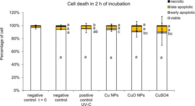
After two hours of incubation of rainbow trout spermatozoa in solutions containing Cu NPs, CuO NPs and CuSO4, a significant increase (p < .05) in the percentage of early apoptotic cells was found (1.82%, 1.56%, and 1.71%, respectively), compared to the negative control (0.14%). Changes in the percentage of viable cells, late apoptotic cells, and necrotic cells did not differ among the groups (p > .05) (Fig. 1).
Fig. 1.
The influence of two hours incubation in copper compounds Cu NPs, CuO NPs and CuSO4, at a concentration of 50 mg Cu L−1 on cell death. Different superscripts indicate a significant difference in a given stage of cell death among the experimental groups. Median and interquartile range (n = 17, Kruskal-Wallis test, p < .05).
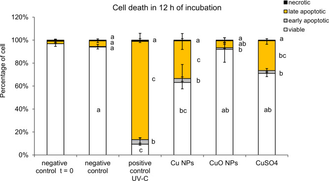
After a 12 h incubation of spermatozoa with copper products, the percentage of early-apoptotic cells remained significantly higher in solutions containing Cu NPs, CuO NPs and CuSO4 (3.32, 2.17, 3.27%, respectively) compared to the control (0.46%) (Fig. 2). Additionally, a significant increase (p < .05) in the percentage of late apoptotic cells compared to the control occurred after incubation with Cu NPs and CuSO4 (4.80% in the control, 32.77% in Cu NPs, and 30.77% in CuSO4, respectively). The percentage of necrotic cells was similar in all the groups. At the same time, there was a significant decrease in the percentage of live cells with Cu NPs (68.23%) compared to the control (93.57%). In the case of CuO NPs and CuSO4, the percentage decrease was not significant (84.40% and 85.22%, respectively) (Fig. 2).
Fig. 2.
The influence of 12 h incubation with copper compounds Cu NPs, CuO NPs and CuSO4, at a concentration of 50 mg Cu L−1 on cell death. Different superscripts indicate a significant difference in a given stage of dying cells among experimental groups. Median and interquartile range (n = 17, Kruskal-Wallis test, p < .05).
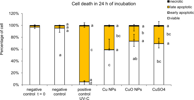
Following a 24 h incubation period in copper-containing compounds, the number of late apoptotic cells increased significantly (p < .05) from 3.86% in the control group to 36.43% in Cu NPs, 31.55% in CuSO4 and 23.95% in CuO NPs. The effect of copper compounds did not result in alterations in the proportion of necrotic and early apoptotic cells compared to the control (Fig. 3). The percentage of viable cells significantly decreased in Cu NPs and CuSO4 (from 89.46% in the control to 63.96% and 68.83%, respectively), whereas no significant change was observed in the case of CuO NPs (79.69%). The toxic effect exerted by Cu NPs was found to be greater than that exerted by CuO NPs (p < .05) while for CuSO4 exhibited a similar effect on both NPs (Fig. 3).
Fig. 3.
The influence of 24 h incubation in copper compounds Cu NPs, CuO NPs and CuSO4, at a concentration of 50 mg Cu L−1 on cell death. Different superscripts indicate a significant difference in a given stage of dying cells among experimental groups. Median and interquartile range (n = 17, Kruskal-Wallis test, p < .05).
Caspase 8 and 9 activations
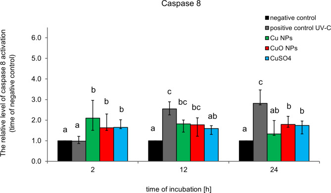
After 2 h of incubation, the relative level of caspase 8 activation in rainbow trout sperm was higher in all copper-containing compounds than in the negative control (Fig. 4). After 12 and 24 h of incubation, the level of caspase 8 activation fluctuated slightly, remaining similar or higher in the copper treatment compared to the control, with a range of 1.3 to 2.1 times that of the control (Fig. 4).
Fig. 4.
The relative level of caspase 8 activation following incubation with copper-containing compounds at a concentration of 50 mg Cu L−1 including Cu NPs, CuO NPs, and CuSO4, was determined as a fold change compared to the negative control. Different superscripts indicate significant differences among experimental groups at a given incubation time. Median and interquartile range (n = 9, Kruskal-Wallis test, p < .05).
After 2 h of incubation, the relative levels of caspase 9 were similar (CuO NPs and CuSO4, p > .05) or significantly lower (Cu NPs, p < .05) compared to the control (0.69-fold control) (Fig. 5). After 12 h of incubation in solutions containing copper, the level of caspase 9 activation increased significantly. The level was found to be similar and was approximately 1.80 times higher than that in the control group for all copper solutions. After 24 h of incubation, the greatest increase in caspase 9 activation levels was observed in sperm incubated with nanoparticles (3.19-fold control in Cu NPs and 2.98-fold control in CuO NPs). In CuSO4 the level of caspase 9 activation was also significantly higher than that in the control (2.27-fold control), but lower than that in solutions containing nanoparticles (Fig. 5).
Fig. 5.
The relative level of caspase 9 activation following incubation with copper-containing compounds at a concentration of 50 mg Cu L−1 including Cu NPs, CuO NPs, and CuSO4, was determined as a fold-change compared to the control. Different superscripts indicate a significant difference among experimental groups at a given incubation time. Median and interquartile range (n = 21, Kruskal-Wallis test, p < .05).
Reactive oxygen species (ROS)
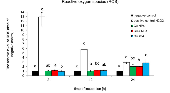
After 2 h of incubation, the ROS level was found to be comparable between the Cu NPs and CuO NPs solutions, while exhibiting a lower level in CuSO4 compared to the control (Fig. 6). A significant increase in ROS levels (1.22-fold control) compared to the control was observed after 12 h incubation of sperm in CuO NPs. After 24 h, increased ROS levels were observed for all the tested copper compounds. In Cu NPs and CuO NPs, ROS levels increased approximately 2.05 times compared to the control, whereas in CuSO4, they increased by 2.86-times (Fig. 6).
Fig. 6.
The reactive level of Reactive Oxygen Species (ROS) in rainbow trout spermatozoa cells was followed by incubation in solutions containing copper compounds Cu NPs, CuO NPs, and CuSO4 at a concentration of 50 mg Cu L−1. Different superscripts indicate a significant difference among experimental groups at a given incubation time. Median and interquartile range (n = 12, Kruskal-Wallis test, p < .05).
Mitochondrial membrane potential (MMP)
After 2 h of incubation in solutions containing copper compounds, the percentage of cells with a high mitochondrial membrane potential remained unchanged in comparison to the control (Fig. 7). After 12 h, a significant (p < .05) increase in the percentage of cells with high MMP was observed in spermatozoa incubated with CuO NPs (98.75%) and CuSO4 (97.59%) compared with the control (87.23%). No significant changes were observed in spermatozoa incubated with Cu NPs (p > .05). However, after 24 h of incubation in solutions containing copper compounds, a decrease in the percentage of cells with a high MMP was observed. A significant decrease was observed for Cu NPs (64.99%) and CuSO4 (74.37%) compared with the control (93.29%). Conversely, the decrease in the percentage of cells with high MMP levels incubated with CuO NPs (79.25%) was not significant (p > .05) when compared to the control. The percentage were similar in the three-copper solution (Fig. 7).
Fig. 7.
Changes in the percentage of rainbow trout spermatozoa with high mitochondrial membrane potential (MMP) following incubation in solutions containing copper compounds Cu NPs, CuO NPs, and CuSO4 at a concentration of 50 mg Cu L−1. Different superscripts indicate a significant difference among experimental groups at a given incubation time. Median and interquartile range (n = 17, Kruskal-Wallis test, p < .05).
Discussion
The increasing production of nanocopper products has highlighted the necessity to gain a deeper understanding of the effects of nanoparticles on aquatic organisms and the pathways through which they exert their effects. However, data concerning the influence of Cu NPs on fish (and particularly on spermatozoa) are limited, and their results are not homogeneous45,61–63.
A previous study31 and the present study demonstrated that copper nanoparticles (Cu NPs) are more toxic to rainbow trout spermatozoa than copper oxide nanoparticles (CuO NPs). The initial study indicated that there was a higher degree of spermatozoa immobilisation, whereas a subsequent study demonstrated a lower proportion of viable cells. In the aforementioned studies, the effects of the Cu NPs were comparable to those of the ionic form of Cu. Similarly, Al-Bairuty et al.64 observed that the toxicity of Cu NPs and CuSO4 to juvenile rainbow trout is comparable. Administration of both substances resulted in the development of pathological conditions in the target organs. In a separate study of this species, a notable distinction was observed in the extent of brain damage, whereby the damage caused by Cu NPs was more pronounced than that induced by the same concentrations of CuSO465. In their investigation of the impact of Cu NPs and CuSO4, Hoseini et al.61 observed that the ionic form induced a more pronounced harmful effect and necrosis in common carp (Cyprinus carpio) liver and kidney cells compared to the nano form61. Similarly, a more harmful effect was observed in the ionic form by Yang et al.30, who investigated the effects on Chinese soft-shelled turtle (Pelodiscus sinensis) spermatozoa and Kowalska-Góralska et al.63, examined the effects on the eggs of rainbow trout (Oncorhynchus mykiss W.).
The preceding CASA study indicated that CuO NPs exhibit a diminished level of toxicity in comparison to CuSO431. This could be attributed to the fact that the initial studies encompassed a longer exposure period of up to 96 h, whereas the current study was conducted for a duration of 24 h. The reduced toxicity of CuO NPs compared to the ionic form of copper (derived from CuCl2) was also demonstrated by Mansano et al.66, who evaluated toxicity in fish gill cells. The study demonstrated that copper ions caused greater increases in the proportion of early apoptotic and late apoptotic/necrotic cells than CuO NPs66.
Analysis of caspase activation within the cell enables the identification of the pathway that directs the cell towards programmed cell death. In the present study, the activation levels of caspase 8 were measured for the extracellular pathway (extrinsic) and caspase 9 for the mitochondrial (intrinsic) pathway of cell death. The level of caspase 8 activition under the influence of the tested copper compounds was observed to be higher from as early as two hours of incubation, indicating that the cell death signal originates externally through death receptors. The studies revealed no significant differences in caspase 8 activity between copper nanoproducts and the ionic form. Conversely, caspase 9 activation was observed to increase with incubation duration. Following a 24-hour incubation period, a significant elevation in caspase 9 activation was observed in spermatozoa, particularly in the presence of nanoproducts. This indicated that the studied nanoproducts induced apoptosis by intensifying the activation of the intrinsic pathway in response to their presence. This finding is in accordance with the results of a study conducted by Sarkar et al.50, who observed the activation of caspase 3 and 8 in kidney tissue under Cu NPs exposure. The same type of nanocopper induced the activity of caspase 3 and caspase 9 in the liver of juvenile fish Takifugu fasciatus67. Caspase 3 is an essential protein. Siddiqui et al.57 observed a significant increase in caspase 3 activity in human hepatocarcinoma cells in the presence of Cu NPs.
Reactive oxygen species (ROS) play a crucial role in cellular signalling and the maintenance of cellular homeostasis. Excess of these substances can result in oxidative stress and subsequent damage to cells. The present study revealed a significant elevation in ROS generation in cells following a 24-hour incubation period with copper products. The findings of the present study are consistent with those previously reported by Yang et al.30, who demonstrated that after 24 h, both Cu NPs and CuSO4 induced a significant increase in ROS production in spermatozoa from Chinese soft-shelled turtles (Pelodiscus sinensis), where CuSO4 markedly elevated ROS production compared to Cu NPs30. Exposure of fish gills of H. eques to Cu ions (from CuCl2) also resulted in a higher increase in ROS production than that observed with CuO NPs66. Similarly, Sarkar et al.50 observed that exposure to Cu NPs elevated ROS production.
ROS generation is associated with impairment in mitochondrial membrane permeability and the respiratory chain. The mitochondrial membrane potential (MMP) serves as a crucial indicator for evaluating the functional capacity of mitochondria, a pivotal organelle in cellular energy production. Modifications of this parameter are associated with a range of physiological and pathological conditions. It is widely acknowledged that during apoptosis, there is a decrease in the mitochondrial membrane potential (MMP) as a result of mitochondrial outer membrane permeabilisation (MOMP). The findings of our study support this hypothesis, as after 24 h of incubation of rainbow trout spermatozoa, a decrease in MMP was observed for both Cu NPs and CuSO4. An anomalous result was observed after 12 h of incubation, whereby an increase in MMP was observed in the copper-containing solutions. This finding is inconsistent with the widely described mechanism of caspase 9 activation which requires earlier MOMP. It is plausible that a 12-hour incubation period with copper products may result in caspase 9 activation through a mechanism that does not involve MOMP. An alternative mechanism has also been described in other studies. Specifically, caspase 9 can be directly activated by caspase 8 without MOMP in the extrinsic apoptotic pathway in cancer HeLa cells68 and murine cells69,70. This temporary increase in MMP may explain the higher spermatozoa movement speed following incubation with the same copper compounds at specific concentrations, as reported in our earlier study31. Furthermore, an increase in spermatozoa velocity under the influence of nanoproduct exposure under certain experimental conditions has also been documented by other researchers48,71. It may be related to the fact that several studies have shown that copper can improve (or inhibit) enzyme function and influence gene expression, affecting some vital functions17,18,72 but further research is required in this case.
The toxicity of NPs depends on a multitude of factors, including their concentration, exposure time, chemical composition, particle size, surface area, dissolution, agglomeration, and other physical properties. Several studies have indicated that the toxicity of copper nanoparticles can be attributed mainly to penetration into cells or migration to various tissues and organs, thereby disrupting their metabolic activities and linked to the release of soluble copper ions11,42,59,60. Some studies have demonstrated that smaller nanoparticles have a greater capacity to exaggerate toxic effects than larger ones46,47. In the present study, it was observed that the average size of the Cu NPs was approximately twice smaller that of CuO NPs (Fig. S1). Additionally, its positive zeta potential was four times smaller in respect to CuO NPs (Table S1). It is plausible that these parameters contributed to enhanced cell penetration, resulting in a greater degree of toxicity. What is more, from optical study it was observed that the CuO nanoparticles released more Cu2+ to the artificial seminal plasma solution within 24 h incubation test (Fig. S3). This could indicate that the harmful effect of CuO NPs can be related to the released of Cu2+ instead of the solid CuO NPs. It also explains the reason why the toxicity of CuSO4 is placed between Cu NPs and CuO NPs. Therefore, basing on our research one can conclude that copper based compounds increase their harmful effect on the studied cells in the following order: Cu NPs > Cu 2+> CuO NPs.
Conclusion
The study demonstrated that the diverse copper based nanoparticles and Cu2+ subjected to examination exhibited disparate toxic effects on rainbow trout spermatozoa. The Cu NPs were observed to induce cell death by apoptosis to a greater extent than the CuO NPs. The toxic effect of CuSO4 was found to be situated between the other two compounds. The physico-biochemical variables were found to be similar, with the exception of caspase 9 activation, which was observed to be higher under the influence of the nanoparticles. Different effects exerted by NPs on spermatozoa, apart from their chemical properties, could have been influenced by their different size and zeta potential. The size and zeta potential of Cu NPs were found to be lower than those of CuO NPs. It may be hypothesised that the parameters of Cu NPs contribute to easier penetration into cells resulting in greater toxicity to cells. Nevertheless, further research is recommended to determine what property of the nanoparticles predominantly influences its impact on cells.
Methods
Material
During the artificial spawning season, employees of The Trout Breeding Center “Kuźniczka” in Wieleń (coordinates: 52°57′03.8232″N 16°13′52.0320″E) stripped sperm from rainbow trout (Oncorhynchus mykiss W.) into plastic containers. The containers were then placed on ice (2–4 °C) and transported to the laboratory, which took approximately 2 h. Samples with high milt quality (motility exceeding 80% and curvilinear velocity above 100 μm s-1) were selected for experiments based on computer-assisted sperm analysis (CASA) by Microptic S.L. (Barcelona, Spain), the Sperm Class Analyzer (SCA) v. 4.0.0 (https://www.micropticsl.com/products/sperm-class-analyzer-casa-system/).
To select the best portion of milt for the study, spermatozoa activation was triggered with 30 mM NaCl buffered with 20 mM Tris, pH 9, at 8 oC with 200-fold dilution. The temperature of the solution was maintained using a cooling block (FINEPCR, Seoul, Korea) and microscope table cooling device (Semic Bioelektronika, Kraków, Poland). Half-second films (25 frames) were recorded at 10 s after activation. Activation and film recording was repeated three times. The criterion for defining motility was an average path velocity VAP > 20 μm s−1. Spermatozoa concentration was assessed in a Bürker chamber counting by the SCA at a dilution of 3000x with 0.8% NaCl. The percentage of motile sperm in the samples selected for the study ranged from 80 to 90% and mean curvilinear velocity and linearity were above 100 μm s−1 and 80%, respectively. Spermatozoa concentration ranged from 11.1 to 17.3 × 10⁹ mL−1. The experimental procedures conducted on sperm do not require permission from the Ethics Committee for Animal Experimentation, according to Polish law.
Nanoproducts and experiment description
Cu and CuO nanoproducts and copper sulfate pentahydrate (CuSO4·5H2O) were purchased from Sigma Aldrich. Suspensions with a concentration of 50 mg Cu L−1 were prepared in artificial seminal plasma (ASP31, pH 8.5). The solutions were sonicated in an ultrasonic bath for 30 min. The morphology and dimensions of nanoparticles were examined using transmission electron microscopy (TEM) at the Department of Physicochemistry of Nanomaterials, West Pomeranian University of Technology in Szczecin. The mean diameter of Cu NPs and CuO NPs was 14.3 nm and 24.8 nm, respectively. The morphology of the nanoproducts was presented in the Supplementary material (Fig. S1). XRD diffraction has been conducted to reveal the crystal phase in nanoproducts and copper sulfate. The characteristics are included in the Supplementary material (Fig. S2). Milt was diluted with copper solutions in a ratio of 1:10, respectively. The diluted milt was incubated in a flat-bottomed container, mixture height 5 mm, for 2, 12 and 24 h at 6 °C. Through optical spectroscopy, the qualitative assessment of ion dissolution from Cu and CuO nanoparticles has been conducted during the incubation process and included in the Supplementary material (Fig. S3). To assess the behaviour of the nanoparticles (agglomeration effect) dynamic light scattering has been conducted in the incubation conditions at 0, 2, 12, and 24 h (Supplementary material, Fig. S4). At the same time intervals, cell death (n = 17), mitochondrial membrane potential (MMP) (n = 17), internal reactive oxygen species (ROS) (n = 12), and caspase 8 (n = 9), and 9 (n = 21), activity were measured using cytometry (FACScan, Becton Dickinson, Franklin Lakes, NJ, USA). Fluorochromes for flow cytometry analysis FITC Annexin V Apoptosis Detection Kit I and MitoScreen JC-1 Kit were purchased from Becton Dickinson, 2′,7′-dichlorofluorescein diacetate (2′,7′-DCFH‐DA) was obtained from Merck, CaspGLOW™ Fluorescein Active Caspase-8 Staining Kit from Thermo Fisher Scientific and Caspase‐9 FITC staining kit from Abcam.
Cell death assay
Cell death tests were performed with the use of FITC Annexin V Apoptosis Detection Kit I. The 50 µL of cells diluted in ASP to a concentration 107 were incubated with 2 µL Annexin V FITC and 2 µL of PI detection kit solutions for 15 min, at 6 °C in the dark. Additionally, the cells in a 2 mm layer in a Petri dish exposed in the experiment to UV-C irradiation were used as a positive control (UV-C 70 mJ cm-2, 30 min, 4oC). After the indicated incubation time, 300 µL of ASP was added and flow cytometry (FACScan; Becton Dickinson, Franklin Lakes, NJ, USA) analysis was carried out. The green fluorescence (FL-1) corresponding to the FITC-Annexin V signal was recorded using a laser beam at 530 ± 15 nm (λex = 488 nm) and the red fluorescence (FL-2) corresponding to the PI signal was recorded using a laser beam at 585 ± 21 nm (λex = 488 nm). The measurements were made for 10 × 103 events per sample. The percentages of living, early apoptotic, late apoptotic, and necrotic cells, respectively were determined using BD CellQuest Pro (Becton Dickinson) software ver. 5.2.1 (BD) Bioscience, (Franklin Lakes, NJ, USA) (https://www.bdbiosciences.com/en-pl/products/instruments/flow-cytometers).
Caspase activity assays
The activation of caspase 8 and caspase 9 in the examined cells was measured with the use of FLICA assay commercial kits containing fluorochrome-labelled inhibitors of caspases. The 50 µL of cells diluted in ASP to a concentration of 107 were incubated with 50 µL of the inhibitor of the active form of caspase‐8 (Ile‐Glu‐Thr‐Asp‐fluoromethylketone, IETD‐FMK) or caspase‐9 (Leu-Glu-His-Asp-fluoromethyl ketone, LEHD-FMK), respectively, conjugated with FITC for 30 min at 6 °C, then washed in ASP by mixing and centrifugating 1000 g for 6 min, at 4 °C. After adding 300 µL of ASP, the samples were analysed by flow cytometry (FACScan, Becton Dickinson, Franklin Lakes, NJ, USA). Additionally, the cells in a 2 mm layer in a Petri dish exposed in the experiment to UV-C irradiation were used as a positive control (UV-C 70 mJ cm-2, 30 min, 4oC). The green fluorescence (FL‐1) of 10 × 103 events was recorded using a laser beam at 530 ± 15 nm (λex = 488 nm). The data were analysed using the BD CellQuest Pro (Becton Dickinson) software ver. 5.2.1 (BD) Bioscience, (Franklin Lakes, NJ, USA) (https://www.bdbiosciences.com/en-pl/products/instruments/flow-cytometers).
Intracellular ROS level assay
2’,7’-DCFH-DA easily diffuses through the cell membrane of cells and undergoes non-specific hydrolysis by esterases in the cytoplasm. The product of this reaction is oxidised by hydrogen peroxide (H2O2), hydroxyl radical (HO•) and peroxyl radicals (ROO•) to the fluorescent compound 2’,7’-DCF. The method is commonly used to determine the overall level of ROS in the cell73.
The 50 µL of cells diluted in ASP to a concentration 107 were stained with 4 µL of 0.5 mM 2′,7′-dichlorofluorescein diacetate (2′,7′-DCFH‐DA) for 10 min, at 6 °C in the dark. Thereafter, 300 µL of ASP was added. Additionally, the cells exposed to H2O2 were used as a positive control (1.5% H2O2, 30 min, 4oC). The samples were analysed by flow cytometry (FACScan, Becton Dickinson, Franklin Lakes, NJ, USA). The green fluorescence (FL‐1) of 10 × 103 events per sample was recorded using a laser beam at 530 ± 15 nm (λex = 488 nm). The data were analysed using BD CellQuest Pro (Becton Dickinson) software ver. 5.2.1 (BD) Bioscience, (Franklin Lakes, NJ, USA) (https://www.bdbiosciences.com/en-pl/products/instruments/flow-cytometers).
Mitochondrial membrane potential detection assay kit
Changes in mitochondrial membrane potential (MMP) were detected using the JC-1 MitoScreen Kit. The 50 µL of cells diluted in ASP to a concentration of 107 were stained with 1 µL of JC-1 dye (which was reconstituted according to the manufacturer’s instructions to create a working solution) and incubated for 30 min at 6 °C in the dark. Additionally, the cells in a 2 mm layer in a Petri dish exposed in the experiment to UV-C irradiation were used as a positive control (UV-C 70 mJ cm-2, 30 min, 4oC). After the indicated incubation time, cells were analysed by flow cytometry (FACScan, Becton Dickinson, Franklin Lakes, NJ, USA). The green fluorescence (FL-1) and red fluorescence (FL-2) of 10 × 103 events were recorded using a laser beam at 530 ± 15 nm (λex = 488 nm) and at 581 ± 21 nm (λex = 488 nm), respectively. The data were analysed using the BD CellQuest Pro (Becton Dickinson) software ver. 5.2.1 (BD) Bioscience, (Franklin Lakes, NJ, USA) (https://www.bdbiosciences.com/en-pl/products/instruments/flow-cytometers).
Statistical analysis
The normality of the data distribution was verified with the use of the Shapiro–Wilk test. Significant differences were analysed by ANOVA nonparametric Kruskal-Wallis test using Statistica software (version 13.1) (StatSoft, Kraków, Poland) (https://www.statsoft.pl/statistica_13/) and p < .05 was considered statistically significant. The data were presented as median and interquartile range.
Electronic supplementary material
Below is the link to the electronic supplementary material.
Acknowledgements
We thank The Trout Breeding Center “Kuźniczka” in Wieleń for the opportunity to collect research material.
Author contributions
Conceived and designed the experiments: MGM, AM, KD. Performed the experiments: MGM, AM, KD. Analysed the data: MGM, AM, KD. Contributed reagents/materials/analysis tools: AM, MKG, EM, KZ, MGM, KD. Writing — original draft preparation: MGM, KD. Supplementary material – writing: EM, KZ. All authors have read and agreed to the published version of the manuscript.
Funding
Co-financed by the Minister of Science under the “Regional Excellence Initiative” Program for 2024–2027 (RID/SP/0045/2024/01).
The APC/BPC is financed/co-financed by Wrocław University of Environmental and Life Sciences.
Data availability
Correspondence and requests for materials should be addressed to corresponding author.
Declarations
Competing interests
The authors declare no competing interests.
Footnotes
Publisher’s note
Springer Nature remains neutral with regard to jurisdictional claims in published maps and institutional affiliations.
References
- 1.Applerot, G. et al. Understanding the antibacterial mechanism of CuO nanoparticles: revealing the route of induced oxidative stress. Small. 8, 3326–3337 (2012). [DOI] [PubMed] [Google Scholar]
- 2.Morsi, R. E., Alsabagh, A. M., Nasr, S. A. & Zaki, M. M. Multifunctional nanocomposites of chitosan, silver nanoparticles, copper nanoparticles and carbon nanotubes for water treatment: antimicrobial characteristics. Int. J. Biol. Macromol.97, 264–269 (2017). [DOI] [PubMed] [Google Scholar]
- 3.Sadek, M. E. et al. Antifungal activities of Sulfur and Copper nanoparticles against Cucumber Postharvest diseases caused by Botrytis Cinerea and Sclerotinia Sclerotiorum. J. Fungi 2022. 8, 412 (2022). [DOI] [PMC free article] [PubMed] [Google Scholar]
- 4.Toledo, E. et al. Nanocomposite coatings for the prevention of surface contamination by coronavirus. PLoS One. 17, e0272307 (2022). [DOI] [PMC free article] [PubMed] [Google Scholar]
- 5.Harishchandra, B. D. et al. Copper nanoparticles: a review on synthesis, characterization and applications. Asian Pac. J. Cancer Biology. 5, 201–210 (2020). [Google Scholar]
- 6.Woźniak-Budych, M. J. et al. Copper-gold nanoparticles: fabrication, characteristic and application as drug carriers. Mater. Chem. Phys.179, 242–253 (2016). [Google Scholar]
- 7.Zhong, X. et al. Copper-based nanomaterials for cancer theranostics. Wiley Interdiscip Rev. Nanomed. Nanobiotechnol.14, e1797 (2022). [DOI] [PubMed] [Google Scholar]
- 8.Cohen, D. et al. Evaluation of topically applied copper(II) oxide nanoparticle cytotoxicity in human skin organ culture. Toxicol. In Vitro. 27, 292–298 (2013). [DOI] [PubMed] [Google Scholar]
- 9.Hou, J., Wang, X., Hayat, T. & Wang, X. Ecotoxicological effects and mechanism of CuO nanoparticles to individual organisms. Environ. Pollut. 221, 209–217 (2017). [DOI] [PubMed] [Google Scholar]
- 10.Llorens, A., Lloret, E., Picouet, P. A., Trbojevich, R. & Fernandez, A. Metallic-based micro and nanocomposites in food contact materials and active food packaging. Trends Food Sci. Technol.24, 19–29 (2012). [Google Scholar]
- 11.Yang, L. & Wang, W. X. Comparative contributions of copper nanoparticles and ions to copper bioaccumulation and toxicity in barnacle larvae. Environ. Pollut.249, 116–124 (2019). [DOI] [PubMed] [Google Scholar]
- 12.Parks, A. N. et al. Assessing the release of copper from nanocopper-treated and conventional copper-treated lumber into marine waters I: concentrations and rates. Environ. Toxicol. Chem.37, 1956–1968 (2018). [DOI] [PMC free article] [PubMed] [Google Scholar]
- 13.Wu, F., Harper, B. J., Crandon, L. E. & Harper, S. L. Assessment of Cu and CuO nanoparticle ecological responses using laboratory small-scale microcosms. Environ. Sci. Nano. 7, 105–115 (2020). [DOI] [PMC free article] [PubMed] [Google Scholar]
- 14.Adeleye, A. S., Oranu, E. A., Tao, M. & Keller, A. A. Release and detection of nanosized copper from a commercial antifouling paint. Water Res.102, 374–382 (2016). [DOI] [PubMed] [Google Scholar]
- 15.Tran, T. K. et al. Review on fate, transport, toxicity and health risk of nanoparticles in natural ecosystems: emerging challenges in the modern age and solutions toward a sustainable environment. Sci. Total Environ.912, 169331 (2024). [DOI] [PubMed] [Google Scholar]
- 16.Bashir, I. et al. Concerns and threats of contamination on aquatic ecosystems. Bioremediat. Biotechnol.110.1007/978-3-030-35691-0_1 (2020).
- 17.Kumar, N. et al. Nanocopper enhances thermal efficiency and stimulates gene expression in response to multiple stresses in Pangasianodon hypophthalmus (striped catfish). Aquaculture. 564, 739059 (2023). [Google Scholar]
- 18.Kumar, N., Thorat, S. T., Gite, A. & Patole, P. B. Nano-copper enhances Gene Regulation of non-specific immunity and Antioxidative Status of Fish reared under multiple stresses. Biol. Trace Elem. Res.201, 4926–4950 (2023). [DOI] [PubMed] [Google Scholar]
- 19.Kowalska-Góralska, M., Dziewulska, K. & Kulasza, M. Effect of copper nanoparticles and ions on spermatozoa motility of sea trout (Salmo trutta m. trutta L). Aquat. Toxicol.211, 11–17 (2019). [DOI] [PubMed] [Google Scholar]
- 20.Habas, K., Demir, E., Guo, C., Brinkworth, M. H. & Anderson, D. Toxicity mechanisms of nanoparticles in the male reproductive system. Drug Metab. Rev.53, 604–617 (2021). [DOI] [PubMed] [Google Scholar]
- 21.Dietrich, G. J. et al. Exposure of rainbow trout milt to mercury and cadmium alters sperm motility parameters and reproductive success. Aquat. Toxicol.97, 277–284 (2010). [DOI] [PubMed] [Google Scholar]
- 22.Shaliutina, O., Materiienko, A., Shaliutina-Kolešová, A. & Gazo, I. Using fish spermatozoa in in vitro toxicity tests: a potential toxicology tool. Aquaculture. 539, 736647 (2021). [Google Scholar]
- 23.Lopes, F. M. et al. Effect of glyphosate on the sperm quality of zebrafish Danio rerio. Aquat. Toxicol.155, 322–326 (2014). [DOI] [PubMed] [Google Scholar]
- 24.Billard, R. & Roubaud, P. The effect of metals and cyanide on fertilization in rainbow trout (Salmo gairdneri). Water Res.19, 209–214 (1985). [Google Scholar]
- 25.Shaw, B. J. & Handy, R. D. Physiological effects of nanoparticles on fish: a comparison of nanometals versus metal ions. Environ. Int.37, 1083–1097 (2011). [DOI] [PubMed] [Google Scholar]
- 26.Baker, T. J., Tyler, C. R. & Galloway, T. S. Impacts of metal and metal oxide nanoparticles on marine organisms. Environ. Pollut.186, 257–271 (2014). [DOI] [PubMed] [Google Scholar]
- 27.Chyb, J., Kime, D., Mikołajczyk, T., Szczerbik, P. & Epler, P. The influence of zinc on sperm motility of common carp - a computer assisted studies. Archives Pol. Fisheries8, 5–14 (2000).
- 28.Ebrahimi, M. Effects of in vivo and in vitro zinc and cadmium treatment on sperm steroidogenesis of the African catfish Clarias Gairepinus. Pak. J. Biol. Sci.10, 2862–2867 (2007). [DOI] [PubMed] [Google Scholar]
- 29.Sarosiek, B., Pietrusewicz, M., Radziwoniuk, J. & Glogowski, J. The effect of copper, zinc, mercury and cadmium on some sperm enzyme activities in the common carp (Cyprinus carpio L). Reprod. Biol.9, 295–301 (2009). [DOI] [PubMed] [Google Scholar]
- 30.Yang, L. et al. Effect of copper nanoparticles and ions on Epididymis and Spermatozoa viability of Chinese soft-shelled turtles Pelodiscus sinensis. Coatings. 12, 110 (2022). [Google Scholar]
- 31.Garncarek, M., Dziewulska, K. & Kowalska-Góralska, M. The effect of copper and copper oxide nanoparticles on Rainbow Trout (Oncorhynchus mykiss W.) Spermatozoa Motility after incubation with contaminants. Int. J. Environ. Res. Public. Health19, 8486 (2022). [DOI] [PMC free article] [PubMed]
- 32.Vajargah, M. F., Yalsuyi, A. M., Hedayati, A. & Faggio, C. Histopathological lesions and toxicity in common carp (Cyprinus carpio L. 1758) induced by copper nanoparticles. Microsc Res. Tech.81, 724–729 (2018). [DOI] [PubMed] [Google Scholar]
- 33.Vajargah, M. F., Yalsuyi, A. M., Sattari, M., Prokić, M. D. & Faggio, C. Effects of Copper Oxide nanoparticles (CuO-NPs) on Parturition Time, Survival Rate and Reproductive Success of Guppy Fish, Poecilia reticulata. J. Clust Sci.31, 499–506 (2020). [Google Scholar]
- 34.Garncarek, M., Kowalska-Góralska, M., Senze, M. & Czyż, K. The influence of available cu and au nanoparticles (NPs) on the survival of water fleas (Daphnia pulex). Int. J. Environ. Res. Public. Health16, 3617 (2019). [DOI] [PMC free article] [PubMed]
- 35.Khoshnood, R., Jaafarzadeh, N., Jamili, S., Farshchi, P. & Taghavi, L. Nanoparticles ecotoxicity on Daphnia magna. Transylv. Rev. Systematical Ecol. Res.18, 29–38 (2016). [Google Scholar]
- 36.Liu, J. et al. Effects of ZnO, CuO, au, and TiO2 nanoparticles on Daphnia magna and early life stages of zebrafish Danio rerio. Environ. Prot. Eng.40, 139–150 (2014).
- 37.Boyle, D., Clark, N. J. & Handy, R. D. Toxicities of copper oxide nanomaterial and copper sulphate in early life stage zebrafish: effects of pH and intermittent pulse exposure. Ecotoxicol. Environ. Saf.190, 109985 (2020). [DOI] [PubMed] [Google Scholar]
- 38.Aruoja, V., Dubourguier, H. C., Kasemets, K. & Kahru, A. Toxicity of nanoparticles of CuO, ZnO and TiO2 to microalgae Pseudokirchneriella subcapitata. Sci. Total Environ.407, 1461–1468 (2009). [DOI] [PubMed] [Google Scholar]
- 39.El-Kassas, H. Y. & Okbah, M. A. E. A. Phytotoxic effects of seaweed mediated copper nanoparticles against the harmful alga: Lyngbya majuscula. J. Genetic Eng. Biotechnol.15, 41–48 (2017). [DOI] [PMC free article] [PubMed] [Google Scholar]
- 40.Manusadžianas, L. et al. Toxicity of copper oxide nanoparticle suspensions to aquatic biota. Environ. Toxicol. Chem.31, 108–114 (2012). [DOI] [PubMed] [Google Scholar]
- 41.Fang, R. et al. The combined toxicity and mechanism of multi-walled carbon nanotubes and nano copper oxide toward freshwater algae: Tetradesmus Obliquus. J. Environ. Sci.112, 376–387 (2022). [DOI] [PubMed] [Google Scholar]
- 42.Kumar, N., Gismondi, E. & Reddy, K. S. Copper and nanocopper toxicity using integrated biomarker response in Pangasianodon Hypophthalmus. Environ. Toxicol.39, 1581–1600 (2024). [DOI] [PubMed] [Google Scholar]
- 43.Boyle, D. & Goss, G. G. Effects of silver nanoparticles in early life-stage zebrafish are associated with particle dissolution and the toxicity of soluble silver. NanoImpact 12, 1–8 (2018).
- 44.Griffitt, R. J., Luo, J., Gao, J., Bonzongo, J. C. C. & Barber, D. S. Effects of particle composition and species on toxicity of metallic nanomaterials in aquatic organisms. Environ. Toxicol. Chem.27, 1972–1978 (2008). [DOI] [PubMed] [Google Scholar]
- 45.Shaw, B. J., Al-Bairuty, G. & Handy, R. D. Effects of waterborne copper nanoparticles and copper sulphate on rainbow trout, (Oncorhynchus mykiss): physiology and accumulation. Aquat. Toxicol.116–117, 90–101 (2012). [DOI] [PubMed] [Google Scholar]
- 46.Garncarek-Musiał, M., Dziewulska, K. & Kowalska-Góralska, M. Effect of different sizes of nanocopper particles on rainbow trout (Oncorhynchus mykiss W.) spermatozoa motility kinematics. Sci. Total Environ.941, 173763 (2024). [DOI] [PubMed] [Google Scholar]
- 47.Hua, J., Vijver, M. G., Ahmad, F., Richardson, M. K. & Peijnenburg, W. J. G. M. Toxicity of different-sized copper nano- and submicron particles and their shed copper ions to zebrafish embryos. Environ. Toxicol. Chem.33, 1774–1782 (2014). [DOI] [PubMed] [Google Scholar]
- 48.Özgür, M. E. et al. Investigation of toxic effects of amorphous SiO2 nanoparticles on motility and oxidative stress markers in rainbow trout sperm cells. Environmental Science and Pollution Research 26, 15641–15652 (2019). [DOI] [PubMed]
- 49.Keller, A. A. et al. Comparative environmental fate and toxicity of copper nanomaterials. NanoImpact. 7, 28–40 (2017). [Google Scholar]
- 50.Sarkar, A., Das, J., Manna, P. & Sil, P. C. Nano-copper induces oxidative stress and apoptosis in kidney via both extrinsic and intrinsic pathways. Toxicology. 290, 208–217 (2011). [DOI] [PubMed] [Google Scholar]
- 51.Fent, K. Ecotoxicology of engineered nanoparticles. in Nanoparticles in the Water Cycle: Properties, Analysis and Environmental Relevance doi: (2010). 10.1007/978-3-642-10318-6_11
- 52.Wang, X. & Wang, W. X. Cu(I)/Cu(II) released by Cu nanoparticles revealed Differential Cellular Toxicity related to mitochondrial dysfunction. Environ. Sci. Technol.57, 9548–9558 (2023). [DOI] [PubMed]
- 53.Fahmy, B. & Cormier, S. A. Copper oxide nanoparticles induce oxidative stress and cytotoxicity in airway epithelial cells. Toxicol. Vitro. 23, 1365–1371 (2009). [DOI] [PMC free article] [PubMed] [Google Scholar]
- 54.Berntsen, P. et al. Biomechanical effects of environmental and engineered particles on human airway smooth muscle cells. J. R Soc. Interface. 7, S331 (2010). [DOI] [PMC free article] [PubMed] [Google Scholar]
- 55.Srikanth, K., Pereira, E., Duarte, A. C. & Rao, J. V. Evaluation of cytotoxicity, morphological alterations and oxidative stress in Chinook salmon cells exposed to copper oxide nanoparticles. Protoplasma. 253, 873–884 (2016). [DOI] [PubMed] [Google Scholar]
- 56.Akhtar, M. J. et al. Dose-dependent genotoxicity of copper oxide nanoparticles stimulated by reactive oxygen species in human lung epithelial cells. Toxicol. Ind. Health. 32, 809–821 (2016). [DOI] [PubMed] [Google Scholar]
- 57.Siddiqui, M. A. et al. Copper Oxide nanoparticles Induced Mitochondria mediated apoptosis in human Hepatocarcinoma cells. PLoS One. 8, e69534 (2013). [DOI] [PMC free article] [PubMed] [Google Scholar]
- 58.Thit, A., Selck, H. & Bjerregaard, H. F. Toxic mechanisms of copper oxide nanoparticles in epithelial kidney cells. Toxicol. In Vitro. 29, 1053–1059 (2015). [DOI] [PubMed] [Google Scholar]
- 59.Volland, M. et al. Synthesis methods influence characteristics, behaviour and toxicity of bare CuO NPs compared to bulk CuO and ionic Cu after in vitro exposure of Ruditapes philippinarum hemocytes. Aquat. Toxicol.199, 285–295 (2018). [DOI] [PubMed] [Google Scholar]
- 60.Zhang, Y. J. et al. Transcriptional responses and mechanisms of copper nanoparticle toxicology on zebrafish embryos. J. Hazard. Mater.344, 1057–1068 (2018). [DOI] [PubMed] [Google Scholar]
- 61.Hoseini, S. M., Hedayati, A., Taheri Mirghaed, A. & Ghelichpour, M. Toxic effects of copper sulfate and copper nanoparticles on minerals, enzymes, thyroid hormones and protein fractions of plasma and histopathology in common carp Cyprinus carpio. Exp. Toxicol. Pathol.68, 493–503 (2016). [DOI] [PubMed] [Google Scholar]
- 62.Tunçsoy, M. & Erdem, C. Copper Accumulation in tissues of Oreochromis niloticus exposed to copper oxide nanoparticles and Copper Sulphate with their effect on antioxidant enzyme activities in liver. Water Air Soil. Pollut. 229, 1–10 (2018). [Google Scholar]
- 63.Kowalska-Góralska, M., Senze, M., Łuczyńska, J. & Czyż, K. Effects of the ionic and nanoparticle forms of Cu and Ag on these metals’ Bioaccumulation in the Eggs and Fry of Rainbow Trout (Oncorhynchus mykiss W). Int. J. Environ. Res. Public. Health. 17, 6392 (2020). [DOI] [PMC free article] [PubMed] [Google Scholar]
- 64.Al-Bairuty, G., Shaw, B., Handy, R. & Henry, T. Histopathological effects of waterborne copper nanoparticles and copper sulphate on the organs of rainbow trout (Oncorhynchus mykiss). Aquat. Toxicol.126, 104–115 (2013). [DOI] [PubMed] [Google Scholar]
- 65.Taylor, L. N., Wood, C. M. & McDonald, D. G. An evaluation of sodium loss and gill metal binding properties in rainbow trout and yellow perch to explain species differences in copper tolerance. Environ. Toxicol. Chem.22, 2159–2166 (2003). [DOI] [PubMed] [Google Scholar]
- 66.Mansano, A. S. et al. Toxicity of copper oxide nanoparticles to neotropical species Ceriodaphnia silvestrii and Hyphessobrycon eques. Environ. Pollut. 243, 723–733 (2018). [DOI] [PubMed] [Google Scholar]
- 67.Wang, T. et al. Copper nanoparticles induced oxidation stress, cell apoptosis and immune response in the liver of juvenile Takifugu fasciatus. Fish. Shellfish Immunol.84, 648–655 (2019). [DOI] [PubMed] [Google Scholar]
- 68.Ogura, K. et al. Characterization of cholix toxin-induced apoptosis in HeLa cells. J. Biol. Chem.286, 37207–37215 (2011). [DOI] [PMC free article] [PubMed] [Google Scholar]
- 69.Ashkenazi, A. Targeting the extrinsic apoptosis pathway in cancer. Cytokine Growth Factor. Rev.19, 325–331 (2008). [DOI] [PubMed] [Google Scholar]
- 70.McDonnell, M. A., Wang, D., Khan, S. M., Vander Heiden, M. G. & Kelekar, A. Caspase-9 is activated in a cytochrome c-independent manner early during TNFα-induced apoptosis in murine cells. Cell Death & Differentiation 10, 1005–1015 (2003). [DOI] [PubMed]
- 71.Ozgur, M. E. et al. Investigation of toxicity properties of flower-like ZnO nanoparticles on cyprinus carpio sperm cells using computer-assisted sperm analysis (CASA). Turk. J. Fish. Aquat. Sci.18, 771–780 (2018). [Google Scholar]
- 72.Kumar, N., Thorat, S. T. & Chavhan, S. R. Multifunctional role of dietary copper to regulate stress-responsive gene for mitigation of multiple stresses in Pangasianodon hypophthalmus. Scientific Reports 14, 1–23 (2024). [DOI] [PMC free article] [PubMed]
- 73.Gomes, A., Fernandes, E. & Lima, J. L. F. C. fluorescence probes used for detection of reactive oxygen species. J. Biochem. Biophys. Methods. 65, 45–80 (2005). [DOI] [PubMed] [Google Scholar]
Associated Data
This section collects any data citations, data availability statements, or supplementary materials included in this article.
Supplementary Materials
Data Availability Statement
Correspondence and requests for materials should be addressed to corresponding author.


