Abstract
The avian coronavirus Infectious bronchitis virus (IBV), like other coronaviruses, expresses several small nonstructural (ns) proteins in addition to those from gene 1 (replicase) and the structural proteins. These coronavirus ns genes differ both in number and in amino acid similarity between the coronavirus groups but show some concordance within a group or subgroup. The functions and requirements of the small ns gene products remain to be elucidated. With the advent of reverse genetics for coronaviruses, the first steps in elucidating their role can be investigated. We have used our reverse genetics system for IBV (R. Casais, V. Thiel, S. G. Siddell, D. Cavanagh, and P. Britton, J. Virol. 75:12359-12369, 2001) to investigate the requirement of IBV gene 5 for replication in vivo, in ovo, and ex vivo. We produced a series of recombinant viruses, with an isogenic background, in which complete expression of gene 5 products was prevented by the inactivation of gene 5 following scrambling of the transcription-associated sequence, thereby preventing the expression of IBV subgenomic mRNA 5, or scrambling either separately or together of the translation initiation codons for the two gene 5 products. As all of the recombinant viruses replicated very similarly to the wild-type virus, Beau-R, we conclude that the IBV gene 5 products are not essential for IBV replication per se and that they are accessory proteins.
Avian Infectious bronchitis virus (IBV), a group 3 member of the genus Coronavirus (order Nidovirales, family Coronaviridae), is a highly infectious pathogen of domestic fowl that replicates primarily in the respiratory tract but also in epithelial cells of the gut, kidney, and oviduct (10, 13, 15). Genetically very similar coronaviruses cause disease in turkeys and pheasants (11, 12). Coronaviruses are enveloped viruses that replicate in the cell cytoplasm and contain an unsegmented, 5′-capped and 3′-polyadenylated, single-stranded, positive-sense RNA genome of 28 to 32 kb (22, 32, 46). During the replication cycle, coronaviruses produce a 3′-coterminal nested set of subgenomic (sg) mRNAs synthesized by a discontinuous transcription process during the synthesis of the negative strand (44, 45). The sg mRNAs contain identical 5′ ends due to the addition of a leader sequence derived from the 5′ end of the genomic RNA. Preceding the body sequence of each sg mRNA sequence on the genomic RNA is a consensus sequence, the transcription-associated sequence (TAS) (29), involved in the acquisition of the leader sequence.
The IBV Beaudette gene 5 TAS consists of two canonical consensus sequences (CUUAACAA), TAS-1 and TAS-2 (closest to the initiation codon of the first gene 5 product), in a tandem repeat and has been used to express the chloramphenicol acetyltransferase (CAT) and luciferase reporter genes (24, 51) and chicken gamma interferon (26) from IBV defective RNAs (D-RNAs). Previous work has demonstrated that either of the two TASs, in the absence of the other sequence, can direct the transcription of an sg mRNA from a D-RNA for the expression of a reporter gene and that TAS-2 was preferentially used for the expression of sg mRNA 5 from the Beaudette genome (51). Other IBV strains contain only a consensus sequence equivalent to Beaudette TAS-2.
The sg mRNAs, although mainly polycistronic, normally express only the open reading frame (ORF) at the 5′ end generating the structural proteins present in all coronavirus virions: spike glycoprotein (S), integral membrane protein (M), and nucleocapsid protein (N). In addition, a fourth structural protein, the small membrane protein (E), also present in all coronavirus envelopes, is either expressed from a specific sg mRNA or coexpressed with one or more gene products from an sg mRNA.
All coronaviruses generate some sg mRNAs carrying genes capable of expressing two or more small nonstructural (ns) gene products. The IBV genome, which has the gene order replicase-S-3-M-5-N, contains two genes, 3 and 5, that expresses three (3a, 3b, and 3c [E]) and two (5a and 5b) small gene products, respectively. The role of the coronavirus small ns genes is unknown (for reviews, see references 7, 32, and 34); however, with the advent of the coronavirus reverse genetics systems (1, 8, 9, 53, 55-57) or targeted recombination (28, 31), the requirement for the small ns gene products for replication of recombinant viruses can be investigated. Recent work has shown that the ns gene product ORF 7 for the porcine group 1 coronavirus Transmissible gastroenteritis virus (TGEV) was not essential for replication in vitro and in vivo, but its loss resulted in a loss of pathogenicity of the virus in pigs (38). In addition, the 3a and 3b products of the ns gene 3 of TGEV are not essential for replication (20, 48). Deletion of the feline group 1 coronavirus Feline infectious peritonitis virus ns gene clusters 3abc and 7ab resulted in recombinant viruses that replicated well in vitro but showed attenuated pathogenicity in cats (27). Similarly, the ns gene products 2a, 4, and 5a for the murine group 2 coronavirus Murine hepatitis virus (MHV) were not essential for replication in vitro but led to attenuation of the pathogenicity of the recombinant MHVs in mice (21).
In this paper, we describe the generation of recombinant viruses by site-specific mutagenesis to investigate the requirement for ns gene 5 in vitro, in ovo, and ex vivo for the group 3 avian coronavirus IBV.
MATERIALS AND METHODS
Cells and viruses.
The growth of IBV in 11-day-old embryonated specific-pathogen-free domestic fowl eggs and in chick kidney (CK) cells was as described previously (39, 40, 52). All IBV isolates were titrated in CK cells. Beau-R was originally recovered from a full-length cDNA derived from Beaudette-CK (9). Recombinant vaccinia viruses (rVVs) were generated and titrated using monkey kidney fibroblast cells (CV-1) grown in Dulbecco's modified Eagle medium supplemented with 0.37% (wt/vol) sodium bicarbonate, l-glutamine, 10% fetal calf serum, and antibiotics. Baby hamster kidney (BHK-21) cells were grown in Glasgow medium supplemented with 0.37% (wt/vol) sodium bicarbonate, tryptose phosphate broth, l-glutamine, 10% fetal calf serum, and antibiotics and used for the propagation of vaccinia viruses for isolation of virus DNA. Fowlpox virus rFPV/T7 (fpEFLT7pol) (6), a recombinant expressing the bacteriophage T7 RNA polymerase under the direction of the vaccinia virus P7.5 early/late promoter, was grown in chicken embryo fibroblast cells in medium 199 (M199) supplemented with 2% newborn calf serum (24).
Oligonucleotides.
Oligonucleotides used in this work were obtained from MWG-Biotech, Invitrogen, or Sigma and are listed in Table 1.
TABLE 1.
Oligonucleotides used for the generation and sequence analysis of rIBVs
| Oligonucleotide | Sequence (5′ to 3′)a | Polarity | Positionb |
|---|---|---|---|
| Gene5Kpnl-1c | TAAGTAAGGTACCTCTTATAAAAATAAG | − | 25436-25463 |
| Gene5Kpnl-2c | TAAGAGGTACCTTACTTAACAAAAAC | + | 25446-25471 |
| 5ST-ST-1d | CTTTACGTGATCGTGAACTCGTGATACGGACGATGAAATGGCTGA | + | 25456-25500 |
| 5ST-ST-2d | CTAGTCAGCCATTTCATCGTCCGTATCACGAGTTCACGATCACGTAAAGGTAC | − | 25452-25504 |
| 5ST-T-1d | CTTTACGTGATCGTACTTAACAAATACGGACGATGAAATGGCTGA | + | 25456-25500 |
| 5ST-T-2d | CTAGTCAGCCATTTCATCGTCCGTATTTGTTAAGTACGATCACGTAAAGGTAC | − | 25452-25504 |
| 5a-AUG-1d | CTTACTTAACAAAAACTTAACAAATACGGACGTAAAAATTGCTGA | + | 25456-25500 |
| 5a-AUG-2d | CTAGTCAGCAATTTTTACGTCCGTATTTGTTAAGTTTTTGTTAAGTAAGGTAC | − | 25452-25504 |
| ORF5bScal-1c | GGATTATCAGTACTTTAATTTATGCCAGCGATTGGGTGGGCG | − | 25663-25704 |
| ORF5bScal-2c | GCTGGCATAAATTAAAGTACTGATAATCCTTTTCGCGGAG | + | 25676-25715 |
| BG-51e | GCAGTTATTGTTAACGAG | + | 24441-24458 |
| BG-146e | TCTAACACTCTAAGTTGAG | − | 25549-25567 |
| BG-52e | GAATGGTGTTCTTTATTG | + | 24945-24962 |
| BG-147e | AATCTAATCCTTCTCTCAGA | − | 25741-25760 |
| BG-70e | CAAATACGGACGATGAAATG | + | 25476-25495 |
| BG-149e | TTCCACTCCTACCACGGTTC | − | 26352-26371 |
| BG-152e | TAGCGCCTCTGTTTTAAGAGC | + | 25221-25241 |
| BG-148e | ATGTTTTCGTTATCAGGAAC | − | 26038-26057 |
| Leader 1e | CTATTACACTAGCCTTGCGC | + | 26-45 |
| GPT-forwe | ATGAGCGAAAAATACATCGTC | + | 1-21f |
| GPT-rev1e | TTAGCGACCGGAGATTGGC | − | 441-459f |
Nucleotides in bold represent the substituted nucleotides, and double underlining represents nucleotides forming an introduced restriction endonuclease site.
The positions of the oligonucleotides are with respect to the genome of IBV Beau-R (9) accession no. AJ311317.
Oligonucleotides used in overlapping PCR mutagenesis.
Oligonucleotides used as adapters for introduction of mutations.
Oligonucleotides used for PCR and sequence analysis.
Nucleotide positions refer to the 459-nt GPT sequence.
Recombinant DNA techniques.
Recombinant DNA techniques were done using standard procedures (2, 43) or according to the manufacturers' instructions. All IBV-related nucleotide and amino acid residue numbers refer to the positions in the IBV Beau-R genome (9), accession no. AJ311317. All plasmids were amplified in Escherichia coli DH5α (Invitrogen), except plasmids requiring amplification in a dam mutant strain, in which case INV110 (Invitrogen) E. coli cells were used.
Construction of modified IBV cDNAs.
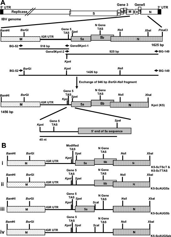
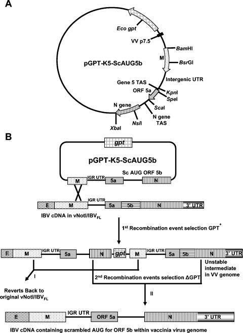
All of the modifications were based on the Beau-R sequence. A 1,625-bp BamHI-PmaCI fragment (Fig. 1A) corresponding to nucleotides 24794 to 26419 of the Beau-R genome was inserted into plasmid pZSL1190 (39), resulting in pBeau-BamHI-PmaCI, and used for modification of the Beau-R gene 5 sequence. Initially, a KpnI restriction endonuclease site was introduced, for subsequent manipulation purposes, 3 nt proximal to TAS-1 of the Beaudette gene 5 TAS by substitution of three nucleotides, 25454GTT to AAC, using overlapping PCR mutagenesis. Two PCR products (518 bp and 925 bp) were generated using oligonucleotides BG-52, Gene5KpnI-1, Gene5KpnI-2, and BG-149 (Table 1; Fig. 1A), in which oligonucleotides Gene5KpnI-1 and Gene5KpnI-2 introduced the three nucleotide substitutions. A third PCR product (1,426 bp) was generated from the two initial PCR products using oligonucleotides BG-52 and BG-149. A 946-bp BsrGI-NsiI fragment from the 1,426-bp PCR product, containing the introduced KpnI site, was used to replace the corresponding sequence in pBeau-BamHI-PmaCI, resulting in pGene5-KpnI. Plasmid pGene5-KpnI, containing the 1,456-bp BamHI-XbaI fragment with the introduced KpnI site (cDNA KpnI [K5]) (Fig. 1A) was used for introducing further modifications into the Beaudette gene 5 sequence. To modify the gene 5 TAS and scramble the ORF 5a initiation codon, three adapters were produced. Equal amounts of complementary pairs of oligonucleotides (5ST-ST-1 and 5ST-ST-2, 5ST-T-1 and 5ST-T-2, and 5a-AUG-1 and 5a-AUG-2) were heated to 100°C for 5 min and hybridized by cooling to room temperature to produce the three adapters. The three adapters were used to replace the 45-bp KpnI-SpeI fragment in pGene5-KpnI (Fig. 1A), resulting in the three plasmids containing the modified cDNAs: K5-ScTScT, K5-ScTTAS, and K5-ScAUG5a (Fig. 1B). A fifth modified cDNA was produced using overlapping PCR mutagenesis to scramble the ORF 5b initiation codon and introduce a ScaI site distal to the modified ATG to act as an identifiable marker. Two PCR products (759 bp and 695 bp) were generated using the oligonucleotides BG-52, ORF5bScaI-1, ORF5bScaI-2, and BG-149, in which oligonucleotides ORF5bScaI-1 and ORF5bScaI-2 introduced the required nucleotide substitutions. A third PCR product (1,426 bp) was generated from the two initial PCR products using oligonucleotides BG-52 and BG-149, from which a 467-bp SpeI-NsiI fragment was used to replace the corresponding sequence in pGene5-KpnI, resulting in pGene5-ORF5b-ScAUG, containing cDNA K5-ScAUG5b (Fig. 1B) with the ORF 5b initiation codon scrambled. The 467-bp SpeI-NsiI fragment was also used to replace the corresponding fragment in cDNA K5-ScAUG5a, resulting in cDNA K5-ScAUG5ab (Fig. 1B) with both ORF 5a and 5b initiation codons scrambled. The modified sequences are summarized in Table 2. Plasmids containing the 1,456-bp BamHI-XbaI cDNAs (K5, K5-ScTScT, K5-ScTTAS, K5-ScAUG5a, K5-ScAUG5b, and K5-ScAUG5ab) were transformed into a dam mutant strain of E. coli to allow removal of the BamHI-XbaI fragments for insertion into pGPTNEB193rev (a gift from M. Skinner, Institute for Animal Health) (4), resulting in a series of plasmids containing the guanine phosphoribosyltransferase (GPT) gene (37) for modifying the IBV Beaudette full-length cDNA in vNotI/IBVFL (5). An example of one such plasmid, pGPT-K5-ScAUG5b, is shown in Fig. 2A.
FIG. 1.
Schematic diagrams for the construction of the modified IBV gene 5 cDNAs. (A) A KpnI restriction endonuclease site was introduced by changing nucleotides 25454GTT to ACC proximal to the IBV Beaudette gene 5 TAS using overlapping PCR mutagenesis. This cDNA (K5) was used for introducing modifications into the gene 5 TAS (ScTScT and ScTTAS) or to scramble the ORF 5a ATG (ScAUG5a) by replacing the 45-nt KpnI-SpeI fragment with adapters containing the required nucleotide substitutions. The ORF 5b ATG was scrambled, and a ScaI restriction endonuclease site was introduced using overlapping PCR mutagenesis. The modified cDNA with the initiation codons of both ORF 5a and ORF 5b scrambled, ScAUG5ab, was constructed by replacing the SpeI-NsiI of ScAUG5a with the corresponding fragment from ScAUG5b. The overlapping PCR mutagenesis step for introduction of the KpnI restriction endonuclease site, in which the Beaudette 946-bp BsrGI-NsiI cDNA fragment was replaced with the corresponding PCR fragment containing the KpnI site, is shown. (B) Diagrams of the modified cDNAs that together with the K5 cDNA were used to produce the rIBVs. The positions of the IBV gene products M, 5a, 5b, and N are shown as labeled bars. However, the coding sequences for ORFs 5a and 5b are shown as black lines, following scrambling of the initiation codon, to indicate that the sequences are retained but that translation of the gene product is lost. IGR UTR, intergenic untranslated region.
TABLE 2.
Summary of the modifications to the Beaudette genome
| Name | Description of modification | Nucleotide changesa |
|---|---|---|
| K5 | Introduction of Kpnl siteb | 25441TTTTATAAGAGGTGTTTTACTTAACAAAAA to 25441TTTTATAAGAGGTAACTTACTTAACAAAAA |
| K5-ScTTAS | Scrambled TAS-1 | 25451GGTAACTTACTTAACAAAAA to 25451GGTAACTTTACGTGATCGTA |
| K5-ScTScT | Scrambled TAS-1 and TAS-2 | 25451GGTAACTTACTTAACAAAAACTTAACAAATAC to 25451GGTAACTTTACGTGATCGTGAACTCGTGATAC |
| K5-ScAUG5a | (i) Scrambled 5a ATG (ii) Modification of out-of-frame ATG | 25471CTTAACAAATACGGACGATGAAATGGCTGA to 25471CTTAACAAATACGGACGTAAAAATTGCTGA |
| K5-ScAUG5b | (i) Scrambled 5b ATG with retention of 5a termination codon | 25675CGCTGGCATGAATAATAGTAAAGATAATCC to 25675CGCTGGCATAAATTAAAGTACTGATAATCC |
| (ii) Introduction of in-frame termination codon | ||
| (iii) Introduction of Scal site | ||
| K5-ScAUG5ab | Combination of modifications in K5-ScAUG5a and K-5ScAUG5b |
The nucleotides in bold represent the substituted nucleotides, and positions of the nucleotides are with respect to the genome of Beau-R (9). Nucleotides corresponding to the gene 5 canonical TASs and 5a and 5b initiation codons are underlined. The introduced restriction endonuclease sites are double underlined.
The Kpnl site is present in all of the modified sequences.
FIG. 2.
Schematic diagrams showing the transient dominant selection process for modifying the full-length cDNA within the genome of vaccinia virus vNotI/IBVFL. The modified cDNAs shown in Fig. 1B were introduced as a 1,456-bp BamHI-XbaI fragment into the TDS GPT transfer/recombination vector pGPTNEB193rev. (A) Representation of one of the modified cDNAs, K5-ScAUG5b, in pGPTNEB193rev. (B) Outline of the two-step process for modifying the IBV cDNA within vNotI/IBVFL. In step 1, the complete plasmid DNA is integrated into the full-length IBV cDNA by a single-step homologous recombination event; a potential recombination event is shown. The resultant rVV has a GPT+ phenotype, allowing selection in the presence of mycophenolic acid. Removal of mycophenolic acid results in two types of spontaneous intramolecular recombination events, I and II, due to the instability of the IBV cDNA with tandem repeats of similar sequences; I results in reversion to the Beaudette genotype (no introduced modifications), and II results in introduction of the modification(s). Both recombination events result in the loss of GPT. The example shown outlines the introduction of the scrambled 5b AUG and associated nucleotide substitutions.
Generation of rVVs containing the modified IBV cDNAs.
Our IBV reverse genetics system is based on the use of vaccinia virus as a vector for the IBV full-length cDNA (9). The modified IBV cDNAs in the GPT-containing plasmids were used to modify the IBV Beaudette full-length cDNA in vNotI/IBVFL by homologous recombination using the transient dominant selection (TDS) method (5, 25) as outlined in Fig. 2B. CV-1 cells (70% confluent monolayers) were infected with vNotI/IBVFL at a multiplicity of infection (MOI) of 0.2 and transfected with 5 μg of the appropriate GPT-containing plasmid using Lipofectin (Invitrogen) at 2 h postinfection. Several rVVs expressing GPT, following recombination with each of the six modified IBV cDNAs, were selected by three rounds of plaque purification in CV-1 cells using growth medium containing 25 μg/ml mycophenolic acid (MPA), 250 μg/ml xanthine, and 15 μg/ml hypoxanthine. MPA-sensitive vaccinia viruses, potentially containing the modified IBV cDNAs, were then generated from the MPA-resistant vaccinia viruses following the spontaneous loss of the GPT gene by three rounds of plaque purification in the absence of MPA (5).
Analysis of vaccinia virus DNA.
Vaccinia virus DNA, isolated from CV-1 cells previously infected with the rVVs, was analyzed by PCR for the presence or absence of the gpt gene using the oligonucleotides GPT-forw and GPT-rev1 (Table 1). DNA samples that lacked the gpt gene were subjected to three overlapping PCRs corresponding to a 1,930-nt region, inclusive of the gene 5 sequence, of the IBV cDNA within the rVVs to confirm, by sequence analysis, the presence/absence of the gene 5 modifications. The PCRs used the following pairs of oligonucleotides: BG-51 (within ORF 3c [E]) and BG-146 (within ORF 5a), BG-52 (within M) and BG-147 (within ORF 5b), and BG-70 (start of ORF 5a) and BG-149 (within N).
Transfection and recovery of infectious IBV.
Recovery of recombinant IBVs (rIBVs) from vaccinia virus DNA was done as described previously (9). CK cells grown to 50% confluence were infected with rFPV/T7 (6) at an MOI of 10 and at 1 h postinfection were transfected with 10 to 20 μg of vaccinia virus DNA containing the modified IBV cDNAs, previously digested with AscI, and 5 μg of pCi-Nuc using 30 μl of Lipofectin (Invitrogen). The cells (P0 cells) were incubated at 37°C for 2.5 days posttransfection, and culture medium, potentially containing rIBV (V1), was removed, centrifuged for 3 min at 2,500 rpm, filtered through a 0.22-μm filter to remove any rFPV/T7 (24), and used for serial passage on CK cells. Stocks of each rIBV were prepared from P3 CK cells, titrated, and used in subsequent experiments.
Isolation and analysis of IBV-derived RNAs.
Total cellular RNA was extracted from IBV-infected CK cells using RNeasy (QIAGEN) and analyzed for (i) the presence of the desired modifications by reverse transcription (RT)-PCR and (ii) IBV-specific RNAs by Northern blot analysis. To confirm that the gene 5 modifications were present in the rIBV genomes, RT-PCRs (Ready-To-Go RT-PCR beads, Amersham Pharmacia Biotech) were carried out using the oligonucleotides BG-152 (within the intergenic untranslated region) and BG-148 (within the N gene). The resultant 836-bp RT-PCR products were purified using the QIAquick (QIAGEN) method and sequenced. For Northern blot analysis, RNA was electrophoresed into denaturing 1% agarose-2.2 M formaldehyde gels and transferred onto Hybond XL nylon membranes (Amersham). IBV-derived RNAs were detected nonisotopically using a mixture of two probes, a 666-bp IBV 3′ untranslated region probe corresponding to the last 666 nt at the 3′ end of the IBV genome and a 430-bp probe corresponding to nt 25941 to 26371 in the IBV N gene, covalently labeled with psoralen-biotin (BrightStar, Ambion) (24). Hybridized probes were detected using streptavidin-alkaline phosphatase conjugate in the presence of alkaline phosphatase 1,2-dioxetane chemiluminescent substrate (CDPStar, BrightStar Biodetect, Ambion) and exposed to film at room temperature (24, 26).
Confirmation that BeauR-K5-ScTScT did not produce sg mRNA 5 was carried out by RT-PCR using the oligonucleotides Leader1 (within the leader sequence) and BG-147 (within ORF 5b), which results in two potential RT-PCR products. An RT-PCR product of 1,371 bp was indicative for sg mRNA 4 and one of 332 bp for sg mRNA 5.
Serial passage of rIBVs.
CK cells (106) were infected with the rIBVs (107 PFU), and medium from the infected cells 24 h postinfection was diluted 1:100 and used to infect CK cells; this was repeated to P30. Total cellular RNA was extracted from the P30 CK cells and analyzed by RT-PCR using oligonucleotides BG-152 and BG-148 to confirm the identity of the rIBV by sequence analysis.
Generation of antibodies against 5a and 5b.
Four peptides, two corresponding to each gene 5 product, were synthesized by 9-fluorenylmethoxy carbonyl chemistry using an automatic peptide synthesizer and purified by reverse-phase high-performance liquid chromatography: PEP-5A-1 (22QLRVLDRLILDHGLLRVLTC41) and PEP-5A-2 (41CSRRVLLVQLDLVYRLAYTPTQSLA65) from 5a and PEP-5B-1 (53CEEHIHNNNLLSWQA67) and PEP-5B-2 ([C]68VKQLEKQTPQRQSLN82) (which included an extra amino-terminal cysteine for coupling purposes) from 5b. The two peptides representing a gene 5 product were coupled in equal amounts to maleimide-activated keyhole limpet hemocyanin (mcKLH; Pierce) using an EZ antibody production and purification kit (Pierce). The KLH-conjugated peptides were used to immunize three rats per conjugate (Eurogentec).
Western blot analysis of cell extracts.
CK cells (5 × 10⁶) were infected with Beau-R or the rIBVs (105 PFU), and 24 h postinfection the cells were washed with phosphate-buffered saline (PBS), lysed using RIPA buffer (50 mM Tris-HCl, pH 7.5, 150 mM NaCl, 1% Nonidet P-40, 0.5% sodium deoxycholate, and 0.1% sodium dodecyl sulfate [SDS]) containing X1 broad-spectrum protease inhibitor cocktail (Complete protease inhibitor cocktail, Boehringer) at 4°C, and centrifuged at 14,000 × g for 10 min at 4°C. The proteins in the supernatants were denatured with SDS lysis buffer for 5 min at 100°C, fractionated on Tris-Tricine-buffered (2) 15% SDS-polyacrylamide gels, and transferred onto 0.2-μm polyvinylidene difluoride membranes (Invitrogen) in Tris-glycine (pH 8.4)-20% methanol transfer buffer for 2 h at room temperature. The membranes were blocked using enhanced chemiluminescence detection (ECL) Advance (Amersham Bioscience) blocking agent. The proteins were analyzed by ECL using rat antiserum diluted 1:25,000 in PBS, 0.1% Tween 20, and 2% (wt/vol) ECL blocking agent (PBS/Tween blocking agent) raised against the KLH-conjugated 5b peptides and rabbit antiserum diluted 1:100,000 in PBS/Tween blocking agent raised against the IBV M protein (a gift from C. E. Machamer, The Johns Hopkins School of Medicine) followed by goat anti-rat and goat anti-rabbit immunoglobulin G conjugated to horseradish peroxidase, both diluted 1:100,000 in PBS/Tween blocking agent, and exposure to film.
Immunofluorescence assay.
Vero cells (70% confluent) on 13-mm-diameter glass coverslips (Western Laboratories Service), infected with Beau-R and the rIBVs at 3 × 10⁶ PFU/ml or uninfected (controls), were fixed 20 h postinfection for 30 min with 4% paraformaldehyde in PBS, washed in PBS, permeabilized using 0.5% Triton X-100 for 15 min, and washed with PBS at room temperature. Nonspecific binding of antibody was blocked using 0.5% bovine serum albumin (BSA) in PBS. The fixed cells were immunolabeled by incubation with rat antiserum SK71 raised against KLH-conjugated IBV 5b peptides and diluted 1:100 in PBS/BSA and with mouse monoclonal antibody IE7 (T. Hodgson et al., unpublished result) raised against the IBV E protein and diluted 1:10 in PBS/BSA for 1 h at room temperature followed by three PBS washes. Primary antibodies were detected with either Alexa 488-labeled donkey anti-rat antibody or Alexa 568-labeled goat anti-mouse antibody (Molecular Probes), diluted 1:150 or 1:200, respectively, in PBS/BSA for 1 h at room temperature, and washed three times in PBS. Nuclei were labeled with ToPro 3 (Molecular Probes) diluted 1:3,000 in PBS/BSA for 5 min. Preimmune serum from rat SK71 diluted 1:100 in PBS/BSA was used as a control on Beau-R-infected Vero cells. Uninfected control cells were labeled as described for infected cells. Coverslips were mounted in Vectashield (Vector Laboratories), sealed with nail varnish, and analyzed using a Leica TCS SP2 confocal microscope.
Multiple-step growth curves of rIBVs in CK cells.
Confluent monolayers of CK cells in 35-mm dishes (approximately 1.8 × 10⁶ cells) were infected with each rIBV (5.4 × 10⁴ PFU). After 1 h of adsorption, cells were washed three times with PBS and 3 ml of medium was added, followed by incubation at 37°C. Cell medium from the infected cells was taken at 1, 20, 30, and 48 h postinfection and assayed, in triplicate, by plaque titration on CK cells. Total cellular RNA was extracted from the CK cells after the 48 h postinfection time point and analyzed by RT-PCR using oligonucleotides BG-152 and BG-148 to confirm the identity of the rIBV by sequence analysis.
Growth kinetics of rIBVs in chick embryos.
Three 11-day-old embryonated eggs for each time point were inoculated in the allantoic cavity with 0.1 ml of a low-dose rIBV (0.4 PFU) and incubated at 37°C in an egg incubator (Octagon, Brinsea). Allantoic fluid from the infected embryos was harvested at 15.5, 19.5, 24, 29, and 38.5 h postinfection and assayed by plaque titration on CK cells. RNA was extracted from allantoic fluid after the 38.5 h postinfection time point and analyzed by RT-PCR using oligonucleotides BG-152 and BG-148 to confirm the identity of the rIBV by sequence analysis.
Growth kinetics of rIBVs in TOCs.
Chicken tracheal organ cultures (TOCs) were prepared from 19-day-old specific-pathogen-free Rhode Island Red chicken embryos (16). Three groups of five TOCs were inoculated with 0.5 ml of medium containing 2.7 × 10⁴ PFU of the rIBVs Beau-R, BeauR-K5-1, BeauR-K5-ScTScT-1, or BeauR-K5-ScTScT-2. After 1 h at 37°C the medium was removed, and the TOCs were washed three times with 5 ml of prewarmed PBS and incubated in 1 ml of medium (16). At selected time points (1.5, 17, 24, 48, 72, and 96 h postinfection), medium from the three groups of five TOCs corresponding to each rIBV was removed and analyzed by plaque titration on CK cells. RNA was extracted from the TOCs after the 96 h postinfection time point or from virus in the TOC medium using the RNeasy method and analyzed by RT-PCR using oligonucleotides BG-152 and BG-148 to confirm the identity of the rIBV by sequence analysis.
Assessment of the pathogenicity of rIBVs in 11-day-old chick embryos.
Two independent assessment experiments were carried out. In the first experiment, groups of five 11-day-old embryonated eggs were inoculated in the allantoic cavity with 0.1 ml of medium containing 8, 80, or 800 PFU of each rIBV. After inoculation, the embryos were incubated at 37°C and observed at 24, 48, 72, and 87 h postinfection for lack of movement or absence of large blood vessels, indicators of IBV pathogenicity. In the second experiment, groups of five 11-day-old embryonated eggs were inoculated with 0.1 ml of medium containing 0.008, 0.08, or 0.8 PFU of each rIBV and monitored for pathogenic signs as in the first experiment. Titers of the rIBV were expressed as median egg lethal dose (ELD50), calculated by the Reed-Muench method (41), as an indication of the pathogenicity of the rIBV for 10-day-old embryos. RNA extracted from allantoic fluid after the 87 h postinfection time point was analyzed by RT-PCR using oligonucleotides BG-152 and BG-148 to confirm the identity of the rIBV by sequence analysis.
Sequence analysis.
Sequence analysis of plasmid DNA, PCR products from IBV cDNA sequences within the rVVs, and RT-PCR products from rIBV RNAs was determined using either an ABI prism BigDye terminator cycle sequencing ready reaction kit (Applied Biosystems) or a CEQ DTCS quick start kit (Beckman Coulter). Sequence reactions were analyzed on an Applied Biosystems 377 DNA sequencer or a CEQ 8000 capillary sequencer. PREGAP4 and GP4 of the Staden Sequence Software Programs (3) were used for sequence entry, assembly, and editing.
RESULTS
Generation of gene 5 rIBVs.
To determine whether IBV gene 5 is required for a successful IBV infectious cycle, a series of rIBVs were generated in which either the gene 5 TAS was modified to prevent the transcription of sg mRNA 5 or the initiation codons of the gene 5 products, 5a and 5b, were scrambled to prevent translation. We decided to modify the IBV genome by site-specific mutagenesis with limited nucleotide substitutions rather than deletion of various sequences to rule out the possibility of destabilizing the genome or polar effects on gene expression as a result of deleting regions of the genome. All of the modifications were introduced into the same isogenic background of IBV Beaudette (Beau-R). The modifications are summarized in Table 2.
Initially, a KpnI restriction endonuclease site was introduced by the substitution of nucleotides 25454GTT to AAC 3 nt proximal to the gene 5 TAS (Fig. 1A; Table 2) in the Beau-R genome. The KpnI site was introduced to allow modification of the gene 5 TAS and scrambling of the ORF 5a initiation codon by using adapters to replace the 45-nt region of the Beaudette genome between the introduced KpnI site and a SpeI site 9 nt distal to the ORF 5a ATG (Fig. 1A).
Two modifications were made to the gene 5 TAS; the first involved scrambling the TAS-1 sequence 25459ACTTAACAAAA to TACGTGATCGT (Fig.1Bi; Table 2). A similar modification, introduced into the gene 5 TAS controlling the CAT reporter gene in an IBV D-RNA, did not affect the expression of CAT; sequence analysis of the D-RNA-derived CAT sg mRNA indicated that TAS-2 was utilized (51). Other IBV strains contain only a consensus sequence equivalent to Beaudette TAS-2, and we have shown that TAS-2 is used for the synthesis of Beaudette sg mRNA 5 in IBV-infected cells (51). Our second modification to the gene 5 TAS involved scrambling of the complete Beaudette gene 5 TAS (TAS-1 and TAS-2), from 25459ACTTAACAAAAACTTAACAA to TACGTGATCGTGAACTCGTG (Fig.1Bi; Table 2), which resulted in the loss of CAT expression from a D-RNA (51).
The ORF 5a modifications 25488ATG to TAA and 25495G to T resulted in scrambling of the ORF 5a initiation codon and modification of a second out-of-frame 25493ATG 2 nt distal to the ORF 5a ATG (Fig.1Bii; Table 2). The ORF 5b initiation codon was modified by overlapping PCR mutagenesis, and the use of a 467-bp SpeI-NsiI restriction fragment allowed the scrambled ORF 5b ATG to be introduced into the IBV cDNA either with an authentic ORF 5a sequence (Fig.1Biii) or containing the scrambled ORF 5a ATG (Fig.1Biv). Modification of the ORF 5b initiation codon involved 5-nt substitutions. The first substitution, T25684GA to TAA, served two functions: scrambling of the ORF 5b initiation codon and modification but retention of the ORF 5a termination codon, which overlaps the initiation codon of ORF 5b. The scrambling of the ORF 5b initiation codon involved substitution of only a single nucleotide. To ensure that ORF 5b could not be expressed if the single nucleotide substitution reverted, two further substitutions, 25688AA25690T to TAA, were introduced to convert the third codon of ORF 5b to an in-frame termination codon. In addition, a ScaI site was introduced by modifying the fifth codon of ORF 5b, A25695A25696A to ACT, to act as a marker to identify the presence of the ORF 5b substitutions (Table 2).
The modifications to the gene 5 TAS were introduced to determine whether a single TAS had any effect on the replication of IBV Beaudette and to prevent transcription of sg mRNA 5 to investigate whether the loss of sg mRNA 5 and the loss of both ORF 5a and 5b gene products affected virus replication. The ORF 5a and 5b initiation codons were scrambled to determine whether the absence of the gene products affected replication per se, without the possibility that the loss of synthesis of sg mRNA had some other affect, either up- or down-regulation, on the synthesis of the other IBV sg mRNAs, which could affect replication. The scrambling of the ORF 5a and 5b initiation codons also ensured that the gene 5 products could not be expressed from any other IBV sg mRNA without the necessity of deleting all or part of the coding sequences.
The modified IBV cDNA sequences were introduced into the Beaudette full-length cDNA in vaccinia virus vNotI/IBVFL (9) by homologous recombination using the TDS method (5) and a series of GPT-based transfer vectors (Fig. 2). PCR analysis, using GPT-specific primers, on rVV DNAs extracted from infected CV-1 cells confirmed the absence of the E. coli GPT gene following the second recombination event (Fig. 2B). The presence of gene 5 modifications was initially confirmed by restriction endonuclease analysis on PCR products generated between nt 24441 (E) and nt 26371 (N) of the IBV cDNAs. The PCR products were analyzed for the presence of the KpnI site, present in all of the modified sequences, and ScaI, the marker site for the introduction of the ORF 5b modifications. As a result, 12 rVVs, representing pairs of viruses from independent transfection reactions and containing the six IBV gene 5 modifications, were obtained. Sequence analysis of the IBV cDNAs within the rVV DNAs, representing the BamHI and XbaI fragments from the GPT-based plasmids (Fig. 2A), and at least 100 nt proximal and distal to the two restriction sites confirmed that the modifications were as expected and that no other changes had been introduced as a result of the homologous recombination events.
Six pairs of rIBVs, corresponding to the six modifications, were rescued in CK cells using DNA isolated from the 12 rVVs (5, 8, 9). The rIBVs were BeauR-K5-1 and -2, which contained the KpnI site proximal to the gene 5 TAS that was also present in all of the rIBVs; BeauR-K5-ScTScT-1 and -2, in which both canonical TASs were scrambled; BeauR-K5-ScTTAS-1 and -2, in which the TAS-1 was scrambled; BeauR-K5-ScAUG5a-1 and -2, in which the ORF 5a initiation codon was scrambled; BeauR-K5-ScAUG5b-1 and -2, in which the ORF 5b initiation codon was scrambled; and BeauR-K5-ScAUG5ab-1 and -2, in which both the ORF 5a and 5b initiation codons were scrambled. Pairs of rIBVs were rescued from the independently isolated rVVs to counteract or identify any other fortuitous mutation(s) that may have been acquired during the recombination events, during the selection of the rVVs, or in the rescue and passage of the rIBVs.
The rIBVs were passaged three times in CK cells and the resulting virus stocks titrated and used for further characterization. The genotypes of the rIBVs were verified using three overlapping RT-PCR products generated from RNA extracted from the infected P3 CK cells. The RT-PCR products corresponding to nt 24441 (E) to 26371 (N) of the IBV genome were sequenced and confirmed the presence of the desired gene 5 modifications (data not shown). No other nucleotide changes were observed apart from a single nucleotide, 25179A to C, in one of the two BeauR-K5 viruses, representing a silent mutation within the last amino acid (threonine) of the M gene, which arose upon rescue of the rIBV. The observation that all of the gene 5 rIBVs were rescued in CK cells indicated that none of the introduced modifications had had a deleterious effect on the replication of IBV in vitro and indicated that the IBV gene 5 products, 5a and 5b, were not essential for replication of the virus in cell culture.
RNA synthesis by gene 5 rIBVs.
To examine the patterns of RNA synthesis by the rIBVs, identical amounts of total cellular RNA isolated from CK cells infected with the same virus titers were separated by electrophoresis in denaturing agarose gels, transferred to nylon membranes, and probed nonradioactively using two IBV-specific probes. Both independent isolates of each rIBV were analyzed. As can be seen from Fig. 3, all of the rIBVs except BeauR-K5-ScTScT-1 and -2 (Fig. 3, lanes 5 and 6) synthesized sg mRNAs 4, 5, and 6, with no observable differences between the pairs of rIBVs. The two BeauR-K5-ScTScT rIBVs did not, as anticipated, produce sg mRNA 5.
FIG. 3.
Northern blot analysis of IBV sg mRNAs 4 to 6 following infection of CK cells with the rIBVs. CK cells were infected with 3 × 10⁶ PFU of Beau-R or of each rIBV, and total cellular RNA was extracted at 24 h postinfection, separated by electrophoresis in 1% denaturing formaldehyde-agarose gels, and transferred to nylon membranes. The IBV-derived RNAs were detected nonisotopically with a mixture of two IBV-specific probes, a 666-bp IBV 3′ UTR probe and a 430-bp IBV N gene probe. RNAs from mock-infected CK cells (lane 1) or CK cells infected with Beau-R (lane 2), BeauR-K5-1 and -2 (lanes 3 and 4), BeauR-K5-ScTScT-1 and -2 (lanes 5 and 6), BeauR-K5-ScTTAS-1 and -2 (lanes 7 and 8), BeauR-K5-ScAUG5a-1 and -2 (lanes 9 and 10), BeauR-K5-ScAUG5b-1 and -2 (lanes 11 and 12), or BeauR-K5-ScAUG5ab-1 and -2 (lanes 13 and 14) were analyzed. The RNA species detected below sg mRNA 6 are observed routinely for all strains of IBV, as originally identified (50), and are of unknown origin.
There were no observable differences in the amounts of the sg mRNAs produced by Beau-R (Fig. 3, lane 2) and BeauR-K5 (Fig. 3, lanes 3 and 4), indicating that the introduction of the KpnI site had not affected sg mRNA 5 production. The level of expression of sg mRNA 4 by BeauR-K5-ScTScT (Fig. 3, lanes 5 and 6) appeared slightly higher, possibly due to the fact sg mRNA 5 was no longer synthesized; the levels of the other sg mRNAs from this rIBV did not appear to be up-regulated (data not shown). Scrambling of the TAS-1 site as in the two BeauR-K5-ScTTAS rIBVs (Fig. 3, lanes 7 and 8) did not affect the synthesis of sg mRNA 5 when compared to BeauR-K5 or Beau-R, indicating, as previously observed (51), that this canonical TAS does not play a significant role in the synthesis of Beaudette sg mRNA 5. Unexpectedly, the level of sg mRNA 5 synthesized by the two BeauR-K5-ScAUG5ab rIBVs (Fig. 3, lanes 13 and 14) was less than observed for BeauR-K5 and Beau-R or any of the rIBVs except BeauR-K5-ScTScT. As there was no similar decrease in the amounts of sg mRNA 5 from the rIBVs in which the initiation codons of only 5a or only 5b were scrambled, it would appear that the decrease in the amount of sg mRNA 5 from BeauR-K5-ScAUG5ab was associated with the loss of both gene 5 products. A potential reason for the reduction in the amount of sg mRNA 5 from the BeauR-K5-ScAUG5ab rIBVs could be an increased instability of the sg mRNA resulting from a lack of translation, which should not occur, as observed, if only one initiation codon is scrambled.
To further verify that BeauR-K5-ScTScT did not produce sg mRNA 5, an mRNA-specific RT-PCR was carried out using oligonucleotides Leader1 and BG-147, which result in the production of a 332-bp product from sg mRNA 5. RT-PCR analysis of RNA isolated from CK cells infected with both isolates of BeauR-K5-ScTScT did not result in a 332-bp PCR product, confirming that sg mRNA 5 was absent, but did result in a 1,371-bp product resulting from the presence of sg mRNA 4 (data not shown).
Stability of the introduced mutations.
IBV gene 5 was modified by a series of nucleotide substitutions either within the TAS or by scrambling the initiation codons of the gene 5 products. A potential consequence of such modifications is nucleotide reversion following rescue and passage of the rIBVs, resulting in restoration of the gene 5 products. Therefore, to investigate whether the introduced mutations were stably maintained during replication of the rIBVs, one isolate of each rIBV was serially passaged from P3 to P30 in CK cells every 24 h. Total RNA was extracted from the P30 CK cells, and the gene 5 region of each rIBV was RT-PCR amplified for sequence analysis. Sequence analysis, between the BsrGI (nt 25021 within the M gene) (Fig. 1A) and NsiI (nt 25967 within the N gene) (Fig. 1A) sites, confirmed that the modifications were still present following 30 passages of the rIBVs. No other nucleotide changes were identified within the region analyzed when compared to the rIBVs isolated at P3 or to the Beau-R sequence. These results showed that the gene 5 modifications did not revert during in vitro passage, indicating they were not under any selective pressure for restoration to the parental (Beau-R) sequence. Therefore, we conclude that neither transcription of sg mRNA 5 nor expression of 5a and 5b is essential for the replication of IBV in vitro in CK cells.
Analysis of the rIBVs for production of the gene 5 products.
Initial analysis of the rat antisera raised against the KLH-conjugated 5a- and 5b-derived peptides showed that only rats immunized with the 5b-derived peptides produced antisera against the corresponding gene 5 product. Analysis of in vitro transcription/translation-derived products using a rabbit reticulocyte lysate-coupled transcription and translation cell-free protein synthesis system (Promega) (23) and a plasmid, pCi-5b, with the 5b sequence under the control of a cytomegalovirus promoter and of a cell lysate from Beau-R-infected CK cells with rat antiserum SK71 raised against the two 5b-derived peptides conjugated to KLH, identified a protein of 9.7 kDa, representing the 5b product (data not shown). Therefore, rat antiserum SK71 was used to investigate, by Western blot analysis, cell lysates derived from CK cells infected with Beau-R and the rIBVs for the presence of 5b. As can be seen from Fig. 4, all of the IBV-infected cell lysates contained a reference protein of 34 kDa, representing the IBV M protein detected using the rabbit anti-M serum. In addition, the CK cells infected with Beau-R (Fig. 4A and B, lane 2) and rIBVs BeauR-K5-1 and -2 (Fig. 4A, lanes 3 and 4), BeauR-K5-ScTTAS-1 and -2 (Fig. 4A, lanes 7 and 8), or BeauR-K5-ScAUG5a-1 and -2 (Fig. 4B, lanes 3 and 4) also contained a protein of 9.7 kDa, representing the 5b product. Cells infected with rIBVs BeauR-K5-ScTScT-1 and -2 (Fig. 4A, lanes 5 and 6), BeauR-K5-ScAUG5b-1 and -2 (Fig. 4B, lanes 5 and 6), or BeauR-K5-ScAUG5ab-1 and -2 (Fig. 4B, lanes 7 and 8) did not contain the 9.7-kDa protein, indicating that they did not, as expected, express the 5b protein.
FIG. 4.
Western blot analysis for the detection of IBV 5b following infection of CK cells with the rIBVs. CK cells were infected with Beau-R or with the two isolates of each type of rIBV. Proteins derived from the infected cell lysates were separated on Tris-Tricine 15% SDS-polyacrylamide gels, transferred to polyvinylidene difluoride membranes, and analyzed for the presence of IBV M and 5b proteins by ECL. The M protein was detected using a rabbit anti-M serum, and 5b was detected using rat anti-serum SK21 followed by goat anti-rabbit and goat anti-rat immunoglobulins conjugated to horseradish peroxidase. Panel A shows the analysis of cell lysates from uninfected CK cells (lane 1) or CK cells infected with Beau-R (lane 2), BeauR-K5-1 and -2 (lanes 3 and 4), BeauR-K5-ScTScT-1 and -2 (lanes 5 and 6), and BeauR-K5-ScTTAS-1 and -2 (lanes 7 and 8). Panel B shows the analysis of cell lysates from uninfected CK cells (lane 1) or CK cells infected with Beau-R (lane 2), BeauR-K5-ScAUG5a-1 and -2 (lanes 3 and 4), BeauR-K5-ScAUG5b-1 and -2 (lanes 5 and 6), or BeauR-K5-ScAUG5ab-1 and -2 (lanes 7 and 8). The proteins representing the IBV M protein (34 kDa) and the 5b product (9.7 kDa) are shown. Molecular weights were determined using broad-range SDS-polyacrylamide gel electrophoresis standard proteins (Bio-Rad).
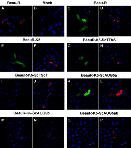
Vero cells infected with Beau-R and the rIBVs were analyzed by indirect immunofluorescence for the expression of 5b and coexpression of 3c (E protein), the latter as an indicator for IBV-infected cells. As can be seen from Fig. 5, the IBV 5b product was detected in Vero cells infected with Beau-R, BeauR-K5-1, BeauR-K5-ScTTAS-1, and BeauR-K5-ScAUG5a-1, in which only IBV-infected cells, identified by the presence of 3c (E) mainly as a Golgi-associated protein (17-19) using the anti-E monoclonal antibody 1E7, contained the 5b product. However, 5b was not detected in cells infected with rIBVs either lacking the expression of sg mRNA 5 (BeauR-K5-ScTScT-1) or with a scrambled initiation codon for 5b (BeauR-K5-ScAUG5b-1 and BeauR-K5-ScAUG5ab-1) (Fig. 5). These observations confirm that the lack of transcription of sg mRNA 5 or scrambling of the 5b initiation codon resulted in the loss of 5b.
FIG. 5.
Detection of 5b in infected Vero cells by indirect immunofluorescence. Vero cells at 70% confluency were infected with Beau-R or with one isolate of each type of rIBV and fixed 20 h postinfection using 4% paraformaldehyde. The infected cells were analyzed by indirect immunofluorescence using either rat SK21 5b anti-peptide sera raised against two KLH-conjugated peptides derived from 5b or mouse monoclonal antibody IE7 against 3c (E) followed by either Alexa 488-labeled donkey anti-rat antibody or Alexa 568-labeled goat anti-mouse antibody, respectively, and stained with ToPro 3 to visualize nuclear DNA. Panel A shows Vero cells that had been infected with Beau-R and analyzed using preimmune rat SK21 serum and anti-E monoclonal antibody IE7. Panel B shows mock-infected Vero cells analyzed with rat SK21 anti-peptide sera and anti-E monoclonal antibody IE7. The other panels show Vero cells infected with Beau-R (C and D), BeauR-K5-1 (E and F), BeauR-K5-ScTTAS-1 (G and H), BeauR-K5-ScTScT-1 (I and J), BeauR-K5-ScAUG5a-1 (K and L), BeauR-K5-ScAUG5b-1 (M and N), or BeauR-K5-ScAUG5ab-1 (O and P) analyzed with rat SK21 5b anti-peptide sera (C, E, G, I, K, M, and O) or mouse monoclonal antibody IE7 against 3c (D, F, H, J, L, N, and P). The cells were examined using confocal microscopy at ×40 magnification; red corresponds to the presence of IBV E protein and green to the presence of 5b protein.
Characterization of the rIBVs in vitro.
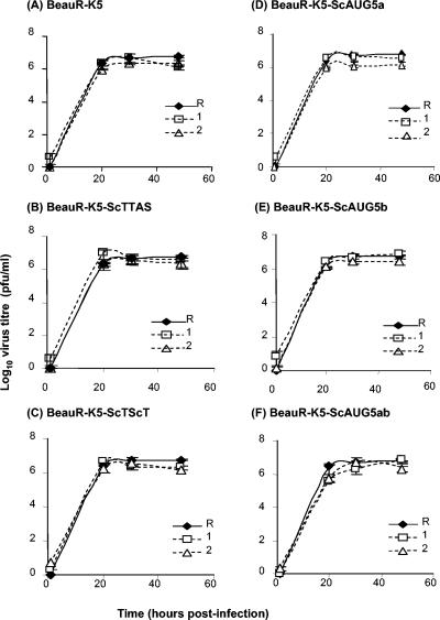
The growth characteristics of the gene 5 rIBVs were investigated in vitro using CK cells infected with 5.4 × 10⁴ PFU (MOI ≈ 0.03) of the two isolates of each rIBV and compared to the growth kinetics of Beau-R. The titers of the progeny viruses were analyzed over a 48-h postinfection period. As can be seen from Fig. 6, all of the rIBVs showed growth profiles similar to that of Beau-R, with maximum titers of 3 × 10⁶ to 5 × 10⁶ PFU/ml by 20 h postinfection. IBV-associated cytopathic effects were observed 30 h postinfection. The observation that the analogous pairs of rIBVs grew to similar titers indicated that no deleterious mutations had been introduced into other regions of the virus genomes. No significant differences were observed in the growth kinetics between Beau-R and the gene 5 rIBVs; however, analysis of virus titers at specific time points indicated that Beau-R had a slightly higher titer at 70% of the time points. No differences were observed in the plaque morphology of the gene 5 rIBVs with respect to Beau-R (data not shown). Sequence analysis of RT-PCR products, using oligonucleotides BG-152 and BG-148, generated from RNA extracted from the CK cells 30 h postinfection confirmed the identity of the viruses used.
FIG. 6.
Comparison of the in vitro growth kinetics of the gene 5 rIBVs in CK cells. Two independent rIBVs with each modification were used. CK cells were infected with 5.4 × 10⁴ PFU (MOI ≈ 0.03) of Beau-R or of each rIBV, and the titer of progeny virus was analyzed by plaque titration assay on CK cells over a period of 48 h. Panels show a comparison of the growth kinetics of each analogous pair (1 [dashed lines with open square] and 2 [dashed lines with triangle]) of the rIBVs with Beau-R (solid line and filled diamond). (A) BeauR-K5, (B) BeauR-K5-ScTTAS, (C) BeauR-K5-ScTScT, (D) BeauR-K5-ScAUG5a, (E) BeauR-K5-ScAUG5b, and (F) BeauR-K5-ScAUG5ab. The error bars represent standard deviations.
Characterization of the rIBVs in ovo.
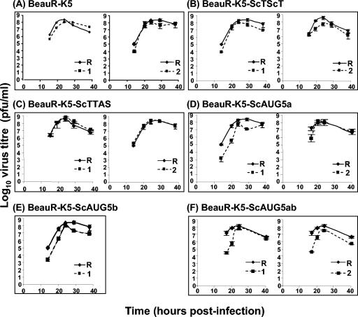
The growth characteristics of the gene 5 rIBVs were investigated in ovo using 11-day-old embryonated eggs. The embryonated eggs were inoculated, in the allantoic cavity, with 0.4 PFU of each rIBV, whose titers had previously been determined on CK cells. Triplicate samples of progeny viruses were isolated from allantoic fluid over a 39-h postinoculation period and titrated on CK cells. As can be seen from Fig. 7, all of the rIBVs showed growth profiles similar to that of Beau-R, with maximum titers of 108 to 109 PFU/ml by 24 h postinoculation. Some of the pairs of rIBVs showed slight variation, within 1 log10 unit of each other, representing variation in sampling, indicating there were no other deleterious mutations in other regions of the genomes of the analogous pairs of rIBVs. Overall, no significant differences were observed in the growth characteristics between Beau-R and the gene 5 rIBVs, indicating that none of the introduced mutations had a detrimental effect on the replication of IBV in ovo. Sequence analysis of RT-PCR products, using oligonucleotides BG-152 and BG-148, generated from RNA extracted from allantoic fluid after the last time point confirmed the identity of the viruses used.
FIG. 7.
Comparison of the in ovo growth kinetics of the gene 5 rIBVs in 11-day-old embryonated eggs. Two independent rIBVs with each modification were used. The embryonated eggs were inoculated with 0.4 PFU of Beau-R or of each rIBV and moved to 4°C at different time points up to 40 h postinfection. Allantoic fluid from the eggs was analyzed for progeny virus by plaque titration assay on CK cells. The panels show the comparisons of the growth kinetics of each analogous pair(1 and 2 [dashed line and filled squares]) (except for BeauR-K5-ScAUG5b, in which only one virus was analyzed) of the rIBVs with Beau-R (solid line and filled diamonds). (A) BeauR-K5, (B) BeauR-K5-ScTScT, (C) BeauR-K5-ScTTAS, (D) BeauR-K5-ScAUG5a, (E) BeauR-K5-ScAUG5b, and (F) BeauR-K5-ScAUG5ab. The error bars represent standard deviations.
Characterization of the rIBVs ex vivo.
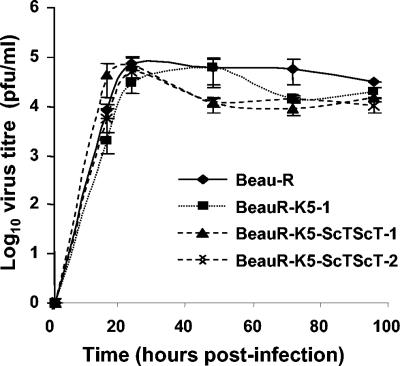
Infection of tracheal epithelial cells is used as a method of growing IBV strains that have not been adapted for growth either in embryonated eggs or in primary cell cultures and results in the cessation of ciliary activity (ciliostasis) of the epithelial cells. Previous work has shown that IBV Beaudette (14) and our recovered rIBV, Beau-R (30), result in ciliostasis of the TOCs with concomitant production of progeny virus. Results described above showed that the gene 5 rIBVs grew in vitro and in ovo. Therefore, we decided to investigate whether gene 5 was required for replication of IBV ex vivo using TOCs. We chose to analyze the growth of rIBVs BeauR-K5-ScTScT-1 and -2, representing inactivation of gene 5. Progeny viruses from TOC medium taken from three groups of TOCs at each time point were titrated by plaque assay on CK cells to investigate the growth kinetics of the rIBVs in TOCs for comparison with those obtained for Beau-R and BeauR-K5-1. As can be seen from Fig. 8, most of the viruses reached maximum titers by 24 h postinfection, except BeauR-K5-1, for which the titers were between 1.7- and 2.6-fold less than the titers observed for other viruses at this time point; however, the titer reached 5.5 × 104 by 48 h postinfection. Interestingly, the titers of the rIBVs gradually declined, by up to eightfold of their maximum titers, compared to Beau-R. This phenomenon was observed in two other experiments with TOCs. However, it should be noted that all of the titers were within 1 log10 unit, indicating that there was little difference in the growth of the viruses. All of the rIBVs investigated caused ciliostasis, resulting in 100% ciliostasis by 72 h postinfection. The fact that the rIBVs grew ex vivo further demonstrated that gene 5 is not essential for replication. Sequence analysis of RT-PCR products, using oligonucleotides BG-152 and BG-148, generated from RNA extracted from TOCs or from virus in the TOC medium after the 96 h postinfection time point confirmed the identity of the viruses used.
FIG. 8.
Ex vivo growth kinetics of the BeauR-K5-ScTScT gene 5 rIBVs in chicken tracheal organ cultures. Groups of five chicken TOCs were infected for 1 h at 37°C with 2.7 × 10⁴ PFU of Beau-R, rIBV BeauR-K5-1, and the two gene 5 rIBVs, BeauR-K5-ScTScT-1 and BeauR-K5-ScTScT-2, that are unable to generate the gene 5 sg mRNA 5. The medium was removed, the TOCs were washed three times with PBS, and incubation continued with 1 ml of medium. At selected time points, progeny viruses from three groups of TOCs were assayed by plaque titration on CK cells. The growth profiles of the four viruses are shown. The error bars represent standard deviations.
Pathogenicity of the gene 5 rIBVs in 11-day-old embryonated eggs.
In the first experiment, groups of five embryos were inoculated with 8, 80, or 800 PFU of each virus. At these doses, all of the embryos were killed; therefore, a second experiment was carried out in which the embryos were inoculated with 0.008, 0.08, or 0.8 PFU of each virus. This allowed the determination of an endpoint titration for the viability of the rIBVs in embryos; the titer for Beau-R was 8.4 log10 ELD50/ml, while the titers for the rIBVs varied between 7.8 and 8.8 log10 ELD50/ml. The ELD50 for Beau-R was 0.07 PFU, and the ELD50 values for the rIBVs were in the range 0.03 to 0.09 PFU, except for BeauR-K5-ScAUG5ab, which had an ELD50 of 0.50 PFU. In conclusion, the inactivation of gene 5 or the lack of production of 5a and 5b had little or no effect on the lethality of Beau-R for 11-day-old embryos. Sequence analysis of RT-PCR products, using oligonucleotides BG-152 and BG-148, generated from RNA extracted from allantoic fluid after the 87 h postinfection time point confirmed the identity of the viruses used.
DISCUSSION
All coronavirus genomes contain a series of genes encoding small ns proteins of unknown function that are a distinguishing feature of the viruses in a particular coronavirus group. The number and genomic location of these genes vary depending on the group of coronaviruses. Most coronaviruses possess at least two small ns genes encoding four to five products. However, the recently isolated coronavirus responsible for an atypical pneumonia in human severe acute respiratory syndrome, SARS-CoV, contains four potential ns genes encoding eight products, of which one product (9b) is encoded as a second product by the N gene (35, 42, 47, 49, 54, 58).
IBV and the coronaviruses isolated from the avian species turkey (11), pheasant (12), peafowl (accession no. AY641576), and partridge (accession no. AY646283) all contain an ns gene 5, transcribed as sg mRNA 5 and located between the M and N genes, that encodes two products, 5a and 5b, of unknown function. No IBV strain that does not contain a potentially functional gene 5 has been identified. In all cases, 5a is situated 9 nt distal to the gene 5 TAS and overlaps the 5b initiation codon. In addition, 5b overlaps the 5′ end of the N gene. The initiation and stop codons of 5a and 5b are conserved. The 5a gene product consists of 65 amino acids (7.5 kDa) with >75% identity among 15 sequences (10 show ≥87% identity), and 5b consists of 82 amino acids (9.5 kDa) with >81% identity among 19 sequences (14 show ≥89% identity). The IBV gene 5 products 5a and 5b have been identified in IBV-infected Vero and CK cells, with the expression of 5b being proposed to result from leaky scanning (33). These observations indicate that IBV and IBV-like viruses isolated from other avian species have conserved the gene 5 sequence, indicating that the gene may play a role in the virus replication cycle. Alternatively, the gene products may be required for some other selective advantage of the virus or the gene sequence per se has to be retained for some RNA secondary structure function. The N gene TAS is 93 nt proximal to the N gene initiation codon and is therefore within the 5b coding sequence, which in turn overlaps the N gene by 58 nt.
The objective of this study was to determine whether IBV gene 5 is required for replication of IBV. We have used our IBV reverse genetics system (5, 8, 9) to produce a series of rIBVs that either lacked transcription of sg mRNA 5 or independently lacked translation of 5a, 5b, or both products as a result of site-specific mutagenesis. Modifications were made to the Beaudette-CK full-length cDNA in rVV vNotI/IBVFL by homologous recombination using the TDS method and a series of plasmids containing the modified IBV cDNA sequences (5). Recombinant IBVs were rescued in CK cells and characterized in vitro, in ovo, and ex vivo. Analogous pairs of rIBVs, derived from independent transfection and selection regimes, were rescued to rule out the possibility that altered phenotypes may have arisen from spontaneous changes in other regions of the IBV genome as a result of the rVV selection processes or in the rescue and subsequent passage of the rIBVs.
The fact that we were able to rescue rIBVs containing the modified IBV gene 5 sequences indicates that gene 5 is not essential for replication of IBV in vitro. Analysis of Beau-R-infected CK cell lysates by Western blot analysis, using anti-5b serum, identified a protein of 9.7 kDa corresponding to 5b, which has a theoretical molecular mass of 9.5 kDa. Western blot and indirect immunofluorescence analysis of cells infected with the rIBVs confirmed that the rIBVs either lacking transcription of mRNA 5 or containing a scrambled 5b AUG initiation codon did not, as anticipated, express 5b. In contrast, the 5b product was detected in the cell lysates or cells infected with rIBVs capable of expressing 5b. We were unsuccessful in raising antibodies against peptides derived from 5a and so were unable to confirm the lack of expression of 5a. However, the modifications to eliminate the expression of 5a were similar to those for 5b; therefore, it is unlikely that 5a was expressed from the relevant rIBVs.
We have further demonstrated that gene 5 is also not required for replication in embryonated eggs or ex vivo using TOCs. The analogous pairs of rIBVs showed similar phenotypes, indicating that the observed growth characteristics were due to the specific modifications and not a result of any fortuitous mutations acquired during the generation of the modified cDNAs or on rescue and passage of the rIBVs.
Pathogenicity experiments using embryonated eggs infected with the rIBVs indicated that inactivation of gene 5 or lack of production of 5a and 5b had little or no effect on the lethality of Beau-R for 11-day-old embryos. The rIBVs described in this work are based on IBV Beaudette-CK, which is apathogenic in chickens due to attenuation of the virus by multiple passages in eggs; therefore, we were unable to determine whether the loss of gene 5 had some effect on pathogenicity in chickens.
Overall, our results support those obtained using two other coronavirus reverse genetics systems for investigating the role of the ns genes for group 1 (27, 38) and group 2 (21) coronaviruses, which showed that some or all of the ns (accessory) genes for these different groups of coronaviruses are not essential for replication. Work involving TGEV (38), MHV (21), and Feline infectious peritonitis virus (27) showed that the loss of the accessory genes did not affect virus replication in vitro but did result in attenuation of the pathogenicity of the viruses for their host species. However, the loss of an accessory gene function does not necessarily lead to attenuation of pathogenicity. A field isolate of porcine coronavirus TGEV that lacked expression of the TGEV accessory gene 3 product 3a as a result of a deletion was still fully virulent in pigs (36). A recombinant TGEV lacking expression of both gene 3 products, 3a and 3b, was still enteropathogenic but with some reduction in enteric tissue growth (48).
We used two alternative ways to modify the IBV genome to affect the expression of the gene 5 products. In the first method, inactivation of the gene was achieved by scrambling the TAS, thereby preventing transcription of sg mRNA 5; the second method involved perturbation of the initiation codons of the two gene 5 products, preventing translation of the products without directly altering transcription of the sg mRNA. Both methods resulted in the rescue of rIBVs, indicating that the gene 5 products are not essential for replication and that the lack of sg mRNA expression was not deleterious to virus replication by some other means. There was no indication that the loss of TAS-1 affected the synthesis of sg mRNA 5, supporting our previous observations that TAS-2 was preferentially used for transcription of this IBV sg mRNA (51).
Our results demonstrate that IBV gene 5 does not play an essential role in the replication of the virus and indicate that at least one of the ns genes of group 3 coronaviruses is dispensable for replication. Therefore, IBV gene 5 can be classified as an accessory gene. Gene 5 of the group 3 coronaviruses is conserved; no natural IBV isolate or other group 3 avian coronavirus has been reported to be deficient in gene 5, strongly indicative of a role in vivo that remains to be elucidated. de Haan et al. (21) proposed that the coronavirus accessory genes might affect host immune responses; it is also possible that the accessory gene products play some other role in the host/pathogen interaction.
Acknowledgments
This work was supported by the Biotechnology and Biological Sciences Research Council (BBSRC) grant no. 201/15836 and the Department of Environment, Food and Rural Affairs (DEFRA) project code OD0712.
We thank L. Hunt of the Institute for Animal Health for synthesizing the peptides used for raising antibodies.
REFERENCES
- 1.Almazán, F., J. M. González, Z. Pénzes, A. Izeta, E. Calvo, J. Plana-Durán, and L. Enjuanes. 2000. Engineering the largest RNA virus genome as an infectious bacterial artificial chromosome. Proc. Natl. Acad. Sci. USA 97:5516-5521. [DOI] [PMC free article] [PubMed] [Google Scholar]
- 2.Ausubel, F. M., R. Brent, R. E. Kingston, D. D. Moore, J. G. Seidman, J. A. Smith, and K. Struhl. 1987. Current protocols in molecular biology. John Wiley and Sons, Inc., New York, N.Y.
- 3.Bonfield, J. K., K. F. Smith, and R. Staden. 1995. A new DNA sequence assembly program. Nucleic Acids Res. 23:4992-4999. [DOI] [PMC free article] [PubMed] [Google Scholar]
- 4.Boulanger, D., P. Green, T. Smith, C.-P. Czerny, and M. A. Skinner. 1998. The 131-amino-acid repeat region of the essential 39-kilodalton core protein of fowlpox virus FP9, equivalent to vaccinia virus A4L protein, is nonessential and highly immunogenic. J. Virol. 72:170-179. [DOI] [PMC free article] [PubMed] [Google Scholar]
- 5.Britton, P., S. Evans, B. Dove, M. Davies, R. Casais, and D. Cavanagh. 2005. Generation of a recombinant avian coronavirus infectious bronchitis virus using transient dominant selection. J. Virol. Methods 123:203-211. [DOI] [PMC free article] [PubMed] [Google Scholar]
- 6.Britton, P., P. Green, S. Kottier, K. L. Mawditt, Z. Pénzes, D. Cavanagh, and M. A. Skinner. 1996. Expression of bacteriophage T7 RNA polymerase in avian and mammalian cells by a recombinant fowlpox virus. J. Gen. Virol. 77:963-967. [DOI] [PubMed] [Google Scholar]
- 7.Brown, T. D. K., and I. Brierly. 1995. The coronavirus nonstructural proteins, p. 191-217. In S. G. Siddell (ed.), The Coronaviridae. Plenum Press, New York, N.Y.
- 8.Casais, R., B. Dove, D. Cavanagh, and P. Britton. 2003. Recombinant avian infectious bronchitis virus expressing a heterologous spike gene demonstrates that the spike protein is a determinant of cell tropism. J. Virol. 77:9084-9089. [DOI] [PMC free article] [PubMed] [Google Scholar]
- 9.Casais, R., V. Thiel, S. G. Siddell, D. Cavanagh, and P. Britton. 2001. Reverse genetics system for the avian coronavirus infectious bronchitis virus. J. Virol. 75:12359-12369. [DOI] [PMC free article] [PubMed] [Google Scholar]
- 10.Cavanagh, D. 2001. A nomenclature for avian coronavirus isolates and the question of species status. Avian Pathol. 30:109-115. [DOI] [PubMed] [Google Scholar]
- 11.Cavanagh, D., K. Mawditt, M. Sharma, S. E. Drury, H. L. Ainsworth, P. Britton, and R. E. Gough. 2001. Detection of a coronavirus from turkey poults in Europe genetically related to infectious bronchitis virus of chickens. Avian Pathol. 30:355-368. [DOI] [PubMed] [Google Scholar]
- 12.Cavanagh, D., K. Mawditt, D. de B. Welchman, P. Britton, and R. E. Gough. 2002. Coronaviruses from pheasants (Phasianus colchicus) are genetically closely related to coronaviruses of domestic fowl (infectious bronchitis virus) and turkeys. Avian Pathol. 31:81-93. [DOI] [PubMed] [Google Scholar]
- 13.Cavanagh, D., and S. Naqi. 2003. Infectious bronchitis, p. 101-119. In Y. M. Saif, H. J. Barnes, J. R. Glisson, A. M. Fadly, L. R. McDougald, and D. E. Swayne (ed.), Diseases of poultry, 11th ed. Iowa State University Press, Ames, Iowa.
- 14.Colwell, W. M., and P. D. Lukert. 1969. Effects of avian infectious bronchitis virus (IBV) on tracheal organ cultures. Avian Dis. 13:888-894. [PubMed] [Google Scholar]
- 15.Cook, J. K., J. Chesher, W. Baxendale, N. Greenwood, M. B. Huggins, and S. J. Orbell. 2001. Protection of chickens against renal damage caused by a nephropathogenic infectious bronchitis virus. Avian Pathol. 30:423-426. [DOI] [PubMed] [Google Scholar]
- 16.Cook, J. K. A., J. H. Darbyshire, and R. W. Peters. 1976. The use of chicken tracheal organ cultures for the isolation and assay of avian infectious bronchitis virus. Arch. Virol. 50:109-118. [DOI] [PubMed] [Google Scholar]
- 17.Corse, E., and C. E. Machamer. 2002. The cytoplasmic tail of infectious bronchitis virus E protein directs Golgi targeting. J. Virol. 76:1273-1284. [DOI] [PMC free article] [PubMed] [Google Scholar]
- 18.Corse, E., and C. E. Machamer. 2003. The cytoplasmic tails of infectious bronchitis virus E and M proteins mediate their interaction. Virology 312:25-34. [DOI] [PMC free article] [PubMed] [Google Scholar]
- 19.Corse, E., and C. E. Machamer. 2000. Infectious bronchitis virus E protein is targeted to the Golgi complex and directs release of virus-like particles. J. Virol. 74:4319-4326. [DOI] [PMC free article] [PubMed] [Google Scholar]
- 20.Curtis, K. M., B. Yount, and R. S. Baric. 2002. Heterologous gene expression from transmissible gastroenteritis virus replicon particles. J. Virol. 76:1422-1434. [DOI] [PMC free article] [PubMed] [Google Scholar]
- 21.de Haan, C. A., P. S. Masters, X. Shen, S. Weiss, and P. J. Rottier. 2002. The group-specific murine coronavirus genes are not essential, but their deletion, by reverse genetics, is attenuating in the natural host. Virology 296:177-189. [DOI] [PMC free article] [PubMed] [Google Scholar]
- 22.de Vries, A. A. F., M. C. Horzinek, P. J. M. Rottier, and R. J. de Groot. 1997. The genome organisation of the Nidovirales: similarities and differences between Arteri-, Toro- and Coronaviruses. Semin. Virol. 8:33-47. [DOI] [PMC free article] [PubMed] [Google Scholar]
- 23.Dove, B., D. Cavanagh, and P. Britton. 2004. Presence of an encephalomyocarditis virus internal ribosome entry site sequence in avian infectious bronchitis virus defective RNAs abolishes rescue by helper virus. J. Virol. 78:2711-2721. [DOI] [PMC free article] [PubMed] [Google Scholar]
- 24.Evans, S., D. Cavanagh, and P. Britton. 2000. Utilizing fowlpox virus recombinants to generate defective RNAs of the coronavirus infectious bronchitis virus. J. Gen. Virol. 81:2855-2865. [DOI] [PubMed] [Google Scholar]
- 25.Falkner, F. G., and B. Moss. 1990. Transient dominant selection of recombinant vaccinia viruses. J. Virol. 64:3108-3111. [DOI] [PMC free article] [PubMed] [Google Scholar]
- 26.Hackney, K., D. Cavanagh, P. Kaiser, and P. Britton. 2003. In vitro and in ovo expression of chicken gamma interferon by a defective RNA of avian coronavirus infectious bronchitis virus. J. Virol. 77:5694-5702. [DOI] [PMC free article] [PubMed] [Google Scholar]
- 27.Haijema, B. J., H. Volders, and P. J. Rottier. 2004. Live, attenuated coronavirus vaccines through the directed deletion of group-specific genes provide protection against feline infectious peritonitis. J. Virol. 78:3863-3871. [DOI] [PMC free article] [PubMed] [Google Scholar]
- 28.Haijema, B. J., H. Volders, and P. J. Rottier. 2003. Switching species tropism: an effective way to manipulate the feline coronavirus genome. J. Virol. 77:4528-4538. [DOI] [PMC free article] [PubMed] [Google Scholar]
- 29.Hiscox, J. A., K. L. Mawditt, D. Cavanagh, and P. Britton. 1995. Investigation of the control of coronavirus subgenomic mRNA transcription by using T7-generated negative-sense RNA transcripts. J. Virol. 69:6219-6227. [DOI] [PMC free article] [PubMed] [Google Scholar]
- 30.Hodgson, T., R. Casais, B. Dove, P. Britton, and D. Cavanagh. 2004. Recombinant infectious bronchitis coronavirus Beaudette with the spike protein gene of the pathogenic M41 strain remains attenuated but induces protective immunity. J. Virol. 78:13804-13811. [DOI] [PMC free article] [PubMed] [Google Scholar]
- 31.Kuo, L., G. J. Godeke, M. J. Raamsman, P. S. Masters, and P. J. Rottier. 2000. Retargeting of coronavirus by substitution of the spike glycoprotein ectodomain: crossing the host cell species barrier. J. Virol. 74:1393-1406. [DOI] [PMC free article] [PubMed] [Google Scholar]
- 32.Lai, M. M., and D. Cavanagh. 1997. The molecular biology of coronaviruses. Adv. Virus Res. 48:1-100. [DOI] [PMC free article] [PubMed] [Google Scholar]
- 33.Liu, D. X., and S. C. Inglis. 1992. Identification of two new polypeptides encoded by mRNA5 of the coronavirus infectious bronchitis virus. Virology 186:342-347. [DOI] [PMC free article] [PubMed] [Google Scholar]
- 34.Luytjes, W. 1995. Coronavirus gene expression: genome organisation and protein synthesis, p. 33-54. In S. G. Siddell (ed.), The Coronaviridae. Plenum Press, New York, N.Y.
- 35.Marra, M. A., S. J. Jones, C. R. Astell, R. A. Holt, A. Brooks-Wilson, Y. S. Butterfield, J. Khattra, J. K. Asano, S. A. Barber, S. Y. Chan, A. Cloutier, S. M. Coughlin, D. Freeman, N. Girn, O. L. Griffith, S. R. Leach, M. Mayo, H. McDonald, S. B. Montgomery, P. K. Pandoh, A. S. Petrescu, A. G. Robertson, J. E. Schein, A. Siddiqui, D. E. Smailus, J. M. Stott, G. S. Yang, F. Plummer, A. Andonov, H. Artsob, N. Bastien, K. Bernard, T. F. Booth, D. Bowness, M. Czub, M. Drebot, L. Fernando, R. Flick, M. Garbutt, M. Gray, A. Grolla, S. Jones, H. Feldmann, A. Meyers, A. Kabani, Y. Li, S. Normand, U. Stroher, G. A. Tipples, S. Tyler, R. Vogrig, D. Ward, B. Watson, R. C. Brunham, M. Krajden, M. Petric, D. M. Skowronski, C. Upton, and R. L. Roper. 2003. The genome sequence of the SARS-associated coronavirus. Science 300:1399-1404. [DOI] [PubMed] [Google Scholar]
- 36.McGoldrick, A., J. P. Lowings, and D. J. Paton. 1999. Characterisation of a recent virulent transmissible gastroenteritis virus from Britain with a deleted ORF 3a. Arch. Virol. 144:763-770. [DOI] [PMC free article] [PubMed] [Google Scholar]
- 37.Mulligan, R., and P. Berg. 1981. Selection for animal cells that express the E. coli gene coding for xanthine-guanine phosphoribosyl transferase. Proc. Natl. Acad. Sci. USA 78:2072-2076. [DOI] [PMC free article] [PubMed] [Google Scholar]
- 38.Ortego, J., I. Sola, F. Almazan, J. E. Ceriani, C. Riquelme, M. Balasch, J. Plana, and L. Enjuanes. 2003. Transmissible gastroenteritis coronavirus gene 7 is not essential but influences in vivo virus replication and virulence. Virology 308:13-22. [DOI] [PMC free article] [PubMed] [Google Scholar]
- 39.Pénzes, Z., K. Tibbles, K. Shaw, P. Britton, T. D. K. Brown, and D. Cavanagh. 1994. Characterization of a replicating and packaged defective RNA of avian coronavirus infectious bronchitis virus. Virology 203:286-293. [DOI] [PMC free article] [PubMed] [Google Scholar]
- 40.Pénzes, Z., C. Wroe, T. D. Brown, P. Britton, and D. Cavanagh. 1996. Replication and packaging of coronavirus infectious bronchitis virus defective RNAs lacking a long open reading frame. J. Virol. 70:8660-8668. [DOI] [PMC free article] [PubMed] [Google Scholar]
- 41.Reed, L. J., and H. Muench. 1938. A simple method of estimating fifty per cent endpoints. Am. J. Hyg. 27:493-497. [Google Scholar]
- 42.Rota, P. A., M. S. Oberste, S. S. Monroe, W. A. Nix, R. Campagnoli, J. P. Icenogle, S. Penaranda, B. Bankamp, K. Maher, M. H. Chen, S. Tong, A. Tamin, L. Lowe, M. Frace, J. L. DeRisi, Q. Chen, D. Wang, D. D. Erdman, T. C. Peret, C. Burns, T. G. Ksiazek, P. E. Rollin, A. Sanchez, S. Liffick, B. Holloway, J. Limor, K. McCaustland, M. Olsen-Rasmussen, R. Fouchier, S. Gunther, A. D. Osterhaus, C. Drosten, M. A. Pallansch, L. J. Anderson, and W. J. Bellini. 2003. Characterization of a novel coronavirus associated with severe acute respiratory syndrome. Science 300:1394-1399. [DOI] [PubMed] [Google Scholar]
- 43.Sambrook, J., E. F. Fritsch, and T. Maniatis. 1989. Molecular cloning: a laboratory manual, 2nd ed. Cold Spring Harbor Laboratory, Cold Spring Harbor, N.Y.
- 44.Sawicki, S. G., and D. L. Sawicki. 1990. Coronavirus transcription: subgenomic mouse hepatitis virus replicative intermediates function in RNA synthesis. J. Virol. 64:1050-1056. [DOI] [PMC free article] [PubMed] [Google Scholar]
- 45.Sawicki, S. G., and D. L. Sawicki. 1998. A new model for coronavirus transcription. Adv. Exp. Med. Biol. 440:215-219. [DOI] [PubMed] [Google Scholar]
- 46.Siddell, S. G. 1995. The Coronaviridae, p. 1-10. Plenum Press, New York, N.Y.
- 47.Snijder, E. J., P. J. Bredenbeek, J. C. Dobbe, V. Thiel, J. Ziebuhr, L. L. Poon, Y. Guan, M. Rozanov, W. J. Spaan, and A. E. Gorbalenya. 2003. Unique and conserved features of genome and proteome of SARS-coronavirus, an early split-off from the coronavirus group 2 lineage. J. Mol. Biol. 331:991-1004. [DOI] [PMC free article] [PubMed] [Google Scholar]
- 48.Sola, I., S. Alonso, S. Zuniga, M. Balasch, J. Plana-Duran, and L. Enjuanes. 2003. Engineering the transmissible gastroenteritis virus genome as an expression vector inducing lactogenic immunity. J. Virol. 77:4357-4369. [DOI] [PMC free article] [PubMed] [Google Scholar]
- 49.Stadler, K., V. Masignani, M. Eickmann, S. Becker, S. Abrignani, H. D. Klenk, and R. Rappuoli. 2003. SARS—beginning to understand a new virus. Nat. Rev. Microbiol. 1:209-218. [DOI] [PMC free article] [PubMed] [Google Scholar]
- 50.Stern, D. F., and S. I. T. Kennedy. 1980. Coronavirus multiplication strategy. I. Identification and characterization of virus-specific RNA. J. Virol. 34:665-674. [DOI] [PMC free article] [PubMed] [Google Scholar]
- 51.Stirrups, K., K. Shaw, S. Evans, K. Dalton, R. Casais, D. Cavanagh, and P. Britton. 2000. Expression of reporter genes from the defective RNA CD-61 of the coronavirus infectious bronchitis virus. J. Gen. Virol. 81:1687-1698. [DOI] [PubMed] [Google Scholar]
- 52.Stirrups, K., K. Shaw, S. Evans, K. Dalton, D. Cavanagh, and P. Britton. 2000. Leader switching occurs during the rescue of defective RNAs by heterologous strains of the coronavirus infectious bronchitis virus. J. Gen. Virol. 81:791-801. [DOI] [PubMed] [Google Scholar]
- 53.Thiel, V., J. Herold, B. Schelle, and S. G. Siddell. 2001. Infectious RNA transcribed in vitro from a cDNA copy of the human coronavirus genome cloned in vaccinia virus. J. Gen. Virol. 82:1273-1281. [DOI] [PubMed] [Google Scholar]
- 54.Thiel, V., K. A. Ivanov, A. Putics, T. Hertzig, B. Schelle, S. Bayer, B. Weissbrich, E. J. Snijder, H. Rabenau, H. W. Doerr, A. E. Gorbalenya, and J. Ziebuhr. 2003. Mechanisms and enzymes involved in SARS coronavirus genome expression. J. Gen. Virol. 84:2305-2315. [DOI] [PubMed] [Google Scholar]
- 55.Yount, B., K. M. Curtis, and R. S. Baric. 2000. Strategy for systematic assembly of large RNA and DNA genomes: transmissible gastroenteritis virus model. J. Virol. 74:10600-10611. [DOI] [PMC free article] [PubMed] [Google Scholar]
- 56.Yount, B., K. M. Curtis, E. A. Fritz, L. E. Hensley, P. B. Jahrling, E. Prentice, M. R. Denison, T. W. Geisbert, and R. S. Baric. 2003. Reverse genetics with a full-length infectious cDNA of severe acute respiratory syndrome coronavirus. Proc. Natl. Acad. Sci. USA 100:12995-13000. [DOI] [PMC free article] [PubMed] [Google Scholar]
- 57.Yount, B., M. R. Denison, S. R. Weiss, and R. S. Baric. 2002. Systematic assembly of a full-length infectious cDNA of mouse hepatitis virus strain A59. J. Virol. 76:11065-11078. [DOI] [PMC free article] [PubMed] [Google Scholar]
- 58.Ziebuhr, J. 2004. Molecular biology of severe acute respiratory syndrome coronavirus. Curr. Opin. Microbiol. 7:412-419. [DOI] [PMC free article] [PubMed] [Google Scholar]


