Abstract
Background
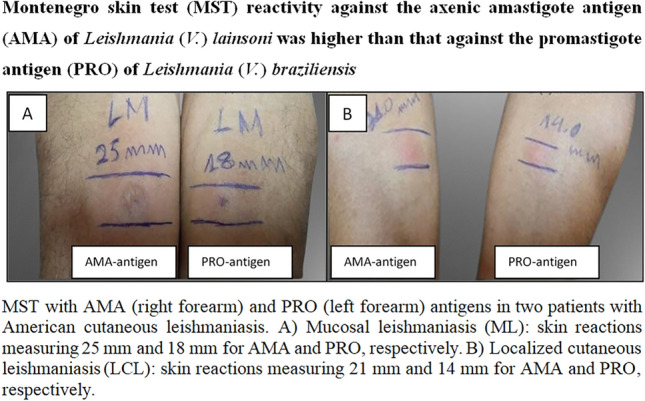
Laboratory diagnosis of American cutaneous leishmaniasis (ACL) requires a tool amenable to the epidemiological status of ACL in Brazil. Montenegro skin test (MST), an efficient immunological tool used for laboratory diagnosis of ACL, induces delayed-type hypersensitivity (DTH) response to the promastigote antigens of Leishmania; however, human immune responses against infection are modulated by the amastigote of the parasite. Leishmania (V.) lainsoni induces strong cellular immunity in humans; therefore, the antigenic reactivity of its axenic amastigote (AMA antigen) to MST was evaluated for the laboratory diagnosis of ACL.
Methods
Among 70 individuals examined, 60 had a laboratory-confirmed diagnosis of ACL; 53 had localized cutaneous leishmaniasis (LCL), and 7 had mucosal leishmaniasis (ML). Patients were treated at the Evandro Chagas Institute’s leishmaniasis clinic, Pará State, Brazil. Ten healthy individuals with no history of ACL (control group) were also examined. Leishmania (V.) braziliensis promastigote antigen (PRO) was used to compare the reactivity with that of AMA antigen. Paired Student’s t-test, kappa agreement, and Spearman test were used to evaluate the reactivity of AMA and PRO.
Results
The mean reactivity of AMA in ACL patients was 19.4 mm ± 13.3, which was higher (P < 0.001) than that of PRO: 12.1 mm ± 8.1. MST reactivity according to the clinical forms revealed that AMA reactivity in LCL and ML, 18.8 mm ± 13.3 and 24.3 mm ± 13.7, was higher (P < 0.001) than that of PRO, 11.8 mm ± 8.2 and 14.6 mm ± 8.4, respectively.
Conclusion
AMA reactivity was higher than that of PRO, indicating that AMA is a promising alternative for optimizing MST in the laboratory diagnosis of ACL.
Graphical abstract

Keywords: Leishmania (V.) lainsoni, Axenic amastigote antigen, Leishmania (V.) braziliensis, Axenic promastigote antigen, Montenegro skin test, Laboratory diagnosis, American cutaneous leishmaniasis
Background
American cutaneous leishmaniasis (ACL) is an infectious, non-contagious disease caused by at least 15 different species of protozoan parasites of the genus Leishmania Ross 1903. Leishmania can be taxonomically classified within the following subgenera: Leishmania (Leishmania) Ross 1903, L. (Viannia) Lainson & Shaw 1987, and L. (Mundinia) Shaw, Camargo & Teixeira 2016. From the clinical-immunopathological perspective, ACL can be considered one of most complex parasitic diseases owing to the interaction of different Leishmania species with the human immune system [1–3].
ACL is characterized by impairment of the cutaneous and/or mucous tissue. Consequently, ulcerated skin lesions are the major clinical manifestation of the disease. Other types of skin lesions, such as papules, tubercles, nodules, infiltrated plaques, and verrucous or keloid-vegetating lesions, have also been observed. Metastatic mucosal lesions are seen on the nose, mouth, or pharynx alone or simultaneously. Nevertheless, these lesions, with an ulcerogranulomatous appearance, are observed more frequently on the nose and can reach the cartilaginous tissue depending on its depth, leading to nasal septum perforation [4–7].
Seven species of Leishmania and a hybrid leishmanial parasite have been recognized as etiological agents of ACL in Brazil, especially in the Brazilian Amazon. This comprises six species of Leishmania (Viannia) subgenus, including L. (V.) braziliensis, L. (V.) guyanensis, L. (V.) shawi, L. (V.) lainsoni, L. (V.) naiffi, L. (V.) lindenbergi, a hybrid parasite, L. (V.) guyanensis/L. (V.) shawi, and only one species of L. (Leishmania) subgenus, L. (L.) amazonensis [8]. The clinical-immunopathological spectrum of ACL resulting from the interaction of these Leishmania species with the human immune system includes four clinical forms: localized cutaneous leishmaniasis (LCL), borderline disseminated cutaneous leishmaniasis (BDCL), mucocutaneous leishmaniasis/mucosal leishmaniasis (MCL/ml), and anergic diffuse cutaneous leishmaniasis (ADCL) [9–11].
Parasitological, histopathological, molecular (mainly the polymerase chain reaction, PCR), and immunological (cellular and/or humoral) methods have been used for the laboratory diagnosis. However, these methods have advantages and disadvantages [12–15]. Parasitological and histopathological methods depend on the parasite load in the skin and/or mucosal lesions. The molecular method requires a complex laboratory infrastructure and highly qualified staff. The immunological method can be used to detect cellular or humoral immune responses. The expression of the cellular immune response is associated with immunological protection in ACL [6, 7]. The immunological method of choice for ACL laboratory diagnosis is based on detecting a specific delayed-type hypersensitivity (DTH) response against Leishmania antigens [16–18]. The Montenegro skin test (MST), conceived in the 1920s [19], is a laboratory tool capable of confirming the presence of a DTH response against Leishmania infection, which generally indicates a good prognosis [9–11, 20, 21].
The Ralph Lainson Leishmaniases Laboratory (RL/LL) of the Parasitology Department, Evandro Chagas Institute, Secretary of Health and Environment Surveillance, Ministry of Health, Ananindeua, Pará State, Brazil, introduced the use of MST for the laboratory diagnosis of ACL using L. (L.) amazonensis promastigote antigen almost 50 years ago owing to its easy adaptation and laboratory cultivation, which facilitated its standardization [22]. This tool was used until the end of the 1990s in a satisfactory and safe manner.
After new findings provided by the lymphocyte proliferation assay in patients with ACL in the Brazilian Amazon showing that L. (V.) braziliensis promastigote antigen was able to induce greater lymphocyte proliferation rates than those by L. (L.) amazonensis promastigote antigen [23], it was decided to replace the L. (L.) amazonensis promastigote antigen used in the MST with the L. (V.) braziliensis promastigote antigen under the same standardized production and conservation procedures [22]. This change has facilitated a greater range (reaction diameter in millimeters) of MST reactivity since L. (Viannia) spp. parasites have been recognized as the main agents of ACL in the region, showing greater affinity and specificity for the L. (L.) braziliensis than L. (L.) amazonensis antigen [3, 9, 10].
The development of new laboratory methods capable of promoting axenic amastigote forms of Leishmania spp. [24–28] has made performing MST using a stage-specific axenic amastigote antigen (AMA) from different Leishmania species feasible and more appropriate. The amastigote form of this parasite is a biological stage that modulates human cellular and humoral immune responses against Leishmania infection. Considering the need for an accessible tool that can be extended to other regions of Brazil, where ACL is predominantly caused by L. (Viannia) spp. [11], using L. (V.) lainsoni AMA in MST may be promising because of not only its easy adaptation to laboratory culture media but also its exceptional capacity to induce a strong person’s cellular immune response [29, 30].
The present study aimed to evaluate the antigenic reactivity of L. (V.) lainsoni AMA for the laboratory diagnosis of ACL in the Brazilian Amazon using MST. Compared with that of the L. (V.) braziliensis promastigote (PRO), the results obtained using the new L. (V.) lainsoni AMA corroborate these aforementioned expectations, representing a new tool for strengthening the laboratory diagnosis of ACL in this region.
Methods
Study site
This prospective study was conducted from August 2020 to July 2022 at the RL/LL of the Parasitology Department, Evandro Chagas Institute, Secretary of Health and Environment Surveillance (Ministry of Health), Ananindeua, Pará State, Amazonian Brazil.
Parasite strains
Two L. (Viannia) spp. strains were used to prepare the two antigens: L. (V.) lainsoni (MHOM/BR/1981/M6426/Benevides/Pará/Brazil) for AMA and L. (V.) braziliensis (MHOM/BR/1999/M17323/Paragominas/Pará/Brazil) for PRO.
Both antigens were obtained from the culture media: 199 culture plates containing a medium for L. (V.) lainsoni and RPMI for L. (V.) braziliensis. The two L. (Viannia) spp. were isolated from LCL [L. (V.) lainsoni] and ML [L. (V.) braziliensis] clinical forms, whose virulence was conserved and stored at the Evandro Chagas Institute biobank.
Study population and clinical evaluation
Seventy individuals, comprising 60 ACL patients and 10 healthy individuals, were enrolled in this study. Patients, mainly from Pará, neighboring Brazilian states and French Guiana, who had visited the leishmaniasis outpatient clinic at the Evandro Chagas Institute, a regional referral center for the laboratory diagnosis and treatment of ACL, comprised the test group. Healthy individuals with no history of ACL residing in a non-endemic area made up the negative control group.
Laboratory investigations, such as direct parasitological and/or culture in Difco B45 medium [31] and serological assay (IFAT-IgG/cut-off: 80) also with L. (V.) lainsoni axenic amastigote antigen, were performed to confirm the diagnosis in all patients with clinical suspicion of ACL. These methods are used routinely at the RL/LL (histopathological and molecular approaches are subject to doubtful cases). Fifty-three patients with LCL and seven with ML were evaluated [6, 7, 17].
Clinical evaluation was performed based on the time course of the disease and type, number, and location of cutaneous and/or mucosal lesions [6, 7].
Identification of Leishmania spp. in the patients
Parasites were preliminarily characterized based on their morphology and behavior in culture medium and in experimentally infected hamsters. Species determination was performed using a PCR-RFLP technique with two target sequences: RNA polymerase II gene, wherein the PCR products were cleaved with the TspRI and HgaI endonucleases, and hsp70 gene, wherein the PCR products were cleaved with HaeIII endonuclease. Restriction profiles were compared with those of the reference strains of the subgenera L. (Viannia) and L. (Leishmania), regarded as ACL agents in the Amazon region [32–34].
Preparation of MST using L. (V.) lainsoni AMA and L. (V.) braziliensis PRO
Leishmania (V.) lainsoni promastigotes were cultured in Difco B45 medium for 7 days at 26 ºC until an optimal level of growth was achieved. It was subsequently seeded into 199 culture plates containing a medium (pH 7.2) supplemented with 0.36 g sodium bicarbonate (NaHCO3), 9.532 g Hepes, 0.1% biotin, 0.1% hemin, 0.5% adenine, 10% fetal bovine serum (FBS), 2% sterile human urine, and 1% penicillin/streptomycin and cultured for 3 days at 26 ºC until the logarithmic phase was reached.
The culture was transferred to 199 culture plates containing a medium (pH 7.2) supplemented with 25% FBS and incubated at 32 ºC (5% CO2) for 24 h.
The promastigote forms were centrifuged at 1562 g for 10 min at room temperature and resuspended in 199 culture plates containing a medium (pH 5.2) supplemented with l-glutamine, tryptone, glucose, 0.1% hemin, 1% adenine, 40% succinate buffer, 25% FBS, 2% sterile human urine, and 1% antibiotic and incubated for 5 days at 32 ºC (5% CO2) for differentiation into amastigote forms. The axenic amastigote forms were maintained at a ratio of one part culture/two parts 199 medium [35, 36].
The axenic amastigotes were washed thrice with sterile PBS after differentiation into 2–3 replicates and centrifuged at 1897 g for 10 min. The sediments containing amastigotes were resuspended, and the cell count was determined using a Neubauer chamber to prepare a final suspension at a concentration of 107 cells/ml in thimerosal solution [ethyl(2-mercaptobenzoate-(2-)-O,S) mercurate(1-) sodium] at a concentration of 1/10,000, as previously used as a preservative and inactivating solution for promastigote antigens of L. (L.) amazonensis or L. (V.) braziliensis [9, 16, 22]. An aliquot of the cell concentrate was extracted before adding the preservative to evaluate (i) the protein concentration present in the AMA using the Bradford Method [37] and (ii) the integrity of axenic amastigote forms using optical microscopy and flow cytometry (BD FACSCANTO II™). The AMA was then aliquoted into 5-ml vials and refrigerated at 4 °C until further use.
Preparation of L. (V.) braziliensis PRO was performed under the same standard procedures and conservation processes as for L. (L.) amazonensis antigen [22] as well as the protein concentration by the Bradford method [37].
MST using L. (V.) lainsoni AMA and L. (V.) braziliensis PRO
An intradermal injection of 0.1 mL AMA was administered on the anterior surface of the right forearm. An intradermal injection of 0.1 ml PRO was administered on the anterior surface of the left forearm. Thimerosal solution was also administered on the anterior surface of the right forearm as a control test to detect possible allergic reactions against the preservative used along with the antigens. Individuals reactive to thimerosal solution were excluded from the study owing to the lack of a definition of the specificity of the response to AMA or PRO.
Hypodermic syringes with 13 × 6.5 mm sterile disposable needles were used to administer the antigens and thimerosal solution intradermally. The tests were performed by two trained personnel with the same amount of experience to minimize individual errors. All individuals were observed for the first 30 min after the intradermal administration of the antigens and the thimerosal solution to detect immediate reactions. The injection sites were examined after 48 h to verify reactivity (induration, infiltration, or papuloerythematous reactions). The residual areas of reactivity were measured using a millimeter ruler, and the contours were marked using a ballpoint pen. Any cutaneous manifestation with a diameter of ≥ 5 mm was considered positive [6, 7].
Data analyses
It was hypothesized that the MST reactivity against L. (V.) lansoni AMA would be greater than that against L. (V.) braziliensis PRO. A paired Student’s t-test was used to test this hypothesis as two dependent variables were present and to evaluate possible differences between the protein concentrations of the two antigens (AMA vs. PRO). The Shapiro-Wilk test was used to test the assumptions of the paired Student’s t-test [38].
Kappa agreement analysis was used [39] to determine the agreement between MST test results for L. (V.) lainsoni (AMA) and L. (V.) braziliensis (PRO). The kappa values were interpreted as follows: K = 0 indicated no agreement, whereas K ≠ 0 indicated agreement [40].
The correlation of the skin reaction diameter, in millimeters, between AMA and PRO antigens was determined using Spearman correlation test to assess their correspondence with the results of the kappa test. The assumptions of the correlation test were also tested using the Shapiro-Wilk test.
All statistical analyses were performed using RStudio software 1.3.3.3. (Rstudio Team, 2020). The “dplyr” [41] and “corrplot” packages were used [42] to analyze the results of the paired Student’s t-test and correlation analysis, whereas the “pacman” package was used to analyze the results of the kappa test [43]. The graphs illustrating the descriptive analyses were created using the “ggplot2” package [44]. The confidence interval was set at 95%, and P ≤ 0.05 indicates statistical significance.
Results
Morphological characteristics and viability of the parasites used to prepare L. (V.) lainsoni AMA and L. (V.) braziliensis PRO
The morphology and integrity of L. (V.) braziliensis and L. (V.) lainsoni were evaluated to ensure that antigens were obtained from intact cells and were analyzed via optical microscopy and flow cytometry. The promastigote forms of L. (V.) braziliensis are characterized by the presence of an elongated and fusiform body, nucleus, evident kinetoplast, and middle flagellum. The promastigote forms of L. (V.) lainsoni are characterized by the presence of a large, elongated, and fusiform cell body; evident nucleus; kinetoplast; and typical long flagellum, which was larger than the body. The axenic amastigote forms of L. (V.) lainsoni presented as small cells without flagella, ovoid bodies, nuclei, or kinetoplasts.
Flow cytometry using propidium iodide (PI) labeling revealed that 72.3% of L. (V.) lainsoni axenic amastigotes were viable.
Protein concentration of L. (V.) lainsoni AMA and L. (V.) braziliensis PRO
Analysis of the protein concentration of L. (V.) lainsoni AMA and L. (V.) braziliensis PRO using the Bradford method revealed that the concentrations of AMA and PRO were 173.3 μg/ml (equivalent to 17.3 ng/ml/antigen dose in 0.1 ml) and 241.2 μg/ml (equivalent to 24.1 ng/ml/dose in 0.1 mL), respectively, indicating that the protein concentrations of both were similar (t-test, t(3) = − 2.6185, P = 0.0588).
Laboratory diagnosis of ACL for the examined patients
Among the 60 patients examined at the RL/LL presenting clinical suspicious of ACL, a direct parasitological and/or culture and serological (IFAT-IgG) diagnosis was confirmed in 46 (76.6%), comprising 43 (81.1%) with LCL and 3 (43%) with ML, while serological diagnosis was confirmed alone in 10 (18.9%) patients with LCL and 4 (57%) with ML.
Personal and clinical characteristics of the study population
Sixty patients (100% male) with a laboratory-confirmed diagnosis of ACL were included in this study. Fifty-three (88.3%) and seven (11.7%) patients presented clinical features of LCL and ML (G1 group), respectively. Ten healthy individuals [4 males (40%) and six females (60%)] with no history of ACL formed the negative control group (G2). The mean age of patients with LCL was 41 years, while in those with ML it was 43; in the control group, it was 38.5.
Clinical evaluation of the examined patients
Average disease duration in the 53 patients with LCL was 4.4 months. Average disease duration in the seven patients with ML was 83 months (approximately 7 years). The predominant type of skin lesion was ulcerated lesions (≥ 98%) in patients with LCL; 2–3 lesions per patient were observed on average, mainly on the upper and lower limbs. In contrast, the mucosal lesion had a granulomatous appearance and affected the nasobuccopharyngeal region in four (57%) of the seven patients with ML.
Identification of Leishmania spp. from the examined patients
Among the 43 LCL patients with a parasitological diagnosis, Leishmania spp. was isolated from 32 samples (one sample from each patient) whose molecular characterization revealed 31 belonging to L. (Viannia) subgenus: L. (V.) braziliensis (n = 9), L. (V.) shawi (n = 6), L. (V.) lainsoni (n = 5), L. (V.) lindenbergi (n = 3), L. (V.) guyanensis (n = 2), and L. (V.) naiffi (n = 1). Five samples could not be defined completely. Leishmania (L.) amazonensis was detected in the last sample. Among the three ML cases with a parasitological diagnosis, L. (V.) braziliensis was isolated from two. Thus, among the 60 patients examined, Leishmania spp. were isolated from 34 (56.6%): L. (Viannia) subgenus (n = 33) and L. (Leishmania) subgenus (n = 1).
Leishmania (V.) lainsoni AMA was more reactive than L. (V.) braziliensis PRO
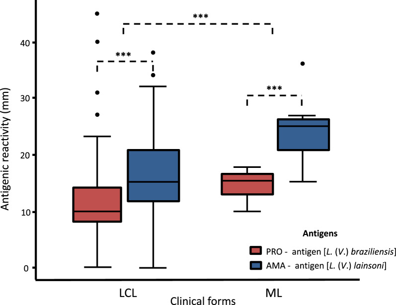
Among the 60 patients with ACL examined in the present study, a positive MST reaction to AMA and PRO was observed in 60 (100%) and 59 (98.3%) patients, indicating similar sensitivity to both antigens. However, comparison of the amplitude of MST antigenic reactivity (diameter of skin reactions in mm) against these antigens revealed that the mean (mean ± standard deviation) for AMA (19.4 mm ± 13.3) was higher than that for PRO (12.1 mm ± 8.1), which was also evident when these results were compared according to the clinical forms of the disease, i.e. LCL (18.8 mm ± 13.3 × 11.8 mm ± 8.2) and ML (24.3 mm ± 13.7 × 14.6 mm ± 8.4), respectively.
Paired Student’s t-test revealed that the mean MST reactivity against AMA differed from that against PRO (t-test, t(63) = − 8.5224, P < 0.001), indicating that the average reactivity against AMA was greater than that against PRO. Evaluation of MST reactivity according to the clinical forms of ACL revealed that patients with LCL and ML exhibited a significantly stronger reaction against AMA (t-test, t(56) = − 7.5619; P < 0.001 and t-test, t(6) = − 4.7532; P = 0.0031), respectively (Table 1; Fig. 1). Individuals in the control group did not exhibit reactivity to AMA or PRO.
Table 1.
Montenegro skin test reactions [mean and standard deviation (SD)] of L. (V.) lainsoni AMA and L. (V.) braziliensis PRO antigens according to ACL clinical forms
| aLCL | bMCL | Total | |
|---|---|---|---|
| PRO | 11.8 (8.2) | 14.6 (8.4) | 12.1 (8.1) |
| AMA | 18.8 (13.3) | 24.3 (13.7) | 19.4 (13.3) |
| P-value | < 0.001 | < 0.001 | < 0.001 |
| t-test | t(56) = − 7.5619 | t(6) = − 4.7532 | t(63) = − 8.5224 |
aLCL: localized cutaneous leishmaniasis
bMCL: mucosal leishmaniasis
Fig. 1.
Montenegro skin test reactions (in millimeters) from ACL patients submitted to laboratory diagnosis. AMA antigen: Leishmania (V.) lainsoni axenic amastigote antigen; PRO antigen: L. (V.) braziliensis promastigote antigen. LCL: localized cutaneous leishmaniasis. ML: mucosal leishmaniasis. ***P < 0.001
Although the 60 ACL patients and ten healthy individuals (control group) had each received three intradermal injections (0.1 ml), two with the AMA and PRO antigens and one with the thimerosal solution, during the first 30 min, none manifested any local sign and/or systemic symptom of significant magnitude, that is, discomfort (irritation or itching) or local pain, general malaise, drowsiness, dizziness, or syncope. Upon return (after 48 h), only three (48%) of the seven ML patients, those with reactions ≥ 15 mm in diameter, reported moderate hyperthermia (up to 37.6 °C) and/or headache lasting up to 24 h at ~ 12 h after the injections; at the time of assessment of intradermal reactions, there were no more complaints of these side reactions.
Diagnostic agreement and correlation between AMA and PRO
Kappa agreement analysis revealed a strong reliability between AMA and PRO antigens [k = 0.94 (95% CI 0.88–1.00)] and a diagnostic agreement of 98.6%. Thus, the antigens were concordant in 69 of the 70 individuals tested (60 patients with ACL and 10 negative controls). One individual was positive only for AMA and negative for PRO (Table 2).
Table 2.
Diagnostic agreement between AMA and PRO antigens used by MST in the laboratory diagnosis of ACL (60 patients) and 10 healthy individuals (control group)
| AMA | ||
|---|---|---|
| Positive | Negative | |
| PRO | ||
| Positive | 59 (0.843) | 0 (0) |
| Negative | 1 (0.014) | 10 (0.143) |
| Agreement | 98.6% | |
| Kappa | 0.94 (IC 95%: 0.88–1.00) | |
| Conclusion | Perfect | |
| P-value (unilateral) | < 0.001 | |
AMA: Leishmania (V.) lainsoni axenic amastigote antigen; PRO: L. (V.) braziliensis promastigote antigen
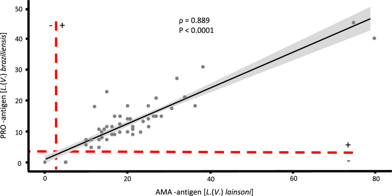
The Spearman test revealed a positive correlation between AMA and PRO (rho = 0.889, P < 0.0001) (Fig. 2).
Fig. 2.
Spearman correlation of Montenegro skin test reactivities (diameter, in millimeters) between AMA and PRO antigens in the test (60 ACL-confirmed patients) and control group (10 healthy individuals)
Discussion
In this study we examined the antigenic reactivity of the AMA of L. (V.) lainsoni, an important etiologic agent of ACL in Latin America [36, 45–49], as an alternative antigen for optimizing MST in the laboratory diagnosis of the disease.
The MST results of the 70 participants tested (60 patients with ACL, comprising 53 with LCL and 7 with ML, and 10 healthy individuals) revealed that the antigenic reactivity of L. (V.) lainsoni AMA was significantly higher than that of L. (V.) braziliensis PRO. This finding was also true in the analysis of the antigenic reactivity according to the clinical forms of ACL, i.e. LCL and ML, respectively. These findings are of great technical and scientific interest, especially since the protein concentrations of both antigens were similar. Thus, the difference in reactivity may be attributed to the greater specificity of L. (V.) lainsoni AMA compared with that of L. (V.) braziliensis PRO, confirming the good stability, such as integrity and cellular viability, of AMA.
Although the antigens used in this study are not species specific, but subgenus specific, i.e. L. (Viannia) lainsoni × L. (Viannia) braziliensis, the above data appear to confirm that the AMAs of the parasite are more specific than those of PROs as the amastigote stage of the parasite interacts directly with the person’s cellular and humoral immune responses following the establishment of the infection [3, 11]. Furthermore, the complete lack of MST response in the 10 healthy individuals with no history of ACL (control group) appears to confirm the specificity of both antigens.
Analysis of the sensitivity of AMA and PRO revealed rates of 100% and 98.3%, respectively, indicating similar rates. This finding highlights the great capacity of these antigens to recognize the specific DTH response observed in patients with ACL. However, the rates were not identical (100% for both), as MST reactivity for L. (V.) braziliensis PRO was not observed in the patient with LCL caused by L. (L.) amazonensis. This finding has already been recorded with high frequency (≥ 80%) for the current L. (V.) braziliensis PRO and a species-specific antigen of L. (L.) amazonensis PRO [7, 9, 10, 16], reinforcing the intriguing ability of L. (L.) amazonensis to escape the human cellular immune response [50–52]. However, this finding also highlights the specificity of L. (V.) lainsoni AMA in recognizing DTH even in patients with LCL caused by L. (L.) amazonensis, although it was observed in only one case examined in the present study (which PRO failed to recognize).
The sensitivity rates of AMA and PRO recorded in the present study are among the highest (≥ 98%) when compared with those recorded by previous studies conducted in Brazil and Argentina (South America), such as Rio de Janeiro, ≥ 90% [53]; São Paulo, 82–89% [12]; Paraná, 84.4% [54]; Ceará, 46.3% [55]; and a retrospective analysis conducted at a reference center in Argentina (also ≥ 90%) [56]. The Department of Surveillance of Communicable Diseases of the Secretariat of Health and Environmental Surveillance of the Ministry of Health of Brazil [57] revealed the reference rate of MST sensitivity is approximately 90%. Thus, the rates observed in the present study for AMA (100%) and PRO (98.3%) indicate more robust results than those reported by different sources.
MST reactivity against AMA [L. (V.) lainsoni] and PRO [L. (V.) braziliensis] in patients with LCL and ML in this study showed that the reactivity against AMA was higher than that against PRO in both clinical forms, LCL and ML; however, stronger MST expression was observed in ML than LCL. This finding can be attributed to the distinct immunopathological mechanisms involved in the development of these clinical forms of ACL. The T-cell immune response, mainly of the CD4+/Th1-type (which is strongly associated with DTH), is more activated in ML than LCL [9, 10, 58, 59]. Furthermore, the duration of disease was longer in ML (approximately 7 years) than LCL (4.4 months), indicating a longer lasting immune stimulus in patients with ML than in those with LCL. This promotes a much stronger MST response in patients with ML. However, this does not seem to remember that, in addition to the action of the parasite’s species-specific antigens [3, 11], human genetic factors [60–62] and even co-infection of L. (V.) braziliensis with Leishmania RNA virus 1 [48, 63] may be involved in modulating these immunopathological mechanisms in ML.
Regarding the higher expression of antigenic reactivity against AMA, notably, diagnostic agreement analysis showed a high rate (98.6%) and Spearman analysis evidenced a strong positive correlation, indicating that these antigens have similar potential for use in the laboratory diagnosis of ACL, i.e. either antigen can be used in the absence of the other. However, this raises a question regarding the advantage of replacing the L. (V.) braziliensis PRO with L. (V.) lainsoni AMA. The advantage is not necessarily in replacing PRO with AMA but in knowing that today a new antigen (AMA) is available that expresses greater reactivity in the laboratory diagnosis of ACL caused by all Leishmania spp. of L. (Viannia) subgenus occurring in Brazil [L. (V.) braziliensis, L. (V.) guyanensis, L. (V.) shawi, L. (V.) lainsoni, L. (V.) lindenbergi, and L. (V.) naiffi] and possibly caused by Leishmania spp. of L. (Leishmania) subgenus, such as L. (L.) amazonensis [8, 11], as shown here (and perhaps in Latin America).
This finding is particularly useful as Brazil and most Latin American countries do not have a similar PRO owing to the lack of industrial infrastructure compatible with that of Good Manufacturing Practices (GMP) for the production of MST antigen and the low commercial profitability of industrial production of antigen [64, 65]. Additionally, this technique has shown potential for detecting ACL due to Leishmania spp. of L. (Leishmania) subgenus, specifically, L. (L.) amazonensis, which has demonstrated great capacity to evade cellular immunity [50–52]. These findings indicate that investing in large-scale AMA antigen productivity, in addition to optimizing MST for laboratory diagnosis of ACL, will aid in addressing the demand from Brazil and neighboring countries.
Even though these findings revealed that AMA antigen represents a promising alternative for optimizing MST in the laboratory diagnosis of ACL, future studies must seek to elucidate the composition and immunogenicity of AMA proteins to develop a synthetic antigen applicable to all epidemiological scenarios of the disease in Latin America. It has been clearly demonstrated that the use of AMA from L. (V.) lainsoni, the most ancestral Leishmania species of L. (Viannia) subgenus [66–68] that carries a protein genetic load common to all Leishmania spp. of this subgenus, would represent a desirable major advance in the laboratory diagnosis of ACL.
The crucial role of MST in defining the clinical-immunopathological spectrum of ACL in the Brazilian Amazon should also be highlighted [2, 3, 9–11, 16, 17], where a moderately positive MST in LCL is caused by Leishmania spp. of L. (Viannia) subgenus, especially L. (V.) braziliensis. This reactivity increases as the infection evolves toward the hyperergic pole (CD4+/Th1-type) of that spectrum represented by ML. In contrast, the reactivity to MST in LCL caused by L. (L.) amazonensis is weak or negative and completely regresses as the infection progresses to the hypoergic pole (CD4+/Th2 type) of the ACL spectrum represented by severe and incurable ADCL. Thus, MST can also be used as a prognostic marker for the treatment of the different clinical forms within the clinical-immunopathological spectrum of ACL, which can be performed with a higher degree of accuracy using AMA antigen as it showed greater fidelity to the cellular immune response (DTH) of patients with LCL and ML examined here.
Lastly, as mentioned before, it has been almost 50 years since the RL/LL introduced the MST as a complementary tool for the laboratory diagnosis of ACL. During this period, two antigenic compositions were used to perform MST, with promastigote of L. (L.) amazonensis [22] and L. (V.) braziliensis [3, 9, 10], without records of any incident (serious side effect) resulting from use of these antigenic compositions. The results presented now demonstrated that, although a different methodology from the previous one was introduced for the production of the new antigen [L. (V.) lainsoni AMA] used to perform MST, no serious side effects, local or systemic, were recorded in 70 individuals tested (60 ACL patients and 10 healthy individuals), confirming the GMP used for the preparation of these antigens.
Conclusions
The studied patients exhibited a higher reactivity to L. (V.) lainsoni AMA than to L. (V.) braziliensis PRO in the MST. This indicates that the L. (V.) lainsoni AMA is a promising alternative for optimizing MST in the laboratory diagnosis of ACL.
Acknowledgements
We thank Raimundo Nonato Pires, Rodrigo Ribeiro Furtado, and Lucivaldo Ferreira for their technical assistance in the laboratory work.
Abbreviations
- ACL
American cutaneous leishmaniasis
- MST
Montenegro skin test
- DTH
Delayed-type hypersensitivity response
- LCL
Localized cutaneous leishmaniasis
- BDCL
Borderline disseminated cutaneous leishmaniasis
- ML
Mucosal leishmaniasis
- ADCL
Anergic diffuse cutaneous leishmaniasis
- AMA
Axenic amastigote antigen
- PRO
Promastigote antigen
- PCR
Polymerase chain reaction
- RL/LL
Ralph Lainson Leishmaniases Laboratory
- RPMI
Roswell Park Memorial Institute
- IFAT
Indirect fluorescent antibody test
- IgG
Immunoglobulin G
- RFLP
Restriction fragment length polymorphism
- RNA
Ribonucleic acid
- hsp
Heat shock protein
- CD4/Th1
Lymphocyte CD4/T-helper type 1
- CD4/Th2
Lymphocyte CD4/T-helper type 2
Author contributions
LVM: Data curation, Formal analysis, Investigation, Resources, Writing–original draft; MBC: Conceptualization, data curation, Formal analysis, Methodology, Writing–original draft; TVS: Resources, Software, Formal analysis, Visualization; PKR: Resources, Software, Formal analysis, Visualization, LVL: Project administration, Formal analysis, Validation, Visualization; FTS: Supervision, Funding acquisition, Project administration, Validation, Visualization, Writing–original draft.
Funding
This work was supported by Evandro Chagas Institute (Surveillance Secretary of Health and Environment, Ministry of Health, Brazil) and Tropical Medicine Nucleus (Federal University of Pará, Brazil).
Availability of data and materials
All data supporting the main findings of this study are found in the manuscript.
Declarations
Ethics approval and consent to participate
All participants were provided with a detailed explanation regarding the objective of the study. Written informed consent (WICF) was obtained from all participants. WICF was obtained from the participant and the guardian [free and informed assent term (FIAT)] for participants aged < 18 years. The study protocol was approved by the Ethics Committee of the Tropical Medicine Nucleus at the Federal University of Pará, Brazil (protocol number: 4306847).
Competing interests
The authors declare no competing interests.
Footnotes
Publisher's Note
Springer Nature remains neutral with regard to jurisdictional claims in published maps and institutional affiliations.
Marliane Batista Campos and Fernando Tobias Silveira contributed equally to this work.
References
- 1.Lainson R, Shaw JJ. New World Leishmaniasis. In: L Collier, A Balows, M Sussman. Topley & Wilson’s Microbiology and Microbial Infections, 10th ed. Parasitol. Arnold, London. 2010; 5: 313–349.
- 2.Campos MB, Lima LVR, de Lima ACS, VasconcelosdosSantos T, Ramos PKS, Gomes CMC, et al. Toll-like receptors 2, 4, and 9 expressions over the entire clinical and immunopathological spectrum of American cutaneous leishmaniasis due to Leishmania (V.) braziliensis and Leishmania (L.) amazonensis. PLoS ONE. 2018;13:e0194383. 10.1371/journal.pone.0194383. [DOI] [PMC free article] [PubMed] [Google Scholar]
- 3.Silveira FT. What makes mucosal and anergic diffuse cutaneous leishmaniases so clinically and immunopathologically different? Trans Roy Soc Trop Med Hyg. 2019;113:505–16. [DOI] [PubMed] [Google Scholar]
- 4.Marsden PD. Mucosal leishmaniasis (“espundia” Escomel, 1911). Trans Roy Soc Trop Med Hyg. 1986;80:859–76. [DOI] [PubMed] [Google Scholar]
- 5.Marsden PD, Llanos-Cuentas EA, Lago EL, Cuba CC, Barreto AC, Costa JM, et al. Human mucocutaneous leishmaniasis in Três Braços, Bahia-Brazil An area of Leishmania braziliensis braziliensis transmission. III. Mucosal disease presentation and initial evolution. Rev Soc Bras Med Trop. 1984;17:179–86. [Google Scholar]
- 6.Silveira FT, Lainson R, Muller SFR, de Souza AAA, Corbett CEP. Leishmaniose tegumentar americana. In: Medicina Tropical e Infectologia na Amazônia. RNG Leão (Ed). Ed. Samauma, 1a ed. Vol. 2, Instituto Evandro Chagas, Belém, Pará, Brasil. 2013;1203–1244.
- 7.Silveira FT, Muller SFR, Laurenti MD, Gomes CMC, Corbett CEP. Leishmaniose Tegumentar Americana. In: Tratado de Dermatología. Belda Junior W, Di Chiacchio N, Criado PR (Ed). 3a Ed. São Paulo, Atheneu. 2018;72:1691–1700.
- 8.Jennings YL, de Souza AAA, Ishikawa EAY, Shaw J, Lainson R, Silveira FT. Phenotypic characterization of Leishmania spp. causing cutaneous leishmaniasis in the lower Amazon region, western Pará state, Brazil, reveals a putative hybrid parasite, Leishmania (Viannia) guyanensis x Leishmania (Viannia) shawi shawi. Parasite. 2014;1:1–11. [DOI] [PMC free article] [PubMed] [Google Scholar]
- 9.Silveira FT, Lainson R, Corbert CEP. Clinical and immunopathological spectrum of American cutaneous leishmaniasis with special reference to the disease in the Amazonian Brazil. Mem Inst Oswaldo Cruz. 2004;99:239–51. [DOI] [PubMed] [Google Scholar]
- 10.Silveira FT, Lainson R, Gomes CM, Laurenti MD, Corbett CEP. Immunopathogenic competences of Leishmania (V.) braziliensis and L. (L.) amazonensis in American cutaneous leishmaniasis. Parasite Immunol. 2009;31:423–31. [DOI] [PubMed] [Google Scholar]
- 11.Silveira FT, Campos MB, Müller SF, Ramos PK, Lima LV, Vasconcelos dos Santos T, et al. From Biology to Disease: Importance of Species-Specific Leishmania Antigens from the subgenera Viannia (L. braziliensis) and Leishmania (L. amazonensis) in the pathogenesis of American cutaneous leishmaniasis. In: Leishmania Parasites; IntechOpen, Croatia. 2023;1–29. 10.5772/intechopen.108967
- 12.Goto H, Lindoso JA. Current diagnosis and treatment of cutaneous and mucocutaneous leishmaniasis. Expert Rev Anti Infect Ther. 2010;8:419–33. 10.1586/eri.10.19. [DOI] [PubMed] [Google Scholar]
- 13.De Vries HJC, Reedijk SH, Schallig HDFH. Cutaneous leishmaniasis: recent developments in diagnosis and management. Am J Clin Dermatol. 2015;16:99–109. 10.1007/s40257-015-0114-z. [DOI] [PMC free article] [PubMed] [Google Scholar]
- 14.Espir TT, Guerreiro TS, Naiff MF, Figueira LP, Soares FV, Silva SS, et al. Evaluation of different diagnostic methods of American cutaneous leishmaniasis in the Brazilian Amazon. Ex Parasitol. 2016;167:1–6. 10.1016/j.exppara.2016.04.010. [DOI] [PubMed] [Google Scholar]
- 15.Pena HP, Belo VS, Xavier-Junior JCC, Teixeira-Neto RG, Melo SN, Pereira DA, et al. Accuracy of diagnostic tests for American tegumentary leishmaniasis: a systematic literature review with meta-analyses. Trop Med Int Heal. 2020;25:1168–81. 10.1111/tmi.13465. [DOI] [PubMed] [Google Scholar]
- 16.Silveira FT, Lainson R, Shaw JJ, de Souza AAA, Ishikawa EAY, Braga RR. Cutaneous leishmaniasis due to Leishmania (Leishmania) amazonensis in Amazonian Brazil, and the significance of a Montenegro skin-test in human infections. Trans Roy Soc Trop Med Hyg. 1991;85:735–8. [DOI] [PubMed] [Google Scholar]
- 17.Silveira FT, Lainson R, Corbett CEP. Further observations on clinical, histopathological and immunological features of borderline disseminated cutaneous leishmaniasis caused by Leishmania (Leishmania) amazonensis. Mem Inst Oswaldo Cruz. 2005;100:525–34. [DOI] [PubMed] [Google Scholar]
- 18.Carstens-Kass J, Paulini K, Lypaczewski P, Matiashewski G. A review of the leishmanin skin test: a neglected test for a neglected disease. PLoS Neglec Trop Dis. 2021;15:1–16. [DOI] [PMC free article] [PubMed] [Google Scholar]
- 19.Montenegro J. Cutaneous reaction in leishmaniasis. Arch Dermatol Syphil. 1926;13:187–94. [Google Scholar]
- 20.Aronson N, Herwaldt BL, Libman M, Pearson R, Lopez-velez R, Weina P, et al. Diagnosis and treatment of leishmaniasis: clinical practice guidelines by the infectious diseases society of America (IDSA) and the American society of tropical medicine and hygiene (ASTMH). Clin Infect Dis. 2016;63:e202–264. 10.1093/cid/ciw670. [DOI] [PubMed] [Google Scholar]
- 21.Skraba CM, PerlesdeMello TF, Pedroso RB, Ferreira EC, Demarchi IG, Aristides SMA, et al. Evaluation of the reference value for the Montenegro skin test. Rev Soc Bras Med Trop. 2015;48:437–44. 10.1590/0037-8682-0067-2015. [DOI] [PubMed] [Google Scholar]
- 22.Shaw J, Lainson R. Leishmaniasis in Brazil: X. some observations on intradermal reactions to different trypanosomatid antigens of patients suffering from cutaneous and mucocutaneous leishmaniasis. Trans Roy Soc Trop Med Hyg. 1975;69:323–35. [DOI] [PubMed] [Google Scholar]
- 23.Silveira FT, Blackwell JM, Ishikawa EA, Shaw J, Quinnell RJ, Soong L, et al. T cell responses to crude and defined leishmanial antigens in patients from the lower Amazon region of Brazil infected with different species of Leishmania of the subgenera Leishmania and Viannia. Parasite Immunol. 1998;20:19–26. [DOI] [PubMed] [Google Scholar]
- 24.Bates PA, Robertson CD, Tetley L, Coombs GH. Axenic cultivation and characterization of Leishmania mexicana amastigote-like forms. Parasitol. 1992;105:202. 10.1017/S0031182000074102. [DOI] [PubMed] [Google Scholar]
- 25.Balanco JM, Pral EM, Silva S, Bijovsky AT, Mortara RA, Alfieri SC. Axenic cultivation and partial characterization of Leishmania braziliensis amastigote-like stages. Parasitol. 1998;116:103–13. 10.1017/s003118209700214x. [DOI] [PubMed] [Google Scholar]
- 26.Gupta N, Goyal N, Rastogi AK. In vitro cultivation and characterization of axenic amastigotes of Leishmania. Trends Parasitol. 2001;17:150–3. 10.1016/S1471-4922(00)01811-0. [DOI] [PubMed] [Google Scholar]
- 27.Debrabant A, Joshi MB, Pimenta PF, Dwyer DM. Generation of Leishmania donovani axenic amastigotes: their growth and biological characteristics. Int J Parasitol. 2004;34:205–17. 10.1016/j.ijpara.2003.10.011. [DOI] [PubMed] [Google Scholar]
- 28.Barak E, Amin-Spector S, Gerliak E, Goyard S, Holland N, Zilberstein D. Differentiation of Leishmania donovani in host-free system: analysis of signal perception and response. Mol Biochem Parasitol. 2005;141:99–108. 10.1016/j.molbiopara.2005.02.004. [DOI] [PubMed] [Google Scholar]
- 29.Eresh S, Bruijn MH, Mendoza-Leon JA, Barker DC. Leishmania (Viannia) lainsoni occupies a unique niche within the subgenus Viannia. Trans R Soc Trop Med Hyg. 1995;89:231–6. [DOI] [PubMed] [Google Scholar]
- 30.Corrêa JR, Santos SG, Araújo MS, Baptista C, Soares MJ, Brazil RP. Axenic promastigote forms of Leishmania (Viannia) lainsoni as an alternative source for Leishmania antigen production. J Parasitol. 2005;91:551–6. [DOI] [PubMed] [Google Scholar]
- 31.Walton BC, Shaw JJ, Lainson R. Observations on the in vitro cultivation of Leishmania braziliensis. J Parasitol. 1997;3:1118–9. [PubMed] [Google Scholar]
- 32.Simon S, Nacher M, Carme B, Basurko C, Roger A, Adenis A, et al. Cutaneous leishmaniasis in French Guiana: revising epidemiology with PCRRFLP. Trop Med Health. 2017;45:5. 10.1186/s41182-017-0045-x. [DOI] [PMC free article] [PubMed] [Google Scholar]
- 33.Gonçalves LP, Vasconcelos dos Santos T, Campos MB, Lima LVR, Ishikawa EAY, Silveira FT, et al. Further insights into the eco-epidemiology of American cutaneous leishmaniasis in the Belem metropolitan region, Pará State Brazil. Rev Soc Bras Med Trop. 2020;53:20200255. 10.1590/0037-8682-0255-2020. [DOI] [PMC free article] [PubMed] [Google Scholar]
- 34.Lima ACS, Gomes CMC, Tomokane TY, Campos BC, Zampieri RA, Jorge CL, et al. Molecular tools confirm natural Leishmania (Viannia) guyanensis/L. (V.) shawi hybrids causing cutaneous leishmaniasis in the Amazon region of Brazil. Gen Mol Biology. 2021;44:e20200123. 10.1590/1678-4685-GMB-2020-0123. [DOI] [PMC free article] [PubMed] [Google Scholar]
- 35.Saar Y, Ransford A, Waldman E, Mazareb S, Amin-Spector S, Plumblee S, et al. Characterization of developmentally-regulated activities in axenic amastigotes of Leishmania donovani. Mol Biochem Parasitol. 1998;95:9–20. 10.1016/S0166-6851(98)00062-0. [DOI] [PubMed] [Google Scholar]
- 36.da Silva TBS, Silveira FT, Tomokane TY, Batista LFS, Nunes JB, da Matta VLR, et al. Reactivity of purified and axenic amastigotes as a source of antigens to be used in serodiagnosis of canine visceral leishmaniasis. Parasitol Int. 2020. 10.1016/j.parint.2020.102177. [DOI] [PubMed] [Google Scholar]
- 37.Bradford MM. A rapid and sensitive method for the quantitation of microgram quantities of protein utilizing the principle of protein-dye binding. Analyt Bioch. 1976;72:248–54. [DOI] [PubMed] [Google Scholar]
- 38.Zar JH. Biostatistical analysis. 4th ed. Upper Saddle River: Prentice-Hall; 1999. p. 931. [Google Scholar]
- 39.Cohen J. A coefficient of agreement for nominal scales. Educat Psychol Measur. 1960;20:37–46. [Google Scholar]
- 40.Mchugh ML. Interrater reliability: the kappa statistic. Bioch Medica. 2012;22:276–82. [PMC free article] [PubMed] [Google Scholar]
- 41.Wickham H, Averick M, Bryan J, Chang W, McGowan L, François R, et al. Welcome to the Tidyverse. J Open Sour Software. 2019;4:1686. [Google Scholar]
- 42.Wei T, Simko V, Levy M, Xie Y, Jin Y, Zemla J, et al. Package ‘corrplot.’ Statistician. 2017;56:4. [Google Scholar]
- 43.Rinker TW, Kurkiewicz D. Pacman: package management for R. version 0.5. 0. Buffalo, New York. 2017. http://github.com/trinker/pacman
- 44.Wickham H, Chang W, Henry L, Takahashi K, Wilke C, Woo K, et al. ggplot2: Create elegant data visualisations using the grammar of graphics. R package version 2.2. 1. Stata Software Package: College Station, TX, USA. 2016.
- 45.Lucas CM, Franke ED, Cachay MI, Tejada A, Carrizales D, Kreutzer RD. Leishmania (Viannia) lainsoni: first isolation in Peru. Am J Trop Med Hyg. 1994;5:533–7. [PubMed] [Google Scholar]
- 46.Martinez E, Le Pont F, Mollinedo S, Cupollilo EA. First case of cutaneous leishmaniasis due to Leishmania (Viannia) lainsoni in Bolivia. Trans Roy Soc Trop Med Hyg. 2001;95:375–7. [DOI] [PubMed] [Google Scholar]
- 47.TojaldaSilva AC, Cupolillo E, Volpini AC, Almeida R, Romero GAS. Species diversity causing human cutaneous leishmaniasis in Rio Branco, state of Acre Brazil. Trop Med Int Health. 2006;11:1388–98. 10.1111/j.1365-3156.2006.01695. [DOI] [PubMed] [Google Scholar]
- 48.Cantanhêde LM, da Silva Júnior CF, Ito MM, Felipin KP, Nicolete R, Salcedo JMV, et al. Further evidence of an association between the presence of leishmania RNA Virus 1 and the mucosal manifestations in tegumentary leishmaniasis patients. PLoS Negl Trop Dis. 2015;9:e0004079. 10.1371/journal.pntd.0004079. [DOI] [PMC free article] [PubMed] [Google Scholar]
- 49.Kato H, Bone AE, Mimori T, Hashiguchi K, Shiguango GF, Gonzales SV, et al. First human cases of Leishmania (Viannia) lainsoni infection and a search for the vector sand flies in ecuador. PLoS Negl Trop Dis. 2016;10:e0004728. 10.1371/journal.pntd.0004728. [DOI] [PMC free article] [PubMed] [Google Scholar]
- 50.Silveira FT. Leishmaniose cutânea difusa na Amazônia, Brasil: aspectos clínicos e epidemiológicos. Gaz Méd Bahia. 2009;79:25–9. [Google Scholar]
- 51.Silveira FT, Müller SFR, de Souza AAA, Lainson R, Gomes CMC, Laurent MD, et al. Revisão sobre a patogenia da leishmaniose tegumentar americana na Amazônia, com ênfase à doença causada por Leishmania (V.) braziliensis e Leishmania (L.) amazonensis. Rev Paraense de Medicina. 2008;22:9–20. [Google Scholar]
- 52.Campos MB, Lima LV, Vasconcelos dos Santos T, Ramos PK, Lima ACS, Silveira F T. Further evidence on the intriguing Leishmania (L.) amazonensis interation with T-cell immune response in American cutaneous leishmaniasis. In: The 7th World Con Leishmaniasis. 2022. Cartagena.
- 53.Souza WJ, Sabroza PC, Santos CS, Sousa E, Henrique MF, Coutinho SG. Montenegro skin tests for American cutaneous leishmaniasis carried out on school-children in Rio de Janeiro, Brazil: an indicator of transmission risk. Acta Trop. 1992;52:111–9. [DOI] [PubMed] [Google Scholar]
- 54.Pontello R Jr, Gon AS, Ogama A. American cutaneous leishmaniasis: epidemiological profile of patients treated in Londrina from 1998 to 2009. An Bras Dermatol. 2013;88:748–53. [DOI] [PMC free article] [PubMed] [Google Scholar]
- 55.Granjeiro CR Jr, Pimentel JV, Teixeira AG Jr, Jesus AF, Galvão TC, Souza LA, et al. American cutaneous leishmaniasis in a northeast Brazilian city: clinical and epidemiological features. Rev Soc Bras Med Trop. 2018;51:837–42. [DOI] [PubMed] [Google Scholar]
- 56.Krolewiecki AJ, Almazan MC, Quilpildor M, Juarez M, Gil JF, Espinosa M, et al. Reappraisal of Leishmanin Skin Test (LST) in the management of American Cutaneous Leishmaniasis: a retrospective analysis from a reference center in Argentina. PLoS Negl Trop Dis. 2017;11:e0005980. [DOI] [PMC free article] [PubMed] [Google Scholar]
- 57.Brasil. Ministério da Saúde (MS), Secretaria de Vigilância da Saúde (SVS), Departamento de Vigilância das Doenças Transmissíveis (DVDT). Manual de vigilância da leishmaniose tegumentar. Brasília. 2017;189.
- 58.Bacellar O, Lessa H, Schriefer A, Machado P, de Jesus AR, Dutra WO, et al. Up-regulation of Th1-type responses in mucosal leishmaniasis patients. Inf Immunity. 2002;70:6734–40. [DOI] [PMC free article] [PubMed] [Google Scholar]
- 59.Gaze ST, Dutra WO, Lessa M, Lessa H, Guimarães LH, de Jesus AR, et al. Mucosal leishmaniasis patients display an activated inflammatory T-cell phenotype associated with a nonbalanced monocyte population. Scand J Immunol. 2006;63:70–8. [DOI] [PubMed] [Google Scholar]
- 60.Blackwell JM. Tumour necrosis factor alpha and mucocutaneous leishmaniasis. Parasitol Today. 1999;15:73–6. [DOI] [PubMed] [Google Scholar]
- 61.Castellucci L, Cheng LH, Araújo C, Guimarães LH, Lessa H, Machado P, et al. Familial aggregation of mucosal leishmaniasis in northeast Brazil. Am J Trop Med Hyg. 2005;73:69–73. [PubMed] [Google Scholar]
- 62.Castellucci L, Menezes E, Oliveira J, Magalhaes A, Guimaraes LH, Lessa M, et al. IL6-174 G/C promoter polymorphism influences susceptibility to mucosal but not localized cutaneous leishmaniasis in Brazil. J Inf Dis. 2006;194:519–27. [DOI] [PubMed] [Google Scholar]
- 63.Ito MM, Catanhêde LM, Katsuragawa TH, Silva Junior CF, Camargo LM, Mattos RG, et al. Correlation between presence of Leishmania RNA virus 1 and clinical characteristics of nasal mucosal leishmaniosis. Braz J Otorhinolaryngol. 2015;81:533–40. [DOI] [PMC free article] [PubMed] [Google Scholar]
- 64.Braz LMA. Tegumentary leishmaniasis diagnosis: what happened with MST (Montenegro Skin Test) in Brazil? Rev Inst Med Trop São Paulo. 2019;61:e17. 10.1590/S1678-9946201961017. [DOI] [PMC free article] [PubMed] [Google Scholar]
- 65.Barbeitas MM. Montenegro skin test distracted by the promise of modernity. J Braz Soc Trop Medicine. 2023;56:492–202. 10.1590/0037-8682-0492-2022. [DOI] [PMC free article] [PubMed] [Google Scholar]
- 66.Harkins KM, Schwartz RS, Cartwright RA, Stone AC. Phylogenomic reconstruction supports supercontinent origins for Leishmania. Inf Gen Evol. 2016;38:101–9. 10.1016/j.meegid.2015.11.030. [DOI] [PubMed] [Google Scholar]
- 67.Kaufer A, Stark D, Ellis J. Evolutionary insight into the Trypanosomatidae using alignment-free Phylogenomics of the Kinetoplast. Pathogens. 2019;8:157. 10.3390/pathogens8030157. [DOI] [PMC free article] [PubMed] [Google Scholar]
- 68.Silveira FT, Sousa Junior EC, Silvestre RV, Costa-Martins AG, Pinheiro KC, Sosa-Ochoa W, et al. Whole-genome sequencing of Leishmania infantum chagasi isolates from Honduras and Brazil. Microbiol Res Ann. 2021;10:e00471-e521. [DOI] [PMC free article] [PubMed] [Google Scholar]
Associated Data
This section collects any data citations, data availability statements, or supplementary materials included in this article.
Data Availability Statement
All data supporting the main findings of this study are found in the manuscript.


