Abstract
Background
Type 2 diabetes mellitus (T2DM) is associated with decreased heart rate variability (HRV) with an unclear intermediate mechanism. This study aimed to conduct mediation analysis to explore the impact of various adipose tissues on the relationship between T2DM and HRV.
Methods
A total of 380 participants were enrolled for analysis, including 249 patients with T2DM and 131 non-diabetic controls. The thicknesses of four adipose tissues (subcutaneous, extraperitoneal, intraperitoneal, and epicardial) were measured by abdominal ultrasound or echocardiography respectively. HRV was assessed by 24-hour Holter for monitoring both frequency domain indices (LF, HF, and LF/HF) and time domain indices (SDNN, SDANN, SDNN index, rMSSD and pNN50). Mediation analysis was used toexamine whether adipose tissues mediated the relationship between T2DM and each index of HRV. Then, a latent variable - HRV burden - was constructed by structural equation model with selected HRV indices to comprehensively assess the whole HRV.
Results
Compared to non-diabetic controls, patients with T2DM exhibited a significant reduction in indices of HRV, and a remarkable increase in the thicknesses of extraperitoneal, intraperitoneal, and epicardial adipose tissues. Mediation analysis found significant indirect effects of T2DM on six indices of HRV, including HF, SDNN, SDANN, SDNN index, rMSSD, and pNN50, which was mediated by epicardial adipose tissue rather than other adipose tissues, with the mediation proportions of 64.21%, 16.38%, 68.33%, 24.34%, 24.10% and 30.51%, respectively. Additionally, epicardial adipose tissue partially mediated the relationship between T2DM and reduced HRV burden (24.26%), which composed by SDNN, SDNN index, rMSSD, and pNN50.
Conclusion
Epicardial adipose tissue partially mediated the relationship between T2DM and reduced HRV, which reinforces the value of targeting heart-specific visceral fat to prevent cardiac autonomic neuropathy in diabetes.
Supplementary Information
The online version contains supplementary material available at 10.1186/s12933-024-02438-1.
Keywords: Adipose tissue, Heart rate variability, Mediation analysis, Structural equation modeling, Type 2 diabetes mellitus
Introduction
Type 2 diabetes mellitus (T2DM) continues to be a major public health problem and remains one of leading causes of heart attacks worldwide. Cardiovascular autonomic neuropathy (CAN), with its hallmark reduced heart rate variability (HRV), is an often overlooked and severe complication of diabetes. Studies have shown that at least 20% of diabetic patients diagnosed with CAN, 65% of whom at growing risk of disease progression with increasing age and duration of diabetes [1]. Indices of HRV are composed of different frequency and time domain components attributed to parasympathetic and sympathetic activity, which can predict the risk of cardiovascular morbidity and mortality [2, 3]. Although the association between CAN and T2DM is well defined, the underlying mechanisms are less understood, resulting in limited methods for prevention and intervention.
Patients with T2DM often experience metabolic disorders, leading to fat deposition [4–6]. As accumulating evidences suggesting that adipose tissue accumulation may also be a potential risk factor in neuropathy, independent of hyperglycemia [7–9], a better understanding of the role of fat deposition in mediating the relationship between diabetes and CAN is crucial. Both visceral and subcutaneous adipose tissues are recognized as emerging cardio-metabolic risk factors that correlate with major adverse cardiovascular events [10]. However, the association between adipose tissues at different locations and cardiac autonomic dysfunction remains unknown, particularly in patients with diabetes. As current studies have shown that tissue-specific rather than systemic metabolic changes are the main factors driving the occurrence and development of diabetic complications [11], a pivotal unanswered question is whether the adverse effect of T2DM on cardiac autonomic nervous system can be mitigated by targeting modifiable adipose tissues.
To address these concerns, we undertook a cross-sectional study to identify the association among patients with T2DM, adipose tissues, and HRV. Our analysis attempted to answer three following inter-related issues: (1) Is T2DM associated with reduced HRV and accumulation of adipose tissues? (2) Are adipose tissues at different locations associated with reduced HRV? (3)Do adipose tissues at different locations mediate the relationship between T2DM and reduced HRV?
Methods
Study population
This was a single-center cross-sectional study conducted at the Third Affiliated Hospital of Sun Yat-sen University from December 2020 to December 2021. Consecutive hospitalized patients in the Department of Endocrinology or Cardiovascular Medicine who underwent abdominal color doppler ultrasound, echocardiography and 12-lead Holter system were enrolled. Participants younger than 18 years old (n = 14); those with overt central nervous system disease and secondary peripheral nerve damage (n = 53); Individuals who began taking medications affecting autonomic nerve activity and heart rate over the past week (n = 58); Individuals who have severe hepatic and renal dysfunction (n = 36); endocrine diseases such as hyperthyroidism, hypothyroidism and electrolyte disturbance (n = 34); individuals with overt heart failure with a New York Heart Association (NYHA) functional classification of III and IV, left ventricular ejection fraction (LVEF) less than 50%, history of primary cardiomyopathy, severe valvulopathy, myocarditis and chronic atrial fibrillation (n = 289); those with uncontrolled hypertension (systolic blood pressure ≥ 150 mmHg, diastolic blood pressure ≥ 95mmHg) with stable antihypertensive drugs for at least 4 weeks (n = 78); patients with cancer (n = 31); individuals with drug abuse (n = 19); those with inadequate information (n = 66); and pregnant women (n = 7) were excluded. Finally, a total of 380 participants was enrolled for analysis, including 249 type 2 diabetic patients diagnosed according to the 1999 criteria of the World Health Organization [12], and 131 non-diabetic patients as the control group. Ethics approval was obtained from the Third Affiliated Hospital of Sun Yat-sen University network ethics committee, in accordance with the ethical guidelines of the 1975 Declaration of Helsinki [13]. Informed consent was obtained from all participants. Power analyses based on our sample size were carried out using the Monte Carlo Methods [14].
Measurement of adipose tissues
Mediators were adipose tissues at four locations, including subcutaneous adipose tissue (SAT), extraperitoneal adipose tissue (EPAT), intraperitoneal adipose tissue (IPAT) and epicardial adipose tissue (EAT), which utilized for analysis to explore the effects of various adipose tissues on HRV. The thicknesses of these adipose tissues were measured using abdominal color Doppler ultrasound or echocardiography. To assess SAT, the abdominal wall fat thickness was measured 1 cm above the umbilicus in a longitudinal section along the line between the xiphoid process and umbilicus. EPAT was evaluated by measuring the distance between the left extrahepatic peritoneum and the abdominal white line under the xiphoid process. The thickness of IPAT was determined by measuring the distance between the posterior wall of the abdominal aorta and the peritoneum, also 1 cm above the umbilicus, in a cross-cut section between the xiphoid process and umbilicus. To measure the thickness of EAT, two-dimensional transthoracic echocardiography was performed on each subject to capture images in standard parasternal and minor axis views. Perpendicular to the aortic annulus for the parasternal long-axis view, perpendicular to the interventricular septum at the mid-chordal and the tip of the papillary muscle level for the parasternal short-axis view was used to assess the maximum thickness of EAT at the end-systole. The thickness of EAT was defined based on the average of three cardiac cycles for each echocardiographic view.
Measurement of heart rate variability
The primary outcome of our study was HRV measured using a 12-lead Holter system (Marquette, USA) over a 24-hour period. Participants were instructed to follow their normal daily activities, avoiding caffeine, alcohol, cigarette and heavy activity during the recording time. Both frequency-domain and time-domain indices were adopted to assess HRV. Frequency-domain indices included the low frequency (LF) band power, high frequency (HF) band power and ratio of LF and HF (LF/HF). LF and LF/HF reflect sympathetic activity whereas the HF and time-domain HRV indices reflect parasympathetic activity and the whole autonomic function. Time-domain HRV measures included standard deviation (SD) of all normal to normal intervals (SDNN, ms), SD deviation of the average sequential 5-minute normal to normal interval means (SDANN, ms), the mean value of the SD of all the normal to normal intervals in every 5-minute interval within 24 h (SDNN index), the root mean square of successive differences (rMSSD, ms) and the number of pairs of adjacent normal to normal intervals differing by > 50 ms in the entire recording divided by the total number of NN intervals (pNN50, %).
Measurements of covariates
Factors known to be associated with diabetes, adipose metabolic and HRV were included in the analyses as covariates, including age, gender, body mass index (BMI), systolic blood pressure (SBP), triglyceride (TG), total cholesterol (TC), lifestyle (smoking and alcohol consumption), self-reported comorbidity (hypertension, coronary artery disease), duration of diabetes and medical history. Other laboratories examination including high-density lipoprotein cholesterol (HDL-C), low-density lipoprotein cholesterol (LDL-C), fasting plasma glucose (FPG), hemoglobin, uric acid (UA), estimated glomerular filtration rate (eGFR) and glycated hemoglobin (HbA1c) were analyzed for both T2DM and non-diabetic group by Hitachi 7180 chemical analyzer. Triglyceride-glucose (TyG) index was calculated as (fasting TG × fasting plasma glucose/2).
Statistical analysis
Descriptive statistics were presented as mean ± SD for continuous variables or as numbers and percentages for categorical variables to compare baseline characteristics between participants with T2DM and those without diabetes.
First, the baseline data, HRV indices and thickness of adipose tissues were compared between T2DM and non-diabetes groups. Categorical variables were compared using χ2 test, while continuous data were assessed using the parametric t test or non-parametric Mann–Whitney U test.
Next, Spearman rank correlation tests were used to examine the relationships between the thickness of various adipose tissue and parameters of HRV. Univariate linear regression was performed to assess the unadjusted relationships between various adipose tissue and HRV. Two multivariate linear regression models were performed to adjust for confounding factors. Model 1 was adjusted for age and gender. Model 2 was additionally adjusted for HbA1c, FPG, BMI, TG, TC, smoke, alcohol, hypoglycemic therapy (OAD, Insulin, OAD + insulin), medications (ACEI/ARB, statin, antiplatelet), hypertension, CAD, and diabetic peripheral neuropathy, as well as the duration of diabetes mellitus.
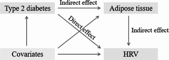
Then, adipose tissues which simultaneously associated with both T2DM and the HRV indices in linear regression model 2 were included in mediation analysis. The shortlisted adipose tissues in mediation analyses recognized as mediators, with T2DM as the independent variable and HRV indices as the dependent variable. Age, gender, HbA1c, FPG, BMI, TG, TC, smoke, alcohol, hypoglycemic therapy (OAD, Insulin, OAD + insulin), medications (ACEI/ARB, statin, antiplatelet), hypertension, CAD, duration of diabetes mellitus were treated as covariates in each model. Figure 1 was employed to enhance the visualization of our statistical approach. A parameter regression approach was used to estimate the total effect, the indirect effect and direct effect of T2DM on HRV. The direct effect is the effect of T2DM (exposure) on HRV (outcome) after adjusting for adipose tissue (mediator). The proportion of mediation represents the fraction of the association of T2DM explained by its association with changes in adipose tissue thickness. A significant disparity in these models would imply that adipose tissue introduces supplementary variability in HRV beyond the impact of T2DM alone. To quantify the magnitude of mediation, the study estimated the proportion of the association mediated by adipose tissue (indirect effect/[direct effect + indirect effect] if the indirect effect is significant.
Fig. 1.

Mediating pathway of the association of type 2 diabetes mellitus (T2DM) with heart rate variability (HRV)
In addition, we constructed a latent variable, HRV burden, to further evaluate overall HRV. Latent variables are variables that cannot be observed directly and need to be measured indirectly by a set of observed variables. Confirmatory factor analysis (also known as a measurement model), which enables exploration of whether a hypothesized latent factor model fits the collected data, was used to evaluate the reliability and validity of latent structure mode (which hence for HRV burden in our research) [15]. Structural validity is mainly used to test the fitness of the overall model, assessing the agreement between the model parameters derived from our data and the parameter values of the theoretical model. Reliability represents the internal stability and aggregation of the model, with high reliability means that the measurement variables under the same potential variable are highly correlated. The latent variable was then incorporated into another series of multiple regressions, modeled within structural equation modeling framework adjusted for covariates to estimate the mediation effect of T2DM on HRV burden via adipose tissues.
Planned additional sensitivity analysis were performed to examine the robustness of study results. First, gender-stratified mediation was estimated. Second, to account for protopathic bias, the analyses excluded all participants diagnosed with T2DM in the 12 months prior to adipose tissues measurement. Third, to mitigate the influence of cardiovascular disease on adipose tissues, individuals diagnosed with coronary artery disease and hypertension were also excluded.
The significance of mediation effect was assessed by computation of bootstrap confidence intervals (CIs). Bootstrapping entailed multiple rounds of sampling from the dataset to gauge the indirect effect in each resampled dataset. Through 5000 iterations of this procedure, an empirical approximation of the sampling distribution for the quantified indirect effect of the independent variable on the dependent variable, mediated by each potential mediator, was established and then utilized to generate CIs for the indirect effect. All reported β’s presented are standardized regression coefficients and were thus directly comparable. All P values were 2-tailed, and values less than 0.05 were considered statistically significant. Analyses were performed using SPSS version 24.0 software for Windows (SPPS Inc, Chicago, IL, USA) and Lavaan packages in R software (version 4.3.0). This mediation analysis was reported in accordance with the Guidelines for Reporting Mediation Analyses in Randomized Trials and Observational Studies (AGReMA) statement [16].
Results
Characteristics of study populations
A flow diagram of the study population is provided in Fig. 2. Of 380 participants eligible for analysis, 249 were diagnosed with T2DM, with a mean age of 62.82 ± 13.91 year. A total of 173 patients with T2DM were treated with hypoglycemic therapy: 22.5% with oral antidiabetic drug only, 16.2% with insulin only and 61.3% with a combining of oral antidiabetic drug and insulin. As for diabetic complications, diabetic nephropathy was present in 23.4% of diabetic patients, diabetic retinopathy in 9.6% and diabetic peripheral neuropathy in 21.7%. Table 1 showed the baseline sociodemographic and clinical characteristics of subjects grouped according to diabetes status. Compared to non-diabetes group, patients with T2DM had a higher reporting in a history of hypertension (59.8% vs. 45%, P = 0.006) and coronary artery disease (40.6% vs. 17.7%, P < 0.001). Higher level of TC (4.35 ± 1.27 vs. 3.89 ± 1.11, P < 0.001), TyG (8.89 ± 0.7 vs. 8.57 ± 0.47, P < 0.001), FPG (6.98 ± 3.26 vs. 5.33 ± 1.06, P < 0.001) and HbA1c (7.66 ± 2.28 vs. 5.22 ± 0.58, P < 0.001) were observed in T2DM group. Additionally, a higher proportion of diabetic patients received treatment with ACEI/ARB, antiplatelet and beta-blockers in diabetic patients when compared to non-diabetic patients (all P < 0.01). Lipid-lowering therapy was also more common among participants in both groups.
Fig. 2.

Flow chart of the study
Table 1.
Baseline characteristics of participants
| Characteristics | NDM | DM | P value |
|---|---|---|---|
| N | 131 | 249 | |
| Age, year, mean ± SD | 57.18 ± 13.31 | 62.82 ± 13.91 | < 0.001 |
| Male, %, | 65 (49.6%) | 130 (52.2%) | 0.631 |
| BMI, kg/m2, mean ± SD | 24.59 ± 2.54 | 24.17 ± 3.76 | 0.227 |
| Hypertension, n (%) | 59 (45%) | 149 (59.8%) | 0.006 |
| CAD, n (%) | 23 (17.7%) | 101 (40.6%) | < 0.001 |
| Smoker, n (%) | 34 (26.4%) | 55 (22.2%) | 0.365 |
| Alcohol, n (%) | 10 (7.6%) | 33 (13.3%) | 0.098 |
| Any albuminuria, n (%) | 18 (13.7%) | 31 (12.4%) | 0.721 |
| Heart rate, bpm, median (IQR) | 71 (66, 76.5) | 71 (64, 79) | 0.799 |
| SBP, mmHg, mean ± SD | 131.86 ± 20.83 | 132.53 ± 20.15 | 0.761 |
| DBP, mmHg, mean ± SD | 78.92 ± 11.18 | 79.96 ± 13.03 | 0.440 |
| HDL-C, mmol/l, mean ± SD | 1.09 ± 0.18 | 0.98 ± 0.29 | < 0.001 |
| LDL-C cholesterol, mmol/l, mean ± SD | 2.50 ± 0.75 | 2.64 ± 1.03 | 0.136 |
| TC, mmol/l, mean ± SD | 3.89 ± 1.11 | 4.35 ± 1.27 | < 0.001 |
| TG, mmol/l, median (IQR) | 1.39 (0.96, 1.68) | 1.32 (0.99, 1.87) | 0.316 |
| TyG index, mean ± SD | 8.57 ± 0.47 | 8.89 ± 0.70 | < 0.001 |
| FPG, mean ± SD | 5.33 ± 1.06 | 6.98 ± 3.26 | < 0.001 |
| HbA1c, %, mean ± SD | 5.22 ± 0.58 | 7.66 ± 2.28 | < 0.001 |
| eGFR, mL/min/1.73 m2, median (IQR) | 82.1 (66.60, 100.15) | 89.35 (68.25, 101.71) | 0.273 |
| UA, umol/l, median (IQR) | 363 (302.5, 423.5) | 361 (301.5, 445.8) | 0.574 |
| diabetic nephropathy, n (%) | 0 (0%) | 58 (23.4%) | < 0.001 |
| diabetic retinopathy, n (%) | 0 (0%) | 24 (9.6%) | < 0.001 |
| diabetic peripheral neuropathy, n (%) | 0 (0%) | 54 (21.7%) | < 0.001 |
| OAD, n (%) | 0 (0%) | 39 (15.7%) | < 0.001 |
| Insulin only, n (%) | 0 (0%) | 28 (11.3%) | < 0.001 |
| OAD + insulin, n (%) | 0 (0%) | 106 (42.9%) | < 0.001 |
| ACEI/ARB, n (%) | 32 (24.4%) | 86 (34.7%) | 0.040 |
| β-blocker, n (%) | 38 (29%) | 74 (30%) | 0.847 |
| Statin, n (%) | 89 (67.9%) | 159 (63.9%) | 0.427 |
| antiplatelet, n (%) | 39 (29.8%) | 109 (43.8%) | 0.008 |
NDM, non-diabetes mellitus; DM, diabetes mellitus; BMI, body mass index; CAD, coronary artery disease; SBP, systolic blood pressure; DBP, diastolic blood pressure; HDL-C, high-density lipoprotein cholesterol; LDL-C, low-density lipoprotein cholesterol; TC, total cholesterol; TG, triglyceride; TyG index, triglyceride-glucose index; FPG, fasting plasma glucose; HbA1c, glycosylated hemoglobin; eGFR, estimated glomerular filtration rate; UA, uric acid; OAD, oral antidiabetic drug; ACEI, angiotensin-converting enzyme inhibitors; ARB, angiotension receptor blocker; EAT, epicardial adipose tissue; SAT, subcutaneous adipose tissue; IPAT, intraperitoneal adipose tissue; EPAT, extraperitoneal adipose tissue; LF, low frequency; HF, high frequency; LF/HF, the ratio of low frequency/high frequency; SDNN, SD of all normal to normal intervals; SDANN, SD deviation of sequential 5-minute normal to normal interval; SDNN index, the mean value of the SD of all the normal normal to normal intervals in every 5-minute interval within 24 h; rMSSD, the root mean square of successive differences; pNN50, the number of pairs of adjacent normal to normal intervals differing by > 50 ms in the entire recording divided by the total number of normal to normal intervals; SD, standard deviation; IQR, interquartile range
The changes of HRV and adipose tissues in type 2 diabetic patients
The changes in HRV indices between patients with or without diabetes were showed in Fig. 3a. Both frequency domain (LF and HF) and time domain (SDNN, SDANN, SDNN index, rMSSD and pNN50) indices were significantly lower in type 2 diabetic patients than those in non-diabetic controls (all P < 0.01). Nevertheless, the ratio of LF/HF was higher in T2DM compared to that in non-diabetic patients (P = 0.024). These findings suggested that patients with T2DM suffer varying degrees of impairment in both their cardiac sympathetic and parasympathetic function.
Fig. 3.
(a) HRV indices in patients with T2DM. T2DM, type 2 diabetes mellitus; HRV, heart rate variability; NDM, non-diabetes mellitus; DM, diabetes mellitus; LF, low frequency; HF, high frequency; LF/HF, the ratio of low frequency/high frequency; SDNN, SD of all normal to normal intervals; SDANN, SD deviation of sequential 5-minute normal to normal interval; SDNN index, the mean value of the SD of all the normal normal to normal intervals in every 5-minute interval within 24 h; rMSSD, the root mean square of successive differences; pNN50, the number of pairs of adjacent normal to normal intervals differing by > 50 ms in the entire recording divided by the total number of normal to normal intervals. *P < 0.05, **P < 0.01 and ***P < 0.001 vs. NDM group. (b) The thicknesses of various adipose tissues in patients with DM. NDM, non-diabetes mellitus; DM, diabetes mellitus; SAT, subcutaneous adipose tissue; EPAT, extraperitoneal adipose tissue; IPAT, intraperitoneal adipose tissue; EAT, epicardial adipose tissue. ***P < 0.001 vs. NDM group
The thicknesses of adipose tissues in various regions were compared between patients with or without diabetes (Fig. 3b). The thickness of EPAT (14.8 ± 2.6 vs. 12.7 ± 3.2 mm), IPAT (72.6 ± 13.4 vs. 65.9 ± 12.1 mm) and EAT (8.6 ± 2.5 vs. 7.4 ± 2.1 mm) were significantly greater in T2DM than in non-diabetic subjects (all P < 0.001), while no significant difference was observed in the thicknesses of SAT (19.2 ± 4.5 vs. 18.9 ± 5.9 mm, P = 0.575) between two groups.
Associations between adipose tissues and HRV indices
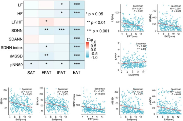
Correlations of various adipose tissues with HRV indices were further analyzed in Fig. 4. The thickness of EAT was significantly correlated with most HRV indicators (all P < 0.001) except for LF/HF. Univariate linear regression analysis demonstrated that changes of EAT were significantly related to all HRV indices (Table 2). When adjusting for age and gender (model 1), these associations remained negative, and they became even stronger after additional adjustments (model 2). IPAT were correlated with HF (P = 0.020) and SDNN (P = 0.012), while no statistical differences were observed between SAT or EPAT and HRV indices after full adjustment.
Fig. 4.
Correlation of different adipose tissues with HRV indices. HRV, heart rate variability; SAT, subcutaneous adipose tissue; EPAT, extraperitoneal adipose tissue; IPAT, intraperitoneal adipose tissue; EAT, epicardial adipose tissue; LF, low frequency; HF, high frequency; LF/HF, the ratio of low frequency/high frequency; SDNN, SD of all normal to normal intervals; SDANN, SD deviation of sequential 5-minute normal to normal interval; SDNN index, the mean value of the SD of all the normal normal to normal intervals in every 5-minute interval within 24 h; rMSSD, the root mean square of successive differences; pNN50, the number of pairs of adjacent normal to normal intervals differing by > 50 ms in the entire recording divided by the total number of normal to normal intervals. *P < 0.05, **P < 0.01 and ***P < 0.001
Table 2.
Correlation regression analysis for the relationship between adipose tissues and HRV indices
| HRV index | EAT | SAT | ||||||||||
|---|---|---|---|---|---|---|---|---|---|---|---|---|
| Univariate | Model 1 | Model 2 | Univariate | Model 1 | Model 2 | |||||||
| β(SE) | P value | β(SE) | P value | β(SE) | P value | β(SE) | P value | β(SE) | P value | β(SE) | P value | |
| LF | -0.248 (0.051) | < 0.001*** | -0.247 (0.051) | < 0.001*** |
-0.22 (0.060) |
< 0.001*** | -0.037 (0.058) | 0.525 | -0.029 (0.059) | 0.622 |
-0.013 (0.069) |
0.854 |
| HF | -0.247 (0.051) | < 0.001*** | -0.238 (0.05) | < 0.001*** | -0.225 (0.057) | < 0.001*** | -0.045 (0.058) | 0.445 | -0.046 (0.058) | 0.424 | -0.021 (0.066) | 0.753 |
| LF/HF | 0.123 (0.052) | 0.018** | 0.113 (0.051) | 0.027* | 0.132 (0.06) | 0.029* | 0.028 (0.06) | 0.646 | 0.040 (0.059) | 0.49 | 0.048 (0.070) | 0.494 |
| SDNN | -0.314 (0.049) | < 0.001*** | -0.302 (0.049) | < 0.001*** | -0.259 (0.051) | < 0.001*** | -0.077 (0.052) | 0.137 | -0.068 (0.052) | 0.191 | -0.064 (0.053) | 0.224 |
| SDANN | -0.289 (0.049) | < 0.001*** | -0.284 (0.05) | < 0.001*** | -0.229 (0.056) | < 0.001*** | 0.036 (0.053) | 0.492 | 0.036 (0.053) | 0.494 | 0.003 (0.059) | 0.959 |
| SDNN index | -0.253 (0.05) | < 0.001*** | -0.250 (0.05) | < 0.001*** | -0.237 (0.056) | < 0.001*** | -0.04 (0.05) | 0.424 | -0.026 (0.05) | 0.601 | -0.032 (0.054) | 0.559 |
| rMSSD | -0.239 (0.05) | < 0.001*** | -0.233 (0.05) | < 0.001*** | -0.243 (0.058) | < 0.001*** | -0.056 (0.051) | 0.268 | -0.048 (0.051) | 0.349 | -0.059 (0.059) | 0.312 |
| pNN50 | -0.206 (0.05) | < 0.001*** | -0.202 (0.051) | < 0.001*** | -0.188 (0.059) | 0.002** | -0.114 (0.052) | 0.029* | -0.114 (0.052) | 0.029* | -0.116 (0.059) | 0.053 |
| HRV indices | IPAT | EPAT | ||||||||||
|---|---|---|---|---|---|---|---|---|---|---|---|---|
| Univariate | Model 1 | Model 2 | Univariate | Model 1 | Model 2 | |||||||
| β(SE) | P value | β(SE) | P value | β(SE) | P value | β(SE) | P value | β(SE) | P value | β(SE) | P value | |
| LF | -0.120 (0.054) | 0.026* | -0.122 (0.055) | 0.026* | -0.133 (0.063) | 0.037 | 0.063 (0.058) | 0.274 | 0.073 (0.058) | 0.212 | 0.113 (0.069) | 0.105 |
| HF | -0.130 (0.054) | 0.014* | -0.103 (0.055) | 0.055 | -0.142 (0.06) | 0.020* | -0.045 (0.058) | 0.437 | -0.033 (0.057) | 0.566 | -0.001 (0.067) | 0.995 |
| LF/HF | 0.079 (0.056) | 0.159 | 0.040 (0.055) | 0.468 | 0.057 (0.065) | 0.379 | 0.090 (0.06) | 0.132 | 0.085 (0.058) | 0.143 | 0.099 (0.071) | 0.163 |
| SDNN | -0.177 (0.052) | 0.001** | -0.165 (0.052) | 0.002** | -0.135 (0.053) | 0.012* | -0.151 (0.052) | 0.004** | -0.142 (0.052) | 0.006** | -0.043 (0.053) | 0.415 |
| SDANN | -0.062 (0.053) | 0.238 | -0.050 (0.053) | 0.346 | -0.065 (0.059) | 0.272 | -0.031 (0.053) | 0.554 | -0.027 (0.053) | 0.607 | -0.004 (0.058) | 0.942 |
| SDNN index | -0.075 (0.05) | 0.13 | -0.078 (0.05) | 0.121 | -0.078 (0.054) | 0.150 | -0.071 (0.05) | 0.154 | -0.066 (0.05) | 0.187 | -0.010 (0.054) | 0.846 |
| rMSSD | -0.055 (0.05) | 0.276 | -0.049 (0.051) | 0.342 | -0.067 (0.058) | 0.257 | -0.097 (0.05) | 0.054 | -0.092 (0.051) | 0.068 | -0.069 (0.058) | 0.236 |
| pNN50 | -0.064 (0.051) | 0.205 | 0.058 (0.052) | 0.259 | -0.052 (0.059) | 0.376 | -0.073 (0.051) | < 0.001*** | -0.07 (0.052) | 0.176 | -0.043 (0.059) | 0.468 |
Model 1 was adjusted for age, gender. Model 2 was adjusted for age, gender, HbA1c, FPG, BMI, TG, TC, smoke, alcohol, hypoglycemic therapy (OAD, Insulin, OAD + insulin), medications (ACEI/ARB, statin, antiplatelet), hypertension, CAD, duration of diabetes mellitus. HRV, heart rate variability; EAT, epicardial adipose tissue; HbA1c, glycosylated hemoglobin; FPG, fasting plasma glucose; BMI, body mass index; TG, triglyceride; TC, total cholesterol; OHA, oral antidiabetic drug; ACEI, angiotensin-converting enzyme inhibitors; ARB, angiotension receptor blocker; CAD, coronary artery disease. LF, low frequency; HF, high frequency; LF/HF, the ratio of low frequency/high frequency; SDNN, SD of all normal to normal intervals; SDANN, SD deviation of sequential 5-min normal to normal interval; SDNN index, the mean value of the SD of all the normal normal to normal intervals in every 5-min interval within 24 h; rMSSD, the root mean square of successive differences; pNN50, the number of pairs of adjacent normal to normal intervals differing by > 50 ms in the entire recording divided by the total number of normal to normal intervals. SE, standard error. *, ** and *** denotes a between group difference at P < 0.05, < 0.01 and < 0.005, respectively
Mediation analysis between adipose tissues and individual HRV index
Based on the results of linear regression above, adipose tissues and HRV indices with significant associations were included in further mediating analysis as mediator and dependent variables, respectively. Finally, ten mediation models were estimated: eight HRV indices (outcomes) conditional on T2DM (independent variable), EAT (mediator), and covariates; and two HRV indices (HF and SDNN) conditional on T2DM, IPAT, and covariates. A brief summary of the mediation effect via EAT or IPAT is provided in Table 3. Mediation effects of EAT were identified in the relationship between T2DM and HF (95% CI [-0.325, -0.058]), SDNN (95% CI [-0.303, -0.062]), SDANN (95% CI [-0.354, -0.083]), SDNN index (95% CI [-0.338, -0.066]), rMSSD (95% CI [-0.317, -0.059]) and pNN50 (95% CI [-0.288, -0.033]). However, LF performed an inconsistent mediation effect in our analysis (direct effect 95% CI [-0.376, 0.443]; indirect effect 95% CI [-0.341, -0.05)). Furthermore, statistical power analyses for all six HRV indices showed that our sample size is adequate to validate our conclusions (almost all estimated power > 0.8) (Supplemental Table 1).
Table 3.
Mediation analysis between T2DM and HRV via adipose tissue
| Mediation variable | Dependent variable | β (Bootstrap 95% CI) | ||
|---|---|---|---|---|
| Crude | Adjusteda | Mediation proportion | ||
| EAT | LF | |||
| Total effect | -0.227 (-0.425, -0.017) | -0.131(-0.551, 0.272) | ||
| Direct effect | -0.108 (-0.314, 0.116) | 0.046 (-0.376, 0.443) | - | |
| Indirect effect | -0.118 (-0.214, -0.050) | -0.177 (-0.341, -0.05) | - | |
| HF | ||||
| Total effect | -0.411 (-0.605, -0.229) | -0.271 (-3.055, 6.553) | ||
| Direct effect | -0.303 (-0.512, -0.104) | -0.097 (-0.477, 0.304) | - | |
| Indirect effect | -0.108 (-0.187, -0.051) | -0.174 (-0.325, -0.058) | 64.21% | |
| LF/HF | ||||
| Total effect | 0.262 (0.020, 0.479) | 0.132 (-0.311, 0.578) | ||
| Direct effect | 0.211 (-0.034, 0.404) | 0.040 (-0.393, 0.460) | - | |
| Indirect effect | 0.051 (-0.001, 0.131) | 0.092 (-0.016, 0.219) | - | |
| SDNN | ||||
| Total effect | -0.973 (-1.171, -0.776) | -1.038 (-1.361, -0.677) | ||
| Direct effect | -0.863 (-1.068, -0.647) | -0.868 (-1.192, -0.526) | - | |
| Indirect effect | -0.110 (-0.196, -0.046) | -0.170 (-0.303, -0.062) | 16.38% | |
| SDANN | ||||
| Total effect | -0.294 (-0.490, -0.094) | -0.300 (-0.670, 0.100) | ||
| Direct effect | -0.155 (-0.371, 0.076) | -0.095 (-0.479, 0.305) | - | |
| Indirect effect | -0.140 (-0.231, -0.068) | -0.205 (-0.354, -0.083) | 68.33% | |
| SDNN index | ||||
| Total effect | -0.527 (-0.768, -0.305) | -0.760 (-1.115, -0.409) | ||
| Direct effect | -0.421 (-0.654, -0.190) | -0.575 (-0.943, -0.208) | - | |
| Indirect effect | -0.105 (-0.180, -0.044) | -0.185 (-0.338, -0.066) | 24.34% | |
| rMSSD | ||||
| Total effect | -0.550 (-0.765, -0.340) | -0.751 (-1.144, -0.353) | ||
| Direct effect | -0.454 (-0.675, -0.241) | -0.571 (-0.950, -0.173) | - | |
| Indirect effect | -0.096 (-0.164, -0.037) | -0.181 (-0.317, -0.059) | 24.10% | |
| pNN50 | ||||
| Total effect | -0.454 (-0.665, -0.243) | -0.508 (-0.899, -0.090) | ||
| Direct effect | -0.370 (-0.598, -0.146) | -0.353 (-0.744, 0.076) | - | |
| Indirect effect | -0.084 (-0.146, -0.028) | -0.155 (-0.288, -0.033) | 30.51% | |
| IPAT | HF | |||
| Total effect | -0.462 (-0.662, -0.284) | -0.273 (-0.657, 0.153) | ||
| Direct effect | -0.412 (-0.642, -0.200) | -0.155 (-0.533, 0.305) | - | |
| Indirect effect | -0.050 (-0.127, 0.025) | -0.118 (-0.287, 0.020) | - | |
| SDNN | ||||
| Total effect | -1.088 (-1.272, -0.906) | -1.090 (-1.388, -0.758) | ||
| Direct effect | -1.057 (-1.265, -0.870) | -1.048 (-1.370, -0.691) | - | |
| Indirect effect | -0.031 (-0.084, 0.026) | -0.042 (-0.148, 0.056) | - | |
aAdjusted for age, gender, HbA1c, FPG, BMI, TG, TC, smoke, alcohol, hypoglycemic therapy (OAD, Insulin, OAD + insulin), medications (ACEI/ARB, statin, antiplatelet), hypertension, CAD, duration of diabetes mellitus. T2DM, type 2 diabetes mellitus; HRV, heart rate variability; EAT, epicardial adipose tissue; HbA1c, glycosylated hemoglobin; FPG, fasting plasma glucose; BMI, body mass index; TG, triglyceride; TC, total cholesterol; OHA, oral antidiabetic drug; ACEI, angiotensin-converting enzyme inhibitors; ARB, angiotension receptor blocker; CAD, coronary artery disease. LF, low frequency; HF, high frequency; LF/HF, the ratio of low frequency/high frequency; SDNN, SD of all normal to normal intervals; SDANN, SD deviation of sequential 5-minute normal to normal interval; SDNN index, the mean value of the SD of all the normal normal to normal intervals in every 5-minute interval within 24 h; rMSSD, the root mean square of successive differences; pNN50, the number of pairs of adjacent normal to normal intervals differing by > 50 ms in the entire recording divided by the total number of normal to normal intervals. CI, confidence interval
Mediation analysis between EAT and comprehensive HRV burden
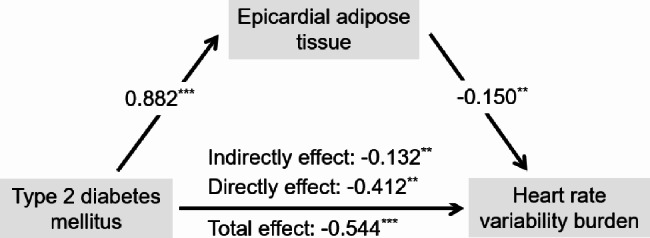
To systemic evaluate the overall HRV, HRV indices were used to construct a latent variable called HRV burden. When comparing the effectiveness of using all HRV indicators versus using SDNN, SDNN index, rMSSD, and pNN50 to construct the latent variable, it was found that the latter combination provided better construct validity, as confirmatory factor analysis confirmed good structural validity and reliability of the HRV burden variable, with fit indices exceeding than 0.9 and standardized factor loadings greater than 0.5 (Supplemental Table 2, Supplemental Tables 3 and Supplemental Fig. 1). The adjusted structural equation modeling-based multiple mediation analysis between T2DM and HRV burden is shown in Fig. 5. The correlation coefficient turned weaker after adding EAT as mediator (total effect: 95% CI [-0.817, -0.266]; direct effect: 95% CI [-0.688, -0.139]), suggesting that the association between T2DM and HRV burden may be partially mediated by the thickness of EAT (indirect effect: 95% CI [-0.244, -0.05]; mediation proportion: 24.26%).
Fig. 5.

Mediation analysis between T2DM and HRV burden via EAT. Adjusted for age, gender, HbA1c, FPG, BMI, TG, TC, smoke, alcohol, hypoglycemic therapy (OAD, Insulin, OAD + insulin), medications (ACEI/ARB, statin, antiplatelet), hypertension, CAD, duration of diabetes mellitus. T2DM, type 2 diabetes mellitus; HRV, heart rate variability; EAT, epicardial adipose tissue; HbA1c, glycosylated hemoglobin; FPG, fasting plasma glucose; BMI, body mass index; TG, triglyceride; TC, total cholesterol; OHA, oral antidiabetic drug; ACEI, angiotensin-converting enzyme inhibitors; ARB, angiotension receptor blocker; CAD, coronary artery disease. *P < 0.05, **P < 0.01 and ***P < 0.001
Sensitivity analysis between T2DM and HRV burden mediated by EAT
Analysis presented in Supplemental Table 4 revealed that females (95% CI [-0.306, -0.023]), rather than male (95% CI [-0.271, 0.008]), showed a significant mediation effect on the association of T2DM with HRV burden. Estimation models that adjusted for protopathic bias (95% CI [-0.214, -0.018]) and those that excluded people with CAD (95% CI [-0.329, -0.058]) and hypertension (95% CI [-0.445, -0.048]) validated the study findings.
Discussion
To the best of our knowledge, this study is the first to investigate the relationship between varied adipose tissue and HRV in individuals with diabetes. Consistent with previous studies [7], we proved that type 2 diabetic patients exhibited an overall decrease in HRV. Additionally, the present study revealed that EAT partially mediated the relationship between T2DM and HRV, suggesting that patients with T2DM may experience impaired regulation of the cardiac autonomic nervous system due to the accumulation of EAT.
CAN is a frequently overlooked yet serious complication of T2DM [17]. Clinical symptoms associated with CAN generally occur late stage of the disease and may include resting tachycardias, myocardial infarction and even cardiac mortality [10]. Evaluation of HRV with a variety of parameters included is deem to be an ideal tool to quantify the impairment or improvement of the cardiovascular autonomic nervous system [18, 19]. Due to the unique prognostic value of each HRV parameter, specific HRV indices are often selected based on the specific experimental design of previous studies. Of note, we introduced a latent variable, HRV burden, to comprehensively assess the cardiovascular autonomic function and avoid selection bias. We believe that the results based on the latent variable model can better reveal the relationship between T2DM and HRV.
Our results focus on the role of adipose tissue in the underlying mechanism of diabetic cardiac autonomic dysfunction. Subcutaneous adipose tissue (SAT) and visceral adipose tissue (VAT) play distinct roles in predicting the risk of cardiovascular complications in diabetes [20, 21]. SAT is associated with a lower risk of metabolic abnormalities due to its higher insulin sensitivity and leptin abundance [22]. Besides, SAT has a lower distribution of nerves compared to other types of adipose tissue [23]. In contrast, VAT showed higher metabolic activity, increased insulin resistance and more steroid hormone receptors compared to SAT [24, 25], thereby posing a greater cardiac risk. This may explain the absence of correlation between SAT thickness and HRV in our study.
Our study interestingly found that EAT partially mediated the relationship between T2DM and cardiac autonomic dysfunction. EAT is a unique fat depot located between the myocardium and the visceral epicardium, containing ganglia, nerves, inflammatory cells, blood vessels, and immune cells and adipocytes. In health, EAT was thought to be brown adipose tissue, thus protecting the heart against cold, and energy storage by supplying free fatty acids to the myocardium [26]. However, EAT shifts toward a pathophysiologic, pro-inflammatory state from its physiological role under conditions like obesity and diabetes, altering its adipokine secretion and promoting cardiovascular disease [27]. The mechanisms by which enlarged EAT partially mediates cardiac autonomic dysfunction in diabetes are yet to be elucidated. There are several potential explanations. First, insulin resistance, a hallmark of T2DM, is associated with inflammation in adipose tissue, leading to a reduction in the number of insulin receptor proteins localized on the cellular membranes of adipose tissue, which diminishes kinase activity and impairs glucose uptake [28]. As GWAS studies indicated that adipose tissue differentiation could in turn affects insulin sensitivity [29], it is possible that the transformation of epicardial brown to white adipose tissue under the disordered glucose metabolism [30] further exacerbates insulin resistance. This transformation may activate the sympathetic nervous system by norepinephrine, resulting in impairment of beta-adrenergic function and nerve endings [31]. Furthermore, inflammation is recognized as a potential contributor to the progression of autonomic dysfunction [32]. As EAT may cause atrial and ventricular fibrosis by secreting pro-inflammatory adipocytokines [33, 34], the decreased release of adiponectin along with increased secretion of inflammatory factors may disturb autonomic regulation of the brain [35], adding to sympathetic activity and cardiovascular stress. Finally, the unbalanced distribution of parasympathetic and sympathetic ganglia in the epicardium may explain the varied correlations between EAT and different HRV indices. Due to the fact that vagus nerves is responsible for 75% of parasympathetic activity and act as the one most frequently damaged by hyperglycemia [36], the initial and primary manifestation of CAN is dysfunction of the parasympathetic nervous system [36]. Our results align with the notion that the damage to the cardiac parasympathetic system tends to be more pronounce in diabetes.
Inflammatory responses contribute to CAN in the course of diabetes [37, 38]. Metformin [39], pioglitazone [40], minocycline [41] and ACEI [42, 43] have been shown to improve cardiac autonomic disorders by inhibiting inflammation, which has not been demonstrated in the diabetic population. Therefore, it is important to further evaluate the effects of lipid-lowering treatment and antidiabetic agents on EAT to attenuate the adverse progression of cardiovascular autonomic function. Moreover, studies have indicated that females tend to show a higher burden of risk factors than males [44]. Besides, female patients were likely to have lower medication adherence and a lower percentage of meeting recommended physical activity goals [44]. These differences seem to explain the different results in the sex-stratified mediation analysis. All in all, further mechanism research is essential for understanding the underlying relationship between EAT and cardiac autonomic dysfunction in patients with T2DM.
The present study has several limitations. First, we did not carry out cardiovascular autonomic reflex test, which is considered as a gold standard for assessing cardiac autonomic neuropathy. Second, considering that HRV might be affected by physical activity, sleep habits as well as emotion, obtaining duplicate measurements of HRV indices or more in-depth information gathering could enhance the reliability of the results. Third, further analysis of the use of lipid-lowering drugs and antidiabetic drugs can better clarify the relationship between diabetes and cardiac autonomic nervous system dysfunction. Last but not least, the observational study can only demonstrate the association and not the causality of the thickness of EAT and HRV indices. Thus, prospective and longitudinal studies are warranted to provide further validation of our findings.
Conclusion
In summary, EAT was identified as a mediator of reduced HRV occurring in T2DM in the present study. This finding reinforces the value of developing effective interventions to prevent CAN in diabetic patients through targeting heart-specific visceral adipose tissue.
Electronic supplementary material
Acknowledgements
Not applicable.
Abbreviations
- CAN
Cardiovascular autonomic neuropathy
- EAT
Epicardial adipose tissue
- EPAT
Extraperitoneal adipose tissue
- HRV
Heart rate variability
- HF
High frequency
- IPAT
Intraperitoneal adipose tissue
- LF
Low frequency
- pNN50
Pairs of adjacent normal to normal intervals differing by > 50 ms in the entire recording divided by the total number of normal to normal interval
- rMSSD
The root mean square of successive difference
- SAT
Subcutaneous adipose tissue
- SDANN
SD deviation of sequential 5-minute normal to normal interval means
- SDNN
SD of all normal to normal interval
- SDNN index
The mean value of the SD of all the normal to normal intervals in every 5-minute interval within 24 h
- T2DM
Type 2 diabetes mellitus
Author contributions
XXT, SHL and YL contributed to discussion and reviewed & edited manuscript. XLOY analyzed data and wrote the manuscript. LP and ZSH revised the manuscript. JFW, HXW, JLZ, BYW and LW collected data. YL and TTW contributed to the visualization of the results. All authors have read and approved the final version of the manuscript. SHL is responsible for the integrity of the work as a whole.
Funding
This study was funded by the Guangdong Basic and Applied Basic Research Foundation (grant numbers 2023A1515012417, 2023A1515010526, 2024A1515011892), the National Natural Science Foundation of China (grant number 82200384), the basic and Applied Basic Research Foundation of the Science and Technology Plan Project of Guangzhou City (grant numbers 202102080388, 2024A03J0175, 2024A04J4783), the Guangdong Provincial Medical Research Fund (grant numbers A2022076, A2022361), the fostering of special funding projects of the National Natural Science Foundation of China in the Third Affiliated Hospital of SYSU (grant number 2021GZRPYQN11) and the Third Affiliated Hospital of Sun Yat-sen University Clinical Medical Research Special Fund ‘Sailing Program’(grant number QHJH202201).
Availability of data and materials
The datasets used and/or analysed during the current study are available from the corresponding author on reasonable request.
Declarations
Competing interests
The authors declare no competing interests.
Ethics approval and consent to participate
Ethics approval was obtained from the Third Affiliated Hospital of Sun Yat-sen University network ethics committee and informed consent was obtained from all participants.
Consent for publication
All authors approved the final manuscript and the submission to this journal.
Footnotes
Publisher’s note
Springer Nature remains neutral with regard to jurisdictional claims in published maps and institutional affiliations.
Xiaolan Ouyang, Long Peng, Zhuoshan Huang, Tongtong Wang have contributed equally to this work.
Contributor Information
Yan Lu, Email: luyan@gzhmu.edu.cn.
Suhua Li, Email: lisuhua3@mail.sysu.edu.cn.
Xixiang Tang, Email: tangxx3@mail.sysu.edu.cn.
References
- 1.Spallone V, Ziegler D, Freeman R, Bernardi L, Frontoni S, Pop-Busui R, et al. Cardiovascular autonomic neuropathy in diabetes: clinical impact, assessment, diagnosis, and management. Diabetes Metab Res Rev. 2011;27:639–53. [DOI] [PubMed] [Google Scholar]
- 2.Ziegler D, Strom A, Strassburger K, Knebel B, Bonhof GJ, Kotzka J, et al. Association of cardiac autonomic dysfunction with higher levels of plasma lipid metabolites in recent-onset type 2 diabetes. Diabetologia. 2021;64:458–68. [DOI] [PMC free article] [PubMed] [Google Scholar]
- 3.Kleiger RE, Miller JP, Bigger JT Jr., Moss AJ. Decreased heart rate variability and its association with increased mortality after acute myocardial infarction. Am J Cardiol. 1987;59:256–62. [DOI] [PubMed] [Google Scholar]
- 4.Boutari C, DeMarsilis A, Mantzoros CS. Obesity and diabetes. Diabetes Res Clin Pract. 2023;202:110773. [DOI] [PubMed] [Google Scholar]
- 5.Lapa C, Arias-Loza P, Hayakawa N, Wakabayashi H, Werner RA, Chen X, et al. Whitening and impaired glucose utilization of brown adipose tissue in a rat model of type 2 diabetes mellitus. Sci Rep. 2017;7:16795. [DOI] [PMC free article] [PubMed] [Google Scholar]
- 6.Fisher VL, Tahrani AA. Cardiac autonomic neuropathy in patients with diabetes mellitus: current perspectives. Diabetes Metab Syndr Obes. 2017;10:419–34. [DOI] [PMC free article] [PubMed] [Google Scholar]
- 7.Benichou T, Pereira B, Mermillod M, Tauveron I, Pfabigan D, Maqdasy S, et al. Heart rate variability in type 2 diabetes mellitus: a systematic review and meta-analysis. PLoS ONE. 2018;13:e0195166. [DOI] [PMC free article] [PubMed] [Google Scholar]
- 8.Houghton D, Zalewski P, Hallsworth K, Cassidy S, Thoma C, Avery L, et al. The degree of hepatic steatosis associates with impaired cardiac and autonomic function. J Hepatol. 2019;70:1203–13. [DOI] [PubMed] [Google Scholar]
- 9.Ulucan S, Katlandur H, Kaya Z. Epicardial fat and liver disease; the contribution of cardio autonomic nervous system function. J Hepatol. 2015;62:1214. [DOI] [PubMed] [Google Scholar]
- 10.Balcioglu AS, Muderrisoglu H. Diabetes and cardiac autonomic neuropathy: clinical manifestations, cardiovascular consequences, diagnosis and treatment. World J Diabetes. 2015;6:80–91. [DOI] [PMC free article] [PubMed] [Google Scholar]
- 11.Wu S, Tan J, Zhang H, Hou DX, He J. Tissue-specific mechanisms of fat metabolism that focus on insulin actions. J Adv Res. 2023;53:187–98. [DOI] [PMC free article] [PubMed] [Google Scholar]
- 12.Alberti KG, Zimmet PZ. Definition, diagnosis and classification of diabetes mellitus and its complications. Part 1: diagnosis and classification of diabetes mellitus provisional report of a WHO consultation. Diabet Med. 1998;15:539–53. [DOI] [PubMed] [Google Scholar]
- 13.Shephard DA. The 1975 declaration of Helsinki and consent. Can Med Assoc J. 1976;115:1191–2. [PMC free article] [PubMed] [Google Scholar]
- 14.Zhang Z. Monte Carlo based statistical power analysis for mediation models: methods and software. Behav Res Methods. 2014;46:1184–98. [DOI] [PubMed] [Google Scholar]
- 15.Conway CC. Clinical applications of confirmatory factor analysis. J Pers Assess. 2020;102:293–5. [DOI] [PubMed] [Google Scholar]
- 16.Lee H, Cashin AG, Lamb SE, Hopewell S, Vansteelandt S, VanderWeele TJ, et al. A Guideline for reporting mediation analyses of randomized trials and observational studies: the AGReMA Statement. JAMA. 2021;326:1045–56. [DOI] [PMC free article] [PubMed] [Google Scholar]
- 17.Svensson MK, Lindmark S, Wiklund U, Rask P, Karlsson M, Myrin J, et al. Alterations in heart rate variability during everyday life are linked to insulin resistance. A role of dominating sympathetic over parasympathetic nerve activity? Cardiovasc Diabetol. 2016;15:91. [DOI] [PMC free article] [PubMed] [Google Scholar]
- 18.Pop-Busui R, Boulton AJ, Feldman EL, Bril V, Freeman R, Malik RA, et al. Diabetic neuropathy: a position statement by the American Diabetes Association. Diabetes Care. 2017;40:136–54. [DOI] [PMC free article] [PubMed] [Google Scholar]
- 19.Chessa M, Butera G, Lanza GA, Bossone E, Delogu A, De Rosa G, et al. Role of heart rate variability in the early diagnosis of diabetic autonomic neuropathy in children. Herz. 2002;27:785–90. [DOI] [PubMed] [Google Scholar]
- 20.Qiu Y, Deng X, Sha Y, Wu X, Zhang P, Chen K, et al. Visceral fat area, not subcutaneous fat area, is associated with cardiac hemodynamics in type 2 diabetes. Diabetes Metab Syndr Obes. 2020;13:4413–22. [DOI] [PMC free article] [PubMed] [Google Scholar]
- 21.Gonzalez N, Moreno-Villegas Z, Gonzalez-Bris A, Egido J, Lorenzo O. Regulation of visceral and epicardial adipose tissue for preventing cardiovascular injuries associated to obesity and diabetes. Cardiovasc Diabetol. 2017;16:44. [DOI] [PMC free article] [PubMed] [Google Scholar]
- 22.Chait A, den Hartigh LJ. Adipose tissue distribution, inflammation and its metabolic consequences, including diabetes and cardiovascular disease. Front Cardiovasc Med. 2020;7:22. [DOI] [PMC free article] [PubMed] [Google Scholar]
- 23.Mittal B. Subcutaneous adipose tissue & visceral adipose tissue. Indian J Med Res. 2019;149:571–3. [DOI] [PMC free article] [PubMed] [Google Scholar]
- 24.Ibrahim MM. Subcutaneous and visceral adipose tissue: structural and functional differences. Obes Rev. 2010;11:11–8. [DOI] [PubMed] [Google Scholar]
- 25.Fang L, Guo F, Zhou L, Stahl R, Grams J. The cell size and distribution of adipocytes from subcutaneous and visceral fat is associated with type 2 diabetes mellitus in humans. Adipocyte. 2015;4:273–9. [DOI] [PMC free article] [PubMed] [Google Scholar]
- 26.Le Jemtel TH, Samson R, Ayinapudi K, Singh T, Oparil S. Epicardial adipose tissue and cardiovascular disease. Curr Hypertens Rep. 2019;21:36. [DOI] [PubMed] [Google Scholar]
- 27.Zhao YX, Zhu HJ, Pan H, Liu XM, Wang LJ, Yang HB, et al. Comparative proteome analysis of epicardial and subcutaneous adipose tissues from patients with or without coronary artery disease. Int J Endocrinol. 2019;2019:6976712. [DOI] [PMC free article] [PubMed] [Google Scholar]
- 28.Greenhill C. Mechanisms of insulin resistance. Nat Rev Endocrinol. 2018;14:565. [DOI] [PubMed] [Google Scholar]
- 29.Camporez JP, Wang Y, Faarkrog K, Chukijrungroat N, Petersen KF, Shulman GI. Mechanism by which arylamine N-acetyltransferase 1 ablation causes insulin resistance in mice. Proc Natl Acad Sci U S A. 2017;114:E11285–92. [DOI] [PMC free article] [PubMed] [Google Scholar]
- 30.Packer M. Epicardial Adipose tissue may mediate deleterious effects of obesity and inflammation on the myocardium. J Am Coll Cardiol. 2018;71:2360–72. [DOI] [PubMed] [Google Scholar]
- 31.Mangmool S, Denkaew T, Parichatikanond W, Kurose H. beta-adrenergic receptor and insulin resistance in the heart. Biomol Ther (Seoul). 2017;25:44–56. [DOI] [PMC free article] [PubMed] [Google Scholar]
- 32.Herder C, Schamarek I, Nowotny B, Carstensen-Kirberg M, Strassburger K, Nowotny P, et al. Inflammatory markers are associated with cardiac autonomic dysfunction in recent-onset type 2 diabetes. Heart. 2017;103:63–70. [DOI] [PubMed] [Google Scholar]
- 33.Grant RW, Dixit VD. Adipose tissue as an immunological organ. Obesity (Silver Spring). 2015;23:512–8. [DOI] [PMC free article] [PubMed] [Google Scholar]
- 34.Gruzdeva OV, Akbasheva OE, Dyleva YA, Antonova LV, Matveeva VG, Uchasova EG, et al. Adipokine and cytokine profiles of epicardial and subcutaneous adipose tissue in patients with coronary heart disease. Bull Exp Biol Med. 2017;163:608–11. [DOI] [PubMed] [Google Scholar]
- 35.Rizzo MR, Fasano R, Paolisso G. Adiponectin and cognitive decline. Int J Mol Sci. 2020;21. [DOI] [PMC free article] [PubMed]
- 36.Axelrod S, Lishner M, Oz O, Bernheim J, Ravid M. Spectral analysis of fluctuations in heart rate: an objective evaluation of autonomic nervous control in chronic renal failure. Nephron. 1987;45:202–6. [DOI] [PubMed] [Google Scholar]
- 37.Grossmann V, Schmitt VH, Zeller T, Panova-Noeva M, Schulz A, Laubert-Reh D, et al. Profile of the immune and inflammatory response in individuals with prediabetes and type 2 diabetes. Diabetes Care. 2015;38:1356–64. [DOI] [PubMed] [Google Scholar]
- 38.Lai YR, Huang CC, Chang HW, Chiu WC, Tsai NW, Cheng BC, et al. Severity of cardiovascular autonomic neuropathy is a predictor associated with major adverse cardiovascular events in adults with type 2 diabetes mellitus: a 6-year follow-up study. Can J Diabetes. 2021;45:155–61. [DOI] [PubMed] [Google Scholar]
- 39.Oliveira PWC, de Sousa GJ, Birocale AM, Gouvea SA, de Figueiredo SG, de Abreu GR, et al. Chronic metformin reduces systemic and local inflammatory proteins and improves hypertension-related cardiac autonomic dysfunction. Nutr Metab Cardiovasc Dis. 2020;30:274–81. [DOI] [PubMed] [Google Scholar]
- 40.Karagiannis E, Pfutzner A, Forst T, Lubben G, Roth W, Grabellus M, et al. The IRIS V study: pioglitazone improves systemic chronic inflammation in patients with type 2 diabetes under daily routine conditions. Diabetes Technol Ther. 2008;10:206–12. [DOI] [PubMed] [Google Scholar]
- 41.Syngle A, Verma I, Krishan P, Garg N, Syngle V. Minocycline improves peripheral and autonomic neuropathy in type 2 diabetes: MIND study. Neurol Sci. 2014;35:1067–73. [DOI] [PubMed] [Google Scholar]
- 42.Marketou ME, Zacharis EA, Koukouraki S, Stathaki MI, Arfanakis DA, Kochiadakis GE, et al. Effect of angiotensin-converting enzyme inhibitors on systemic inflammation and myocardial sympathetic innervation in normotensive patients with type 2 diabetes mellitus. J Hum Hypertens. 2008;22:191–6. [DOI] [PubMed] [Google Scholar]
- 43.Didangelos T, Tziomalos K, Margaritidis C, Kontoninas Z, Stergiou I, Tsotoulidis S, et al. Efficacy of administration of an angiotensin converting enzyme inhibitor for two years on autonomic and peripheral neuropathy in patients with diabetes mellitus. J Diabetes Res. 2017;2017:6719239. [DOI] [PMC free article] [PubMed] [Google Scholar]
- 44.Kautzky-Willer A, Leutner M, Harreiter J. Sex differences in type 2 diabetes. Diabetologia. 2023;66:986–1002. [DOI] [PMC free article] [PubMed] [Google Scholar]
Associated Data
This section collects any data citations, data availability statements, or supplementary materials included in this article.
Supplementary Materials
Data Availability Statement
The datasets used and/or analysed during the current study are available from the corresponding author on reasonable request.


