



ABSTRACT
Traumatic spinal cord injury is characterized by immediate and irreversible tissue loss at the lesion site and secondary tissue damage. Secondary injuries should, in principle, be preventable, although no effective treatment options currently exist for patients with acute spinal cord injury. Traumatized tissues release excessive amounts of adenosine triphosphate and activate the P2X purinoceptor 7/pannexin1 complex, which is associated with secondary injury. We investigated the neuroprotective effects of the blue dye Brilliant Blue FCF, a selective inhibitor of P2X purinoceptor 7/pannexin1 that is approved for use as a food coloring, by comparing it with Brilliant Blue G, a P2X7 purinoceptor antagonist, and carbenoxolone, which attenuates P2X purinoceptor 7/pannexin1 function, in a rat spinal cord injury model. Brilliant Blue FCF administered early after spinal cord injury reduced spinal cord anatomical damage and improved motor recovery without apparent toxicity. Brilliant Blue G had the highest effect on this neurological recovery, with Brilliant Blue FCF and carbenoxolone having comparable improvement. Furthermore, Brilliant Blue FCF administration reduced local astrocytic and microglial activation and neutrophil infiltration, and no differences in these histological effects were observed between compounds. Thus, Brilliant Blue FCF protects spinal cord neurons after spinal cord injury and suppresses local inflammatory responses as well as Brilliant Blue G and carbenoxolone.
Key Words: adenosine triphosphate, spinal cord injuries, receptors, purinergic P2X
INTRODUCTION
Spinal cord injury (SCI) is a disorder that leaves patients with major disabilities, such as tetraplegia and sensory impairment, which drastically reduces patients’ quality of life and is a significant social problem. SCI is a major problem in terms of medical burden because it continues to plague patients, their families, and medical professionals in charge of serious residual disabilities.1,2 SCIs can be broadly classified into primary injuries, which cause direct damage to the spinal cord, and secondary injuries, which are caused by subsequent inflammation.3 Primary injuries are unavoidable owing to their nature, whereas secondary injuries, in which the injured area is aggravated by inflammation that occurs after the injury, are potentially avoidable.
Since the spinal cord itself is difficult to regenerate, it is important to inhibit neuronal cell death associated with secondary SCI. Moreover, several clinical studies have investigated high-dose steroid therapy, riluzole, and minocycline, but these have not yet become established treatments.1,4,5 The secondary injury cascade is a series of changes that begin within just a few hours due to ischemia, edema, or inflammatory changes due to SCI. Inflammation is a key mechanism of spinal cord damage,6,7 and adenosine triphosphate (ATP) release drives inflammation.8,9 Increased ATP release from damaged cells during SCI activates various pathways to produce inflammatory cytokines, which aggravate the injured site. The P2X purinoceptor 7/pannexin1 (Panx1) complex is an ATP release pathway.10 Moreover, when excess ATP is released from injured tissues, the interaction between P2X7 receptors and Panx1 channels aggravates inflammation, providing a theoretical basis for the observation that the blue dye Brilliant Blue G (BBG) acts as an inhibitor of P2X7 to improve motor function11 (Fig. 1).
Fig. 1.
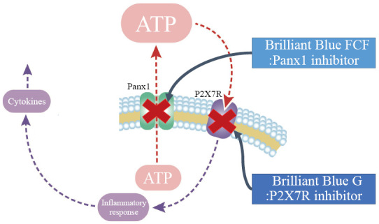
Schema of Brilliant Blue G and Brilliant Blue FCF inhibiting adenosine triphosphate release
Brilliant Blue G inhibits P2X7R, ATP, and inflammation. Brilliant Blue FCF inhibits Panx1, inhibits ATP release, and inhibits inflammation.
ATP: adenosine triphosphate
BBFCF: Brilliant Blue FCF
BBG: Brilliant Blue G
Panx1: P2X purinoceptor 7/pannexin1
Furthermore, the regulation of the P2X7R/Panx1 complex is protective against tissue ischemia. The enhanced ATP signaling during ischemia negatively affects oligodendrocytes and myelin and reduces white matter function in brain ischemia; the ATP signaling during ischemia is mediated by the P2X7R/Panx1 complex.12 In addition, this protective effect is recognized in cardio ischemia. It has been reported that the P2X7R/Panx1 complex is responsible for the release of cardioprotectants, induced by ischemic conditions.13 Thus, the regulation of the P2X7R/Panx1 complex has been shown to be useful against ischemic changes in addition to its anti-inflammatory effects.
The blue dye Brilliant Blue FCF (BBFCF) is a selective inhibitor of Panx1 in the P2X7R/Panx1 complex that is approved for use as a food coloring in humans14 (Fig. 1). BBG is an inhibitor of P2X7R, and it inhibits the inflammatory response. Carbenoxolone (CBX) disodium salt attenuates Panx1 and blocks gap junctions.15 BBG, BBFCF, and CBX are inhibitors of P2X7R/Panx1 complex, although each has different additional targets and varying mechanisms of action. Both BBG and CBX have been shown to be effective for SCI16,17; however, these substances are not used for systemic administration in humans. If BBFCF, which affects the same P2X7R/Panx1 complex, is effective against SCI, its clinical applications can be quickly implemented. Therefore, this study primarily aimed to evaluate the degree of efficacy of BBFCF for SCI, compared with that of BBG and CBX. Our secondary objective was to assess the histological impact of BBFCF on SCI to determine its efficacy. We investigated whether BBFCF is also effective against inflammation.18
MATERIALS AND METHODS
Adult female Sprague-Dawley rats (200–230 g) were used in all animal experiments. All rats were housed in environmentally adequate cages with fewer than three animals. The temperature was maintained at 22 °C, and the room was kept at a 12/12-h light/dark cycle.
Spinal cord injury model
Animals were anesthetized using a combination of medetomidine, midazolam, and butorphanol. Th9 laminectomy was performed, and an SCI model was constructed using a commercially available SCI device (Infinite Horizon Impactor; Precision Systems and Instrumentation) at a force of 200 kDyn to provide a consistent degree of SCI. All injuries included the dorsal corticospinal tract and dorsal gray matter.19 The animals were randomly divided into the following groups: BBG (50 mg/kg of BBG administered intraperitoneally), BBFCF (50 mg/kg of BBFCF administered intraperitoneally), CBX (50 mg/kg of CBX administered intraperitoneally), and vehicle (10 mL/kg of 0.9% sodium chloride administered intraperitoneally) groups.11,20 In addition, we used Sham operation models.
The drug was administered intraperitoneally 10–15 min after the SCI and once daily for the next 3 days (four times in total). After drug administration, the rats were divided into two groups with 5 and 15 rats, respectively. Five rats were examined as one group for immunohistochemical staining on day 7, and 15 rats were treated as one group for tissue damage quantification and immunohistochemical staining on day 42. Randomization and all drug treatments were performed and administered by a blinded laboratory member who did not participate in the behavioral analyses. In addition, 15 sham operation models were evaluated in the behavioral analyses.
Behavioral analyses using open-field Basso, Beattie, and Bresnahan score
The Basso, Beattie, and Bresnahan (BBB) score was measured on days 1, 4, and 7 and then weekly until the end of the observation period (42 days). Two blinded observers scored the animals according to the BBB guidelines.21,22 Animals with BBB scores ≥ 1 on day 1 were excluded from further evaluation. The data were quantified, aggregated and graphed as the average values for the two hind limbs.
Sensory analyses using the von Frey test
To investigate the mechanical sensitivity in animals with neuropathic pain, we used the von Frey filament protocol that is applied to the hind legs in the SCI program at Ohio State University as a touch test. Animals meeting the body weight loading criteria were acclimated to the von Frey test box for at least 15 min. In addition, if the plantar placement was not possible, the stimulation was performed with the plantar hanging from the basket, and a positive result was obtained when a foot-withdrawing motion was observed. Therefore, all cases were included. The center of the plantar surface was then stimulated with a monofilament. If the animal retracted its hind paw, a positive reaction was recorded. Using the same hind limb, 10 trials performed at 30-s intervals were conducted, starting with a monofilament of 5.18. If a positive reaction was observed, the test was gradually changed to a smaller monofilament. Conversely, negative responses prompted testing with progressively larger monofilaments until the monofilament was 6.10. The threshold was set at the lowest monofilament level at which at least 50% of the tests resulted in positive withdrawals.23 All the animals were tested weekly until the end of the observation period.
Immunohistochemistry of the spinal cord
The rats were deeply anesthetized and perfused-fixed transcardially with 4% paraformaldehyde in 0.1 M phosphate buffer. The spinal cord was removed and fixed overnight, incubated with 30% buffered sucrose overnight, and cryoprotected with an appropriate cutting temperature compound (Tissue Freezing Medium, Leica Biosystems, Wetzler, Germany). The tissue was then sliced into 20-μm sections using a cryostat and mounted on a glass slide. The sections were incubated in a citric acid-phosphate-buffered saline solution for 30 min at 90 ºC for antigen retrieval and then incubated in 0.5% Triton X-100 in phosphate-buffered saline for permeabilization. The sections were then blocked in phosphate-buffered saline containing 10% normal donkey serum for immunohistochemistry.
Next, the sections were incubated with primary antibodies against ionized calcium-binding adaptor molecule 1 antibody (019-19741, 1:500, FUJIFILM, Tokyo, Japan), glial fibrillary acidic protein antibody (ab68428, 1:250, Abcam, Cambridge, UK), myeloperoxidase (HP9048-100UG, 1:50, Hycult Biotech, Uden, Netherlands), CD68 antibody (ab125212, 1:500, Abcam), and CD8a (A02236-1 1:500, Boster Bio, Pleasanton, CA, USA). After washing with phosphate-buffered saline, the sections were incubated with secondary antibodies, including Alexa Fluor 488 donkey anti-rabbit immunoglobulin (1:500; Abcam) and Alexa Fluor 594 donkey anti-rabbit immunoglobulin (1:500; Abcam), for 1 h at room temperature.
Finally, the sections were rinsed, mounted with FluorSave (Calbiochem, San Diego, CA, USA) and observed under a fluorescence microscope using a 40× objective lens (BZ9000; Keyence, Osaka, Japan). For ionized calcium-binding adaptor molecule 1 and glial fibrillary acidic protein, the positive fiber area near the tip of the dorsal horn of the coronal sections of the spinal cord was measured, and the percentage of the positive fiber area in the field of view taken with a 40× objective was calculated using the BZ-9000 analyzer software (Keyence). Myeloperoxidase, CD68, CD8a, coronal sections of the spinal cord, and the positive signal near the tip of the anterior horn of the sections were measured, and the percentage of positive signals in the field of view taken with a 40× objective lens was calculated using the BZ-9000 analyzer software (Keyence).
Hematoxylin and eosin staining
The tissue was sliced into 20-μm coronal sections using a cryostat and mounted on a glass slide. Hematoxylin and eosin staining was performed on coronal spinal cord sections to determine the area of tissue injury and atrophy.
The slides were observed under a microscope under bright field illumination using a 4× objective lens (BZ-9000, Keyence) to quantify the area 2 mm from the epicenter of the spinal cord taken for sectioning using the BZ-9000 analyzer software (Keyence). All histological examinations were performed in a blinded manner.
Statistical analyses
Statistical analyses were performed using Prism software for Macintosh, version 9.3.0 (GraphPad Software, San Diego, CA, USA). The Kruskal–Wallis test was used to compare the four groups. Dunn’s multiple comparisons test was used to compare BBB scores and touch test results. Statistical significance was set at p < 0.05. The standard error of the mean is represented by error bars.
Study approval
All animal experiments were performed in accordance with the University Animal Committee Guidelines for the Care and Use of Laboratory Animals and approved by the Nagoya University Institutional Animal Care and Use Committee. All possible efforts were made to minimize pain and suffering.
RESULTS
To evaluate the neuroprotective effects of BBG and BBFCF, functional motor recovery after SCI in the thoracic spinal cord of rats was assessed. The spinal cord was injured at the level of the ninth thoracic vertebra of the rats with a force of 200 kDyn. This contusion model was sufficiently severe to destroy the dorsal column of the spinal cord and the dorsal corticospinal tract. The animals were randomly divided into four groups and systemically administered daily intraperitoneal injections of BBG (50 mg/kg/day), BBFCF (50 mg/kg/day), CBX (50 mg/kg/day), and saline as a control; the first dose was administered immediately after the injury and then for 3 consecutive days for a total of four doses. After intraperitoneal administration of BBG and BBFCF, the color tone of the body surface and eyes promptly changed to blue, confirming that the drugs were transferred to the tissues by intraperitoneal administration.
Brilliant Blue FCF improves behavioral and sensory function recovery
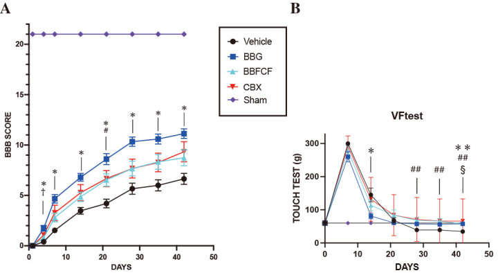
Motor function. We investigated whether BBFCF could promote functional recovery after SCI. Functional recovery was evaluated on days 1, 4, and 7 during the first week and every 7 days until day 42. All animals developed complete paraplegia after SCI, corresponding to a BBB score of 0, although exhibited modest improvements in motor function as early as 2–3 days after injury. The BBB scores in the BBG group showed continuous and significant improvement compared with those in the vehicle group 4 days after SCI. At 21 days after SCI, the mean BBB score in the BBFCF group was 6.53, which was significantly different from that in the vehicle group (4.20; p<0.05, Kruskal–Wallis test). The BBFCF group had a higher mean BBB score after SCI than that in the vehicle group, although no significant difference was observed compared with that in the vehicle group except at 21 days after SCI. Regarding the CBX group, the mean BBB score was higher than that of the vehicle group post-SCI, although no statistically significant difference was observed compared with the vehicle group. BBB scores in the BBFCF group were not significantly different from those in the vehicle group at the final assessment, although a trend towards improvement was observed. BBB scores were significantly higher in the BBG group than those in the BBFCF group only at 4 days after SCI, although no significant differences were observed between BBFCF and BBG groups for the other periods. Furthermore, the BBFCF and CBX groups showed no significant difference in BBB scores throughout the entire period. Thus, BBFCF, although inferior to BBG, restored motor function to the same degree as CBX (Fig. 2A).
Fig. 2.
Functional recovery after spinal cord injury
Fig. 2A: The Basso, Beattie, and Bresnahan score after SCI. The results are presented as the mean ± SEM. Fifteen rats were used and analyzed in each group. Significant differences were observed in Basso, Beattie, and Bresnahan scores between the BBG and vehicle groups at 4 days post-SCI, and the BBFCF group showed significant differences when compared with the vehicle group after 21 days. Significant differences were observed between the BBG and the BBFCF vehicle groups at 4 days post-SCI.
* p<0.05 in the saline-treated and BBG groups.
# p<0.05 in the saline-treated and BBFCF groups.
† p<0.05 in BBG and BBFCF groups using Kruskal–Wallis and post hoc Dunn’s multiple comparison tests.
Fig. 2B: Touch test results after SCI. The results are presented as the mean ± SEM. Only the touch test results for the BBG group were significantly different from those in the vehicle group 14 days after SCI. At 28 days post-SCI, the BBFCF group’s results were significantly different from those in the vehicle group. At 42 days after SCI, the results for the BBG and CBX groups were significantly different from those in the vehicle group.
* p<0.05 and **p<0.01 in the saline-treated and BBG groups.
# p<0.01 in the saline-treated and BBFCF groups.
§ p<0.05 in the saline-treated and CBX groups using Kruskal–Wallis and post hoc Dunn’s multiple comparison tests.
BBFCF: Brilliant Blue FCF
BBG: Brilliant Blue G
CBX: carbenoxolone
SCI: spinal cord injury
SEM: standard error of the mean
Sensory function. At 7 days after SCI, sensory function was analyzed using touch testing with the following results: 300 g in the vehicle group, 260 g in the BBG group, 278.7 g in the BBFCF group, and 292 g in the CBX group, with no significant differences observed. At 14 days after SCI, the touch test result was 81.3 g in the BBG group, which was significantly improved from that in the vehicle group (145 g). The results for the BBFCF and CBX groups were not significantly different from those in the vehicle group. No significant differences were observed among all groups at 21 days after SCI. At 28 and 35 days after SCI, only the results for the BBFCF group were significantly different from those in the vehicle. At 42 days after SCI, the results for the BBG, BBFCF, and CBX groups were significantly different from those for the vehicle group. The results for the BBG, BBFCF, and CBX groups did not differ significantly at any time period (Fig. 2B).
Histological analysis of microglial activation, reactive gliosis, and neutrophil infiltration in the injured spinal cord
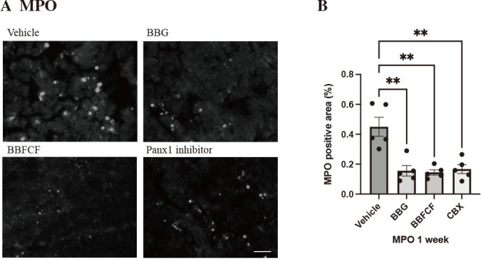
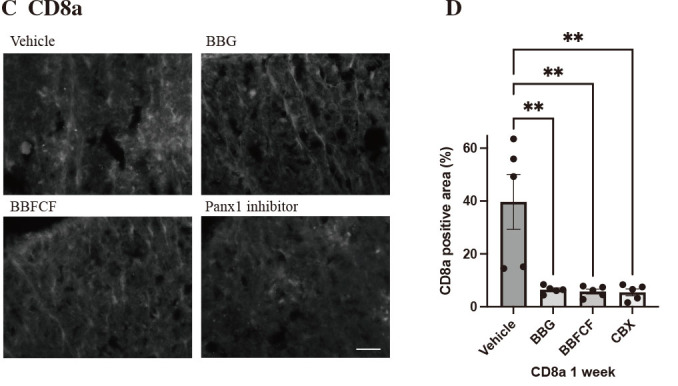
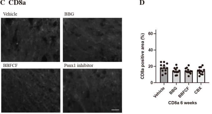
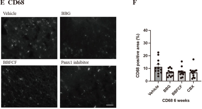
Inflammatory cells. We performed myeloperoxidase staining using spinal cord sections of rats 1 and 6 weeks after SCI to investigate myelomonocytic and neutrophil activation. The area positive for myeloperoxidase staining, which reflects myelomonocytes and neutrophils, was significantly smaller in all drug-treated groups than in the vehicle group 1 week post-SCI, but there was no significant difference 6 weeks post-SCI. There were no significant differences among the drug groups at 1 and 6 weeks post-SCI (Figs. 3A, B and 4A, B). We performed CD8a staining using spinal cord sections of rats 1 and 6 weeks after SCI to investigate T-cell activation. The CD8a staining-positive area, which reflects T cells, was significantly smaller in all drug-treated groups than in the vehicle group 1 week post-SCI, although no significant difference was observed at 6 weeks post-SCI. The p-values for the BBG, BBFCF, and CBX groups were 0.061, 0.060, and 0.058, respectively. The vehicle group indicated a trend, although not a statistical advantage 6 weeks after SCI. No significant differences were observed among the drug groups at 1 and 6 weeks post-SCI (Figs. 3C, D and 4C, D). We performed CD68 staining using spinal cord sections of rats 1 and 6 weeks after SCI to investigate macrophage activation. The CD68 staining-positive area, which reflects macrophages, was significantly smaller in all drug-treated groups than in the vehicle group 1 week post-SCI, but there was no significant difference at 6 weeks post-SCI. The p-values for the BBG, BBFCF, and CBX groups were 0.057, 0.087, and 0.060, respectively, for the vehicle group, indicating a trend, though not a statistical advantage 6 weeks after SCI. No significant differences were observed among the drug groups at 1 and 6 weeks post-SCI (Figs. 3E, F and 4E, F). Thus, inflammatory cells, including myelomonocytes, neutrophils, T cells, and macrophages, were suppressed at 1 week post-SCI due to the administration of these three drugs, which were not significantly different among drugs.
Fig. 3.
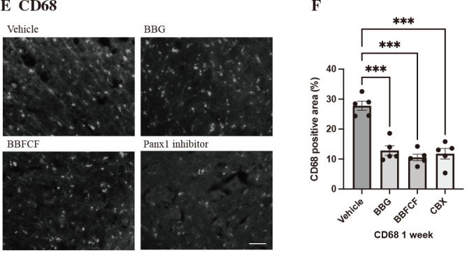
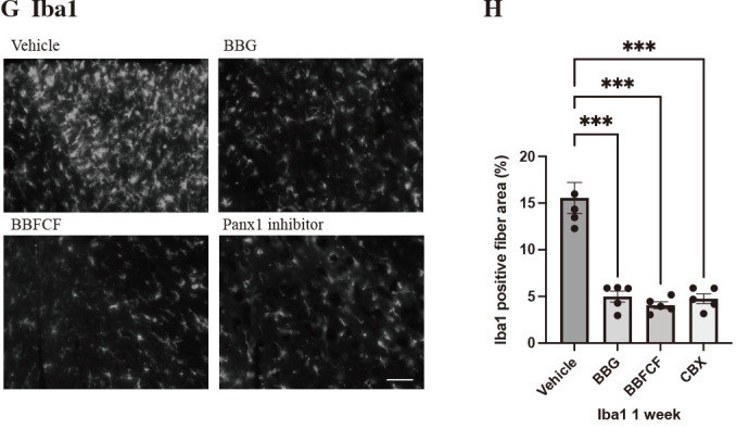
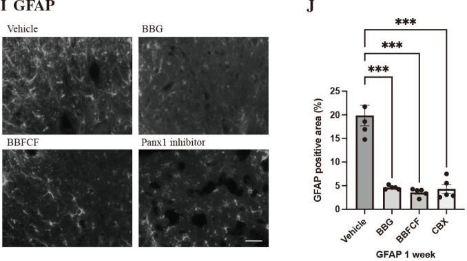
Immunohistochemical analysis of the injured spinal region 7 days after spinal cord injury
Fig. 3A:Immunohistochemical analysis of the myeloperoxidase (MPO)-positive area in the injured spinal region 7 days after SCI. Bar: 40 µm.
Fig. 3B:Quantification of Fig. 3A. The results are presented as the mean ± SEM. **p<0.01 by Student’s t-test. Five rats were used and analyzed in each group.
Fig. 3C:Immunohistochemical analysis of the CD8a-positive area in the injured spinal region 7 days after SCI. Bar: 40 µm.
Fig. 3D:Quantification of Fig. 3C. The results are presented as the mean ± SEM. **p<0.01 by Student’s t-test. Five rats were used and analyzed in each group.
Fig. 3E:Immunohistochemical analysis of the CD68-positive myeloperoxidase-positive area in the injured spinal region 7 days after SCI. Bar: 40 µm.
Fig. 3F:Quantification of Fig. 3E. The results are presented as the mean ± SEM. ***p<0.001 using Student’s t-test. Five rats were used and analyzed in each group.
Fig. 3G:Immunohistochemical analysis of the ionized calcium-binding adaptor molecule 1 (Iba1)-positive area in the injured spinal region 7 days after SCI. Bar: 40 µm.
Fig. 3H:Quantification of Fig. 3G. The results are presented as the mean ± SEM. ***p<0.001 using Student’s t-test. Five rats were used and analyzed in each group.
Fig. 3I:Immunohistochemical analysis of the glial fibrillary acidic protein (GFAP)-positive area in the injured spinal region 7 days after SCI. Bar: 40 µm.
Fig. 3J:Quantification of Fig. 3I. The results are presented as the mean ± SEM. ***p<0.001 using Student’s t-test. Five rats were used and analyzed in each group.
BBFCF: Brilliant Blue FCF
BBG: Brilliant Blue G
CBX: carbenoxolone
Panx1: P2X purinoceptor 7/pannexin1
SCI: spinal cord injury
SEM: standard error of the mean
Fig. 4.
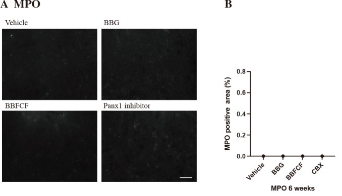
Immunohistochemical analysis of the injured spinal region 42 days after spinal cord injury
Fig. 4A:Immunohistochemical analysis of the myeloperoxidase (MPO)-positive area in the injured spinal region 42 days after SCI. Bar: 40 µm.
Fig. 4B: Quantification of Fig. 4A. The results are presented as the mean ± SEM. Twelve rats were used and analyzed in each group.
Fig. 4C:Immunohistochemical analysis of the CD8a-positive area in the injured spinal region 42 days after SCI. Bar: 40 µm.
Fig. 4D: Quantification of Fig. 4C. The results are presented as the mean ± SEM. Twelve rats were used and analyzed in each group.
Fig. 4E:Immunohistochemical analysis of the CD68-positive area in the injured spinal region 42 days after SCI. Bar: 40 µm.
Fig. 4F: Quantification of Fig. 4E. The results are presented as the mean ± SEM. Twelve rats were used and analyzed in each group.
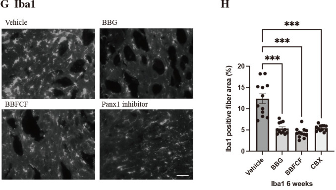
Fig. 4G: Immunohistochemical analysis of the ionized calcium-binding adaptor molecule 1 (Iba1)-positive area in the injured spinal region 42 days after SCI. Bar: 40 µm.
Fig. 4H: Quantification of Fig. 4G. The results are presented as the mean ± SEM. ***p<0.001 by Student’s t-test. Twelve rats were used and analyzed in each group.
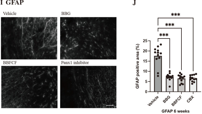
Fig. 4I:Immunohistochemical analysis of the glial fibrillary acidic protein (GFAP)-positive area in the injured spinal region 42 days after SCI. Bar: 40 µm.
Fig. 4J: Quantification of Fig. 4I. The results are presented as the mean ± SEM. ***p<0.001 using Student’s t-test. Twelve rats were used and analyzed in each group.
BBFCF: Brilliant Blue FCF
BBG: Brilliant Blue G
CBX: carbenoxolone
Panx1: P2X purinoceptor 7/pannexin1
N.S.: not significant according to Student’s t-test
SCI: spinal cord injury
SEM: standard error of the mean
Macrophage and glial cells. We performed ionized calcium-binding adaptor molecule 1 staining using spinal cord sections of rats 1 and 6 weeks after SCI to investigate microglial activation. The ionized calcium-binding adaptor molecule 1 staining-positive fiber area, which reflects microglia, was significantly smaller in all drug-treated groups than in the vehicle group at 1 and 6 weeks post-SCI. No significant differences were observed among the drug groups at both 1 and 6 weeks post-SCI (Figs. 3G, H and 4G, H).
We performed glial fibrillary acidic protein staining using spinal cord sections of rats 1 and 6 weeks after SCI to investigate astrocytes and glial scarring. The glial fibrillary acidic protein staining-positive area, which reflects astrocytes and glial scarring, was significantly smaller in all drug-treated groups than in the vehicle group at 1 and 6 weeks post-SCI. There were no significant differences among the drug groups at 1 and 6 weeks post-SCI. Thus, the immunohistological expression of microglia and astrocytes was suppressed by all agents (Figs. 3I, J and 4I, J).
BBFCF reduces the size of the spinal cord injury lesion
To evaluate tissue atrophy after SCI, we compared the area of the most proximal part of the spinal cord and the area 2 mm proximal to the epicenter 6 weeks after SCI. The BBG, BBFCF, and CBX-treated groups showed significantly less atrophy than the vehicle group (p<0.001, analysis of variance). The ratio of the cross-sectional area of the injured site per proximal site of the vehicle group was 60.9%, whereas those of the BBG, BBFCF, and CBX groups were 77.4%, 75.0%, and 75.0%, respectively. No significant differences were observed among the three drugs (Fig. 5).
Fig. 5.

Hematoxylin and eosin staining analysis of the injured spinal region 42 days after spinal cord injury
Fig. 5A: Hematoxylin and eosin staining of the injured area 42 days after SCI. Bar: 500 µm.
Fig. 5B: Quantification of Fig. 5A. The results are presented as the mean ± standard error of the mean. ***p<0.001 (n=15 rats in each group) using Student’s t-test.
Fig. 5C: Schema of a longitudinal section of the spinal cord showing tissue damage, atrophy, and vacuolation after treatment with Brilliant Blue G and Brilliant Blue FCF.
SCI: spinal cord injury
BBFCF: Brilliant Blue FCF
BBG: Brilliant Blue G
CBX: carbenoxolone
Panx1: P2X purinoceptor 7/pannexin1
DISCUSSION
In the current study, BBG was compared to a Panx1 inhibitor to verify the effect of BBFCF on SCI, which is a similar blue food coloring with established safety and potential anti-inflammatory effects mediated by ATP in humans. This study was conducted to determine a new drug that is clinically applicable in terms of safety, is easily administered intravenously, and has potential therapeutic effects in reducing secondary injuries after SCI. Notably, the effectiveness of therapeutic agents, such as steroids, is limited. Although therapies such as stem cell transplantation may be effective, they are difficult to perform immediately after SCI, and even if stem cell transplantation is established in the future, to improve SCI, safe and effective therapeutic agents should be administered in emergency medicine. Therefore, we aimed to identify candidate drugs that are closely related to clinical applications.
The blue dye BBFCF is a selective inhibitor of Panx1, and its safety in humans has been established.14 In the present study, we propose the use of BBFCF, a blue dye with established safety as a food additive, as a possible treatment for the acute stage of SCI. We confirmed that the use of BBFCF, which people consume daily, can reduce the acute severity of SCI.
SCI induces excessive release of ATP, which activates P2X7 receptors and releases inflammatory cytokines, such as interleukin-1, leading to inflammation. BBG can help in the acute recovery from SCI by antagonizing P2X7 receptors and inhibiting the release of excess ATP.11 One of the ATP-mediated pathways is the P2X7 receptor/Panx1 complex, which is involved in various inflammatory pathways.24 Panx1 contributes to inflammation by regulating the release of ATP from the inside of the cell to outside the cell. When tissues are injured, a large amount of ATP is released through Panx1. This large amount of ATP further activates P2X7 receptors, leading to the exacerbation of inflammation. Furthermore, CBX blocks 11beta-hydroxysteroid dehydrogenase, thereby inhibiting the conversion of cortisone to cortisol. This may exhibit anti-inflammatory effects.25
In an ischemic injury model, the P2X7 receptor/Panx1 complex had a role in the deleterious signaling cascade, leading to neuronal and tissue demise; it has also been reported that the blockade of the P2X7 receptor/Panx1 complex plays a significant role in neuroprotection; in particular, blocking Panx1 reduces neural loss.12,13,26 In addition, in a traumatic brain injury model, inhibiting the activity of P2X7R, Panx1, ASC, or caspase prevented inflammation-induced neuronal cell death, and in a colitis model, activating the P2X7R, Panx1, ASC, and caspase pathways caused cell death. The idea that the Panx1-mediated signaling pathway is important in regulating cell death is thus supported.27-29
In the SCI model used in the present study, Panx1 inhibitors suppressed the accumulation of inflammatory cells and reduced inflammation after SCI. Neutrophil invasion releases reactive oxygen species, chemokines, and other determinants of secondary injury after SCI, which may be important for the progressive collapse of the microvasculature and may contribute to atrophy after SCI.30,31 In the present study, we considered that the infiltration of neutrophils in the acute phase after SCI was suppressed, thus preventing atrophy.
Microglial cell activation was strongly inhibited by BBG treatment. This suggests that the blockade of P2X7R may have inhibited the ATP-mediated P2X7R/Panx1 complex and suppressed astrocyte activity and reactive gliosis.32-34 Similarly, we believe that BBFCF also blocks Panx1, thereby inhibiting the excessive release of ATP, suppressing neuronal complexes containing Panx1 and P2X7 receptors, inhibiting microglial cell activation, and suppressing reactive gliosis.35
In behavioral studies, BBG was associated with improved BBB scores compared with BBFCF. BBG may have stronger anti-inflammatory effects than those of BBFCF because BBG has P2X7R-mediated inhibition of Panx1 and connexin, whereas BBFCF inhibits Panx1 but does not inhibit connexin.36 P2X7R is an ion channel activated by extracellular ATP. Its activation triggers several inflammation-related responses, including the release of inflammatory cytokines, induction of cell apoptosis, and activation of macrophages.10,37 Additionally, P2X7R activation has been proposed to open both Panx1 and connexin channels. This could facilitate the release of ATP into the extracellular space, further amplifying the inflammatory response. Connexins create gap junctions, enabling the direct exchange of substances between cells.38 In contrast, Panx1 primarily functions as a channel that supports intercellular communication through ATP release.39 Within the context of inflammation, the activation of these channels might enhance the release of cytokines and other inflammatory agents. Therefore, due to P2X7R’s pivotal role in inflammation, inhibiting P2X7R could offer a more potent anti-inflammatory effect than targeting Panx1 alone. This is because inhibiting P2X7R affects both Panx1 and connexin channels, thereby concurrently suppressing two significant inflammatory pathways. This may be why the BBG showed better functional recovery than the BBFCF. However, histology showed no difference. Furthermore, BBFCF has been evaluated for safety in animal experiments and is used as a food dye. Its safety in humans has been confirmed, making it an easy substance for clinical application.40
However, there are some limitations in applying the present results to human SCI. The administered concentration of BBFCF in the current study might be excessive and further investigation is needed for application in humans. The 50%Inhibitory concentration (IC50) of BBFCF on Panx1 is 0.3 µM, while that of BBG is 3µM, and the Food and Drug Administration established an acceptable daily intake of 12.5 mg/kg/day and the European Food and Safety Authority established a value of 6 mg/kg/day. The same concentration of 50 mg/kg BBFCF as for other compounds was used since the optimal intraperitoneal injection dose of BBFCF was not known; however, further investigation is mandatory. The spinal cord is a difficult place for drugs to reach due to the blood-brain barrier, and BBFCF was administered with the possibility of overdosing for SCI, which is a lethal disorder. No serious organ damage was observed in the present study; however, when actually administering the drug to humans, the optimal concentration should first be considered to determine an appropriate concentration. In addition, the Food and Drug Administration has cautioned about statements indicating that BBFCF can be administered orally and used to track aspiration from feeding tubes, because of its lethality. Therefore, caution should be exercised when administering it to humans. BBB permeability has been reported for BBG,11 although it is not evident for BBFCF. BBG and BBFCF have similar structural formulas and may exhibit similar pharmacokinetics. However, in the case of SCI, since the BBB is disrupted at the injured site, it cannot necessarily be said that the drug does not reach the injured site because of the lack of BBB permeability.
In conclusion, early administration of BBFCF significantly improved functional recovery and reduced the post-traumatic inflammatory response and tissue loss in a rat model of SCI. BBFCF, which is approved for ingestion in humans, has sufficient potential to enhance early functional recovery after traumatic SCI and is a promising substance for future clinical application in humans.
ACKNOWLEDGEMENTS
We greatly appreciate the Departments of Biochemistry and the Division of Neurogenetics, Center for Neurological Diseases and Cancer, Nagoya University Graduate School of Medicine that supported the experiments and investigation of this study.
FUNDING
This research was funded by Fujita Pharmaceutical Co, Ltd. The funder had no role in the study design, data collection, data analysis, decision to publish or preparation of the manuscript of this study.
COMPETING INTERESTS
The authors declare that there is no conflict of interest regarding this paper.
Editorial Note
This article has been selected as an “Editors’ Choice” by the editorial board.
Abbreviations
- ATP
adenosine triphosphate
- BBB
Basso, Beattie, and Bresnahan
- BBFCF
Brilliant Blue FCF
- BBG
Brilliant Blue G
- CBX
carbenoxolone
- Panx1
P2X purinoceptor 7/pannexin1
- SCI
spinal cord injury
REFERENCES
- 1.Hawryluk GW, Rowland J, Kwon BK, Fehlings MG. Protection and repair of the injured spinal cord: a review of completed, ongoing, and planned clinical trials for acute spinal cord injury. Neurosurg Focus. 2008;25(5):E14. doi: 10.3171/FOC.2008.25.11.E14. [DOI] [PubMed]
- 2.Gensel JC, Donnelly DJ, Popovich PG. Spinal cord injury therapies in humans: an overview of current clinical trials and their potential effects on intrinsic CNS macrophages. Expert Opin Ther Targets. 2011;15(4):505–518. doi: 10.1517/14728222.2011.553605. [DOI] [PubMed]
- 3.Borgens RB, Liu-Snyder P. Understanding secondary injury. Q Rev Biol. 2012;87(2):89–127. doi: 10.1086/665457. [DOI] [PubMed]
- 4.Bracken MB, Shepard MJ, Collins WF, et al. A randomized, controlled trial of methylprednisolone or naloxone in the treatment of acute spinal-cord injury. Results of the second national acute spinal cord injury study. N Engl J Med. 1990;322(20):1405–1411. doi: 10.1056/NEJM199005173222001. [DOI] [PubMed]
- 5.Zhu J, Fu Y, Tu G. Role of Smad3 inhibitor and the pyroptosis pathway in spinal cord injury. Exp Ther Med. 2020;20(2):1675–1681. doi: 10.3892/etm.2020.8832. [DOI] [PMC free article] [PubMed]
- 6.Fleming JC, Norenberg MD, Ramsay DA, et al. The cellular inflammatory response in human spinal cords after injury. Brain. 2006;129(Pt 12):3249–3269. doi: 10.1093/brain/awl296. [DOI] [PubMed]
- 7.Bethea JR, Dietrich WD. Targeting the host inflammatory response in traumatic spinal cord injury. Curr Opin Neurol. 2002;15(3):355–360. doi: 10.1097/00019052-200206000-00021. [DOI] [PubMed]
- 8.Wang X, Arcuino G, Takano T, et al. P2X7 receptor inhibition improves recovery after spinal cord injury. Nat Med. 2004;10(8):821–827. doi: 10.1038/nm1082. [DOI] [PubMed]
- 9.North RA. Molecular physiology of P2X receptors. Physiol Rev. 2002;82(4):1013–1067. doi: 10.1152/physrev.00015.2002. [DOI] [PubMed]
- 10.Pelegrin P, Surprenant A. Pannexin-1 mediates large pore formation and interleukin-1beta release by the ATP-gated P2X7 receptor. EMBO J. 2006;25(21):5071–5082. doi: 10.1038/sj.emboj.7601378. [DOI] [PMC free article] [PubMed]
- 11.Peng W, Cotrina ML, Han X, et al. Systemic administration of an antagonist of the ATP-sensitive receptor P2X7 improves recovery after spinal cord injury. Proc Natl Acad Sci U S A. 2009;106(30):12489–12493. doi: 10.1073/pnas.0902531106. [DOI] [PMC free article] [PubMed]
- 12.Cisneros-Mejorado A, Gottlieb M, Cavaliere F, et al. Blockade of P2X7 receptors or pannexin-1 channels similarly attenuates postischemic damage. J Cereb Blood Flow Metab. 2015;35(5):843–850. doi: 10.1038/jcbfm.2014.262. [DOI] [PMC free article] [PubMed]
- 13.Mahi N, Kumar A, Jaggi AS, Singh N, Dhawan R. Possible role of pannexin 1/P2x7 purinoceptor in neuroprotective mechanism of ischemic postconditioning in mice. J Surg Res. 2015;196(1):190–199. doi: 10.1016/j.jss.2015.02.050. [DOI] [PubMed]
- 14.Wang J, Jackson DG, Dahl G. The food dye FD&C Blue No. 1 is a selective inhibitor of the ATP release channel Panx1. J Gen Physiol. 2013;141(5):649–656. doi: 10.1085/jgp.201310966. [DOI] [PMC free article] [PubMed]
- 15.Goncharenko K, Eftekharpour E, Velumian AA, Carlen PL, Fehlings MG. Changes in gap junction expression and function following ischemic injury of spinal cord white matter. J Neurophysiol. 2014;112(9):2067–2075. doi: 10.1152/jn.00037.2013. [DOI] [PMC free article] [PubMed]
- 16.Zhou X, Yang Y, Wu L, et al. Brilliant blue G inhibits inflammasome activation and reduces disruption of blood-spinal cord barrier induced by spinal cord injury in rats. Med Sci Monit. 2019;25:6359–6366. doi: 10.12659/MSM.915865. [DOI] [PMC free article] [PubMed]
- 17.Roh DH, Yoon SY, Seo HS, et al. Intrathecal injection of carbenoxolone, a gap junction decoupler, attenuates the induction of below-level neuropathic pain after spinal cord injury in rats. Exp Neurol. 2010;224(1):123–132. doi: 10.1016/j.expneurol.2010.03.002. [DOI] [PubMed]
- 18.Michalski K, Kawate T. Carbenoxolone inhibits Pannexin1 channels through interactions in the first extracellular loop. J Gen Physiol. 2016;147(2):165–174. doi: 10.1085/jgp.201511505. [DOI] [PMC free article] [PubMed]
- 19.Imagama S, Sakamoto K, Tauchi R, et al. Keratan sulfate restricts neural plasticity after spinal cord injury. J Neurosci. 2011;31(47):17091-17102. doi: 10.1523/JNEUROSCI.5120-10.2011. [DOI] [PMC free article] [PubMed]
- 20.Ma W, Hui H, Pelegrin P, Surprenant A. Pharmacological characterization of pannexin-1 currents expressed in mammalian cells. J Pharmacol Exp Ther. 2009;328(2):409–418. doi: 10.1124/jpet.108.146365. [DOI] [PMC free article] [PubMed]
- 21.Basso DM, Beattie MS, Bresnahan JC. A sensitive and reliable locomotor rating scale for open field testing in rats. J Neurotrauma. 1995;12(1):1–21. doi: 10.1089/neu.1995.12.1. [DOI] [PubMed]
- 22.Basso DM, Beattie MS, Bresnahan JC. Graded histological and locomotor outcomes after spinal cord contusion using the NYU weight-drop device versus transection. Exp Neurol. 1996;139(2):244–256. doi: 10.1006/exnr.1996.0098. [DOI] [PubMed]
- 23.Lang BT, Cregg JM, DePaul MA, et al. Modulation of the proteoglycan receptor PTPσ promotes recovery after spinal cord injury. Nature. 2015;518(7539):404–408. doi: 10.1038/nature13974. [DOI] [PMC free article] [PubMed]
- 24.Poornima V, Madhupriya M, Kootar S, Sujatha G, Kumar A, Bera AK. P2X7 receptor-pannexin 1 hemichannel association: effect of extracellular calcium on membrane permeabilization. J Mol Neurosci. 2012;46(3):585–594. doi: 10.1007/s12031-011-9646-8. [DOI] [PubMed]
- 25.Pagé N, Warriar N, Govindan MV. 11 beta-hydroxysteroid dehydrogenase activity in human lung cells and transcription regulation by glucocorticoids. Am J Physiol. 1994;267(4 Pt 1):L464-L474. doi: 10.1152/ajplung.1994.267.4.L464. [DOI] [PubMed]
- 26.Orellana JA, Froger N, Ezan P, et al. ATP and glutamate released via astroglial connexin 43 hemichannels mediate neuronal death through activation of pannexin 1 hemichannels. J Neurochem. 2011;118(5):826–840. doi: 10.1111/j.1471-4159.2011.07210.x. [DOI] [PMC free article] [PubMed]
- 27.Adamczak S, Dale G, de Rivero Vaccari JP, Bullock MR, Dietrich WD, Keane RW. Inflammasome proteins in cerebrospinal fluid of brain-injured patients as biomarkers of functional outcome: clinical article. J Neurosurg. 2012;117(6):1119–1125. doi: 10.3171/2012.9.JNS12815. [DOI] [PMC free article] [PubMed]
- 28.Gulbransen BD, Bashashati M, Hirota SA, et al. Activation of neuronal P2X7 receptor-pannexin-1 mediates death of enteric neurons during colitis. Nat Med. 2012;18(4):600–604. doi: 10.1038/nm.2679. [DOI] [PMC free article] [PubMed]
- 29.de Rivero Vaccari JP, Lotocki G, Alonso OF, Bramlett HM, Dietrich WD, Keane RW. Therapeutic neutralization of the NLRP1 inflammasome reduces the innate immune response and improves histopathology after traumatic brain injury. J Cereb Blood Flow Metab. 2009;29(7):1251–1261. doi: 10.1038/jcbfm.2009.46. [DOI] [PMC free article] [PubMed]
- 30.Trivedi A, Olivas AD, Noble-Haeusslein LJ. Inflammation and spinal cord injury: infiltrating leukocytes as determinants of injury and repair processes. Clin Neurosci Res. 2006;6(5):283–292. doi: 10.1016/j.cnr.2006.09.007. [DOI] [PMC free article] [PubMed]
- 31.Simard JM, Tsymbalyuk O, Ivanov A, et al. Endothelial sulfonylurea receptor 1-regulated NC Ca-ATP channels mediate progressive hemorrhagic necrosis following spinal cord injury. J Clin Invest. 2007;117(8):2105–2113. doi: 10.1172/JCI32041. [DOI] [PMC free article] [PubMed]
- 32.Kukley M, Barden JA, Steinhäuser C, Jabs R. Distribution of P2X receptors on astrocytes in juvenile rat hippocampus. Glia. 2001;36(1):11–21. doi: 10.1002/glia.1091. [DOI] [PubMed]
- 33.Jabs R, Matthias K, Grote A, Grauer M, Seifert G, Steinhäuser C. Lack of P2X receptor mediated currents in astrocytes and GluR type glial cells of the hippocampal CA1 region. Glia. 2007;55(16):1648–1655. doi: 10.1002/glia.20580. [DOI] [PubMed]
- 34.Yu Y, Ugawa S, Ueda T, et al. Cellular localization of P2X7 receptor mRNA in the rat brain. Brain Res. 2008;1194:45–55. doi: 10.1016/j.brainres.2007.11.064. [DOI] [PubMed]
- 35.Makarenkova HP, Shestopalov VI. The role of pannexin hemichannels in inflammation and regeneration. Front Physiol. 2014;5:63. doi: 10.3389/fphys.2014.00063. [DOI] [PMC free article] [PubMed]
- 36.Bennett MV, Garré JM, Orellana JA, Bukauskas FF, Nedergaard M, Sáez JC. Connexin and pannexin hemichannels in inflammatory responses of glia and neurons. Brain Res. 2012;1487:3–15. doi: 10.1016/j.brainres.2012.08.042. [DOI] [PMC free article] [PubMed]
- 37.Di Virgilio F, Dal Ben D, Sarti AC, Giuliani AL, Falzoni S. The P2X7 Receptor in Infection and Inflammation. Immunity. 2017;47(1):15–31. doi: 10.1016/j.immuni.2017.06.020. [DOI] [PubMed]
- 38.Saez JC, Berthoud VM, Branes MC, Martinez AD, Beyer EC. Plasma membrane channels formed by connexins: their regulation and functions. Physiol Rev. 2003;83(4):1359–1400. doi: 10.1152/physrev.00007.2003. [DOI] [PubMed]
- 39.Penuela S, Gehi R, Laird DW. The biochemistry and function of pannexin channels. Biochim Biophys Acta. 2013;1828(1):15–22. doi: 10.1016/j.bbamem.2012.01.017. [DOI] [PubMed]
- 40.Borzelleca JF, Depukat K, Hallagan JB. Lifetime toxicity/carcinogenicity studies of FD and C Blue No. 1 (brilliant blue FCF) in rats and mice. Food Chem Toxicol. 1990;28(4):221–234. doi: 10.1016/0278-6915(90)90034-k. [DOI] [PubMed]