Abstract
Three-dimensional (3D)-printed models of bones are a convenient and durable alternative to real bone specimens, and they have been used in anatomy laboratories. It is necessary to identify the precise advantages of 3D-printed models from all perspectives; not only the improvement in students’ knowledge of anatomy but also the students’ assessment of such models. Here, students of veterinary medicine and animal science evaluated the reproducibility and effectiveness of 3D-printed models as a learning tool by completing our questionnaires, with a focus on their understanding of the skull-morphological differences among dog breeds. With the COVID-19 pandemic having obliged veterinary universities to provide courses online, we also investigated how the pandemic affected the students’ evaluation of the 3D-printed models. The questionnaire results revealed that the animal science students were satisfied with the reproducibility of the 3D-printed models, but the veterinary students were not (they preferred to use real specimens). The skull differences were well understood by both types of students, indicating that 3D-printed models are effective for learning about rare skeletal specimens. The veterinary students who experienced the COVID-19 pandemic tended to choose real specimens more often than those who did not have this experience. Our results suggest that the use of 3D-printed models as an introduction and the use of real specimens in anatomy laboratory courses can be adequate for veterinary students. Together our findings suggest ways to improve the educational performance of 3D-printed models for veterinary students who need to understand the anatomy of many species.
Keywords: bone specimen, COVID-19, dog breed difference, veterinary anatomy laboratory
One of the important first steps in the study of veterinary and animal anatomy is the observation of bone specimens from several animal species. Anatomy laboratory observations of real specimens from cadaveric dissections (not just bone specimens) are a key component of veterinary and animal science curricula [17, 18]. Real bone specimens are also useful for the study of skeletal structures in veterinary and animal anatomy laboratories, but anatomical specimens including fragile bones such as skulls can be damaged or simply dirty from yearly handling by students over decades, especially in institutions with large student populations [5]. In addition, bone specimens frequently cannot be used by students because the specimens’ preparation required a long time and great effort [1, 23].
Moreover, unlike the study of human medicine, veterinary students are required to know the anatomy of many animal species, including their bone structures. Veterinarians become familiar not only with various species, but also with many within-species breeds, such as those of dogs and cats. The size and morphology of bone structures differ within breeds and among individuals. For example, there are many differences in the size and shape of the skulls of brachycephalic and long-cephalic dogs [12, 20]. It would be ideal to provide students of veterinary medicine with opportunities to become familiar with such differences, but there are few occasions for students to use bone specimens of various dog breeds’ bone specimens because they are scarce.
Tools for anatomical education have changed and improved over the past few decades; anatomy textbooks and handouts had only monotone figures at one time, but they now provide colored, high-quality, and/or three-dimensional (3D) images of specimens [6,7,8, 12]. The development of information technology (IT) has given students the ability to observe anatomical structures with computer-graphic images from various angles on display monitors [9, 16]. Anatomical models can make the characteristics and functions of structures easier to understand, and they help eliminate students’ misunderstanding of anatomy [9, 10, 16].
The progress of IT has brought about the advent of 3D-printers, and 3D-printed models using 3D data of bone specimens can now be made, often at low cost. There have been investigations of whether 3D-printed models are efficient tools in the study of both human and veterinary anatomy, including comparisons of the reproducibility and accuracy of 3D-printed models of many organs (including bones) compared to real specimens [3, 5, 8, 11, 21]. The visualization of affected parts by the construction of 3D-printed models is also useful for clinicians who seek to explain patients’ conditions and treatment techniques [2, 22]. The physical merits of 3D-printed models compared to real specimens include (i) simpler production and preparation; (ii) ease of reproduction, (iii) the ability to make many copies; and (iv) durability [1]. On the other hand, 3D printers have the disadvantage that it is difficult to reproduce bone fine detail such as nutrient foramina [8].
The utility and drawback of 3D-printed models are thus clear, but it would also be informative to obtain students’ evaluations of these models in anatomy laboratories, as this could help improve the models’ quality and the ways that the models can be used for teaching. There is little information about students’ perspective regarding 3D-printed models’ applications as anatomy learning tools compared with conventional tools such as textbooks and other two-dimensional media.
As is the case worldwide, Japan has been dealing with the COVID-19 pandemic since early 2020 [19]. The Japanese government declared a national state of emergency in April 2020 in an attempt to prevent the spread of this disease [4], and universities including schools of medicine and veterinary medicine then moved most of their courses online [13,14,15]. The state of emergency has since been canceled, and lectures and laboratory courses have mostly returned to on-site settings. In Japan as elsewhere, many students enter a university just after graduation from a high school. With the COVID-19 lockdown, the first- and second-year students at schools of veterinary medicine in 2022 had ‘attended’ their veterinary anatomy courses mainly online for one or two years at home, and scarcely on-site.
The present study is a case report to investigate the utility of 3D-printed models in veterinary anatomy laboratories for students. To investigate whether 3D-printed models are accurate and helpful for learning veterinary anatomy, we created 3D-printed models of rare skulls of dog breeds. We then evaluated the quality of the 3D-printed models for use in veterinary anatomy laboratories. To obtain students’ perspective about the models, we asked students of veterinary medicine and animal science who were taking the anatomy course to complete a questionnaire about the skull models. We compared the responses of students who were enrolled in the anatomy course during the COVID-19 lockdown’s online courses with the responses of students who did not have such experience. The further improvement of the educational performance of 3D-printed model for students of veterinary medicine are discussed based on the results of this study.
MATERIALS AND METHODS
Real specimens
To obtain the digital data of skulls to create templates, a computed axial tomography (CT) scanner was used to scan three dog breeds’ skull bone specimens that have been stored at Azabu University (Kanagawa, Japan): Beagle (unknown sex, 8 years old), Shih Tzu (female, 2 years old) and Toy Poodle (male, 10 years old).
Scanning with a CT scanner, 3D editing, and printing 3D-printed models
The real specimens were scanned using a CT scanner, BrightSpeed® (GE HealthCare Technologies, Chicago, IL, USA) in the veterinary hospital of Azabu University with 0.625 mm/pitch, minimum pitch of this CT scanner, to get digital skull data. The CT data were collected in the Digital Imaging and Communications in Medicine (DICOM) data format from scanning, and then the DICOM data were converted to Standard Triangulated Language (STL) format after noise in the surface data was removed and modified with a 3D-data editing application, Volume Extractor® (i-Plants Systems, Takizawa, Japan). Gaps in the surface STL data were detected and fixed with the application Artec Studio® (Artec 3D, Luxembourg, Grand Duchy of Luxembourg). The 3D-printed models were created from STL data by a fused deposition modeling (FDM) 3D printer, UP Box® (Beijing Tiertime Technology, Beijing, China) at 0.2 mm/pitch with acrylonitrile-butadiene-styrene (ABS) resin filament (Beijing Tiertime Technology).
Participants and questionnaire
The contents of the questionnaires about the 3D-printed models and the students’ responses are presented in Tables 1 and 2. In Japan, most veterinary departments provide veterinary anatomy laboratories to students of veterinary medicine during their second year. The timing of animal anatomy laboratories for students of animal science varies widely among departments, depending on each department’s curriculum policy. At Azabu University, the animal functional anatomy laboratory was offered to first-year students at the time the questionnaires were conducted. In 2016, before the COVID-19 pandemic, the questionnaire was completed by both 141 of 142 sec-year students of veterinary medicine (99.3% response rate) and 132 of 150 first-year students of animal science (88.0% response rate). In 2022, with experience with the pandemic’s limitations, 146 of 149 sec-year students of veterinary medicine (98.0% response rate) completed the questionnaire. In the veterinary anatomy laboratory and animal functional anatomy laboratory, real bone specimens, uncolored and colored 3D-printed models of Beagle and other-breed skulls were simultaneously showed and observed by students. They were able to freely touch the specimens and models. The laboratories utilizing 3D-printed models in both 2016 and 2022 were executed uniformly. However, certain content of the veterinary anatomy laboratory in 2022 were provided online. For on-site laboratories, preparatory materials were provided to veterinary students via video on demand, thereby optimizing time management. This adaptation in implementation format of the laboratory was needed to prevent the spread of COVID-19 by a restricted number of on-site laboratories, implemented to mitigate congestion within the facility. The students’ questionnaire responses were collected just after their respective veterinary anatomy and animal functional anatomy laboratories with a digital gadget via the internet using Google Forms, an online tool of Google LLC (Mountain View, CA, USA).
Table 1. Results of the questionnaire from students without COVID-19 pandemic experience (in 2016).
| Veterinary students, n=141 | Animal science students, n=132 | |
|---|---|---|
| Q1. Did you achieve the goal of this anatomy laboratory, that is, “To understand the differences among the skulls of different dog breeds” with 3D-printed models? | ||
| Strongly agree | 63.8% | 87.9% |
| Agree | 34.8% | 11.4% |
| Disagree | 1.4% | 0.8% |
| Strongly disagree | 0% | 0% |
| Q2. How do you evaluate this anatomy laboratory with the 3D-printed models compared to that with skull handouts? | ||
| Better | 81.6% | 80.3% |
| Slightly better | 17.0% | 15.9% |
| Same | 0.7% | 0% |
| Slightly worse | 0.7% | 3.8% |
| Worse | 0% | 0% |
| Q3. On the merits and demerits of each real bone and 3D-printed model, how did you evaluate this laboratory with the 3D-printed models compared to that with real bones of skulls? (Real bone: the merit is its being real, and a demerit is its fragility [and only that]. Three-dimensional-printed models: the merit is durability and reproducible many times, and the demerit is insufficient fidelity for fine details)*. | ||
| Better | 31.9% | 40.2% |
| Slightly better | 35.5% | 38.6% |
| Same | 9.9% | 7.6% |
| Slightly worse | 21.3% | 12.9% |
| Worse | 1.4% | 0.8% |
| Q4. Why did you select it in Q3? Please add the reason in the format shown below (main answers). | ||
| Positive choices: | ||
| Many 3D-printed models are easy to observe (V) | ||
| 3D-printed models can be colored for students to understand (V and A) | ||
| 3D-printed models are securely observed without a risk of breaking (V and A) | ||
| 3D-printed models improve our motivation for learning (A) | ||
| Skulls of many dog breeds can be observed easily (V and A) | ||
| The technology can reduce the number of dogs for specimens (V and A) | ||
| Negative choices: | ||
| It is important to touch real bone specimens for learning (V) | ||
| The reproducibility of the 3D-printed models is not perfect regarding weight and feeling (V) | ||
| It is unprecise and vague to identify detail structures, surface roughness and small canals (V) | ||
| Real specimens will be more carefully treated than 3D-printed models (V) | ||
| 3D-printed models are crude in detail (A) | ||
| 3D-printed model is fake close to the real thing (A) | ||
*There is a case in which the total of the values is not 100% because the values were rounded off to the first decimal place. V: veterinary student, A: animal science student.
Table 2. Results of the questionnaire from veterinary students with COVID-19 pandemic experience (in 2022, n=146).
| Q1. Did you achieve the goal of this anatomy laboratory, that is, “To understand the differences in skulls among dog breeds” with 3D-printed models? | ||
|---|---|---|
| Strongly agree | 46.6% | |
| Agree | 38.4% | |
| Disagree | 13.0% | |
| Strongly disagree | 2.0% | |
| Q2. How do you evaluate this anatomy laboratory with the 3D-printed models compared to that with skull handouts? | ||
| Better | 71.2% | |
| Slightly better | 25.3% | |
| Same | 2.1% | |
| Slightly worse | 1.4% | |
| Worse | 0.0% | |
| Q3. On the merits and demerits of each real bone and 3D-printed model, how did you evaluate this laboratory with the 3D-printed models compared to that with real bones of skulls? (Real bone: the merit is its being real, and a demerit is its fragility [and only that]. Three-dimensional-printed models: the merit is durability and reproducible many times, and the demerit is insufficient fidelity for fine details). | ||
| Better | 30.1% | |
| Slightly better | 33.6% | |
| Same | 11.6% | |
| Slightly worse | 24.7% | |
| Worse | 0.0% | |
| Q4. Why did you select it in Q3? Please add the reason in the format shown below (main answers). | ||
| Positive choices: | ||
| The cranium bones of 3D-printed models can be indicated with colors, which is effective for study | ||
| Many students can observe 3D-printed models simultaneously because of their abundance | ||
| Being reluctant to use real bones that are fragile, it’s easier to use 3D-printed models as a replica | ||
| 3D-printed models don’t limit the observation time | ||
| The color cording of 3D-printed models was very useful to understand skull bones | ||
| Real bones give the texture of bones, whereas real bones may be missing due to destruction. 3D-printed models are effective for beginners studying the correct structures of bones | ||
| 3D-printed models are different weight from real bones, but they are sufficient for the study of the general morphology of the bones | ||
| Negative choices: | ||
| Detailed structures of 3D-printed models such as small foramina are not clearly reproducible | ||
| There is no difference between surface conditions of tuberosity and articular surface | ||
| 3D-printed models are not complete reproductions of the real bones | ||
| The detail structures such as tuberosity are easily recognized by touching the real bones but not 3D-printed models | ||
| Real bones are easier to comprehend than 3D-printed models | ||
RESULTS
The reproducibility of 3D-printed models
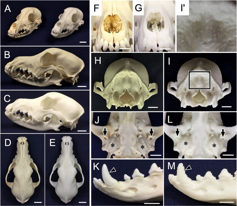
There was little difference in the rough appearance (Fig. 1A) of the ventral and dorsal sites between each real skull bone specimen and the 3D-printed model of it (Fig. 1B–E). However, detailed points and thin and fine structures (e.g., nasal concha) were not reproduced in the 3D-printed models (Fig. 1F, 1G). The minute processes and protuberances of the skull specimens were difficult to replicate and observe in the 3D-printed models as well. For example, the external occipital protuberance was clearly different between the real bone specimen and 3D-printed model (Fig. 1H, 1I), and the protuberance show a rough structure that resembled steps in a magnified image of 3D-printed model (Fig. 1I’). The details were clearly different from those of the real bone specimen. The small foramens, lesser palatine foramen, hypoglossal canal, and retroarticular foramen were unlike those of the skull bone specimen (Fig. 1J) and smaller and unclear in the 3D-printed model (Fig. 1L). The tooth surface of the bone specimen was extremely smooth, and the canines on the real mandible specimen were very smooth (Fig. 1K), whereas those on the 3D-printed model were rough and step-like (Fig. 1M).
Fig. 1.
Comparison of a dog skull specimen and the 3D-printed product of the specimen’s CT data. The appearance of the dog skull and 3D-printed model is shown and compared. A: the real dog skull (left) and the 3D-printed product (right). B, D, F, H, J, K: the real skull. C, E, G, I, L, M: the 3D printer product. The figures show left-lateral views (B, C), dorsal (D, E), rostral (F, G), and caudal (H, I) views. I’: Magnified area of solid-line square in panel I. J, L: Ventral view of the skull base. Arrows: retroarticular foramen. Asterisks: tympanic bulla. K, M: Left-lateral view of mandible rostral part. Arrowheads: canines. Bars: 2 cm in panels A–E, and 1 cm in panels F–M.
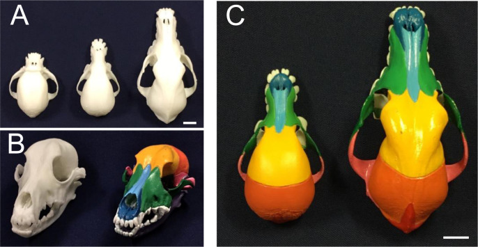
To further examine the qualities of 3D-printed models for a veterinary anatomy laboratory, we created 3D-printed models of the skulls of two rare dog breeds, Shih Tzu and Toy Poodle (Fig. 2A, 2C). The 3D-printed models clearly depicted the morphologically different regions between breeds and provided visual information that made it easy to distinguish developed and undeveloped points (Fig. 2A). For example, although the model of the Toy Poodle skull appeared to be smaller overall than that of the Beagle, the anterior-posterior length of the maxilla was shorter and the left-right width of neural cranium of the Toy Poodle was almost the same size as that of the Beagle, which was emphasized by coloring compared to non-coloring (Fig. 2C).
Fig. 2.
Coloring of 3D-printed models from rare skulls of dogs. The 3D-printed products were painted in different colors according to bones. A: 3D-printed products from skulls of a Shih Tzu (left), Toy Poodle (center) and Beagle (right), before painting. B: 3D-printed products of the Beagle skull before (left) and after (right) painting. C: Painted products of Toy Poodle (left) and Beagle (right) skulls for comparison. Bars: 2 cm in panels A and C.
Questionnaire for students of veterinary medicine and animal science before COVID-19 pandemic
The questionnaire contents and results from students before COVID-19 pandemic are summarized in Table 1. The total percentages of two responses to Q1 in Table 1 (Did you achieve the goal of this anatomy laboratory, that is, “To understand the differences among the skulls of different dog breeds” with 3D-printed models?), i.e., ‘Strongly agree’ and ‘Agree’, were quite high at 98.6% and 99.3% in the veterinary medicine and animal science students, respectively. The responses to Q2 in Table 1 (How do you evaluate this anatomy laboratory with the 3D-printed models compared to that with skull handouts?) were ‘Better’ or ‘Slightly better’ than the handout among 98.6% and 96.2% of the veterinary medicine and animal science students, respectively. Meanwhile, 3.8% of animal science students responded as ‘slightly worse’. Real bone specimens have the merit of being ‘real’ and the demerit of fragility, whereas 3D-printed models have the merit of durableness and reproducibility and the demerit of low fidelity in detail. With their understanding of these merits and demerits, Q3 of Table 1 asked the students to evaluate the 3D-printed models compared to real bone specimens: 67.4% of the veterinary students and 78.8% of the animal science students described the models as ‘Better’ or ‘Slightly better’. However, the percentage of veterinary students who selected ‘Slightly worse’ or ‘Worse’ was 22.7%, which was notably higher than that of the animal science students (13.7%).
The veterinary students also described the reasons for their responses to Q3: for example, ‘many 3D-models are easy to observe and handle without breaking’ and ‘3D-models can be colored for students to understand’ as a positive description. Other veterinary students noted that ‘The reproducibility of the 3D-printed models is not perfect’ and ‘It is important to touch real bone specimens for learning.’ There were only several negative descriptions from animal science students (Table 1, Q4).
Evaluation by the veterinary students during COVID-19 pandemic
For Q1 in Table 2 (Did you achieve the goal of this anatomy laboratory, that is, “To understand the differences in skulls among dog breeds” with 3D-printed models?), some of the veterinary students who experienced the COVID-19 pandemic’s limitations selected ‘Strongly agree,’ which was reduced by 17.2 points compared to the students’ responses before COVID-19 pandemic, and others selected ‘Disagree’ and ‘Strongly disagree,’ which was increased by 13.6 points compared to before COVID-19 pandemic.
For Q2 in Table 1 (How do you evaluate this anatomy laboratory with the 3D-printed models compared to that with skull handouts?), the percentage of the ‘Better’ response from the veterinary students before the COVID-19 pandemic were 81.6%. However, in Table 2, the percentage of students who had experienced the COVID-19 pandemic and selected ‘Better’ was 71.2%, which is 10.4 points lower compared to the responses from students before the pandemic. Some of the veterinary students selected negative options for Q3 described negative comment for Q4 as the reason why they selected, ‘Detailed structures of 3D-printed models such as small foramina are not clearly reproducible’ and ‘Real bones are easier to comprehend than 3D-printed models’. The comments from the pandemic-experienced students were little different from those of non-experienced students although the evaluations of the 3D-printed model for Q3 were clearly lower than those indicated in Table 1. To assess the level of student engagement, we aggregated the number of students who did not provide any comments for Q4 into each positive and negative choice for Q3, because students who provided comments for Q4 are estimated to be more actively engaged in their responses than those who did not. The rates of veterinary and animal science students who did not provide any comments in the total number of positive choices were 11.6% and 3.1%, respectively, and those in the number of negative choices were 3.1% and 11.1%, respectively, in 2016. In 2022, the proportions of veterinary students who did not comment were 3.2% and 0.0% in the total number of positive and negative choices, respectively, both of which were lower than those recorded in 2016.
DISCUSSION
The students’ questionnaire responses confirm their satisfaction with the 3D-printed models. To the best of our knowledge, this is the first investigation focusing on the merits of 3D-printed models for learning the differences in the skulls of typical dog skulls such as Beagle.
The educational performance of 3D-printed model for veterinary and animal anatomy
The most likely reason for the roughness of the 3D-printed surfaces was because we used 0.625 mm/pitch as the CT scanner’s scan slice interval (which was the minimum interval of the CT scanner) in order to obtain the data of detailed surface information from the specimens; however, these data were less adjusted with the PC (personal computer) application so that the differences in figures between each specimen and its 3D-printed model were as small as possible. With a higher-resolution CT scanner, a higher-performance 3D printer, and/or better materials such as resin compared to those used in the present study, finer structures of 3D-printed models without rough surfaces could be manufactured. Since students are required to identify and comprehend fine structures of specimens in a veterinary anatomy course, we have concluded that the 3D-printed models created for the present study are not useful for these courses because of the inadequate quality of the structures’ surfaces, as shown in Fig. 1. The colored 3D-printed models of the skulls made it easy to understand the bones and their connection points.
There are many dog breeds, with significant differences in size and morphology. Although veterinarians need to be familiar with various dog breeds in clinical practice, bone specimens from Beagles are generally used for veterinary and animal anatomy laboratories as standard morphological bone specimens. Rare bone specimens of dog breeds are numerically limited and thus cannot be used in student settings since real bone specimens are fragile, and it is difficult to obtain bone specimens from several breeds [5]. Therefore, for students to understand without 3D-printed models, the only way to explain it is generally by using photographs for veterinary laboratory. An advantage of 3D-printed models is that even rare bone specimens can be recreated and provided to students.
The total percentage of two responses to Q1 in Table 1, the rate of the students that understood the differences among the skulls of different dog breeds, were over 98% in both courses. The percentage of students that responded good evaluation of the 3D-printed models to Q2 in Table 1 was also over 95%. These results demonstrate that the students found the 3D-printed models more helpful to understand the skull differences between dog breeds and more instructive than the handouts, probably because each student was able to touch and examine the models closely.
For Q3 in Table 1, the evaluations from veterinary students were lower than those the animal science students and furthermore there were more negative descriptions as the responses to Q4 from the veterinary students than the animal science students. These results suggest that veterinary students may prefer to study with real bone specimens and gave importance to the ‘realness’ of specimens including detailed structures, probably, because they need this information for their future work as clinical veterinarians. Therefore, 3D-printed models can be adequate for use as an introduction in veterinary anatomy lectures and for preparations in veterinary anatomy laboratories because of their simplicity and 3D visual intelligibility. Bone specimens may be better applied for the direct handling and examinations of specimens by veterinary students in veterinary anatomy settings. In contrast, 3D-printed models may be sufficient for animal science students. These students receive mainly an introduction to animal anatomy, and the minimal negative descriptions they provided on the questionnaire may reflect their academic situation; that is, one of purposes for the animal science students is to widely acquire the knowledge of comparative anatomy among many animal species. Three-dimensional-printed models of bones like scarce animal skulls must help the students to understand comparative anatomical differences between animal species in their anatomy course. Some animal science students, 3.8% (5 students), responded as ‘slightly worse’ to Q2, in which 80% (4 students) of the students described negative comments, ‘3D-printed models are crude in detail’ and ‘3D-printed model is fake close to the real thing’, to Q3. These results suggest that there are a certain number of students who strongly prefer real specimen and they might perceive anything other than real specimens, such as hand outs and 3D-printed models, as equivalent teaching materials.
Evaluation by the veterinary students during COVID-19
This is also the first study to mainly investigate veterinary medical students’ perspectives regarding veterinary anatomy courses equipped with conventional 3D-printed models before and after the beginning of the COVID-19 pandemic and to reveal that the COVID-19 pandemic had increased students’ desire to use real specimens in veterinary anatomy laboratories. We examined this topic because we speculated that the students’ values may have changed with their experiences with online lectures and examinations that involve little human interaction in the new situation of the university during the pandemic.
The responses for Q1, Q2 and Q3 in Table 2 indicated that the evaluation tended to slide towards inferior choices during the COVID-19 pandemic, overall. This difference between the evaluations by the COVID-19 experienced and non-experienced students should result from considering merits and demerits of real specimens and 3D-printed models. Many students of veterinary medicine in Japan enter a university’s school of veterinary medicine right after graduating from high school and do not have experience with on-site university lectures or anatomy laboratories before starting course at their university. During the COVID-19 pandemic, students were unable to go to the university in person until the second year and would have taken lectures online. Attendance at a veterinary anatomy course was unusual even in the second year, and the course partially used online tools, movies, photographs, and computer graphics. To prevent the spread of COVID-19, the veterinary anatomy laboratory sessions were partially conducted online, and the time of the in-person laboratory sessions was shorten utilizing preparatory materials via video on demand. This may have made the students more appreciative of having access to real specimens and may have had an effect on their evaluations. The rates of veterinary students who did not provide comments in the total number of positive or negative choices were lower in 2022 than in 2016. Furthermore, the proportion of veterinary students who did not comment in the total number of negative choices in 2022 was lowest among the rates. Students who provided comments for Q4 are estimated to be more actively engaged in their responses than those who did not. So, these results suggest that the students in 2022 might be more actively engaged in their responses than those in 2019 and tend to prefer real bone specimens. The veterinary students who provided negative comments in 2022 might be most actively engaged and most strongly tend to prefer real bone specimens among the rates because the rate was 0.0%. At present, following the COVID-19 pandemic, all lectures and laboratories, including the veterinary anatomy courses and laboratories, are being conducted on-site as they were before the pandemic. Therefore, the current evaluations from veterinary medical students may revert to the trend observed before the pandemic. To confirm this, a continuous questionnaire survey is probably further needed.
To meet the needs of veterinary medical students, it is necessary to use real specimens and to introduce a 3D printer with good cost-performance for educators. Such a 3D printer will need to have high accuracy and reproducibility of structures, especially for small openings such as a neural foramen. The texture of the 3D-printed models may also be important. For that purpose, reducing the running cost of printers that are capable of advanced reproduction or introducing new printer technology through technological innovation is necessary. In the present situation where we have no choice but to use inexpensive printers, it can be appropriate to use 3D-printed models at the introductory stage of veterinary anatomy courses and to use real specimens in veterinary anatomy laboratories conducted for veterinary medical students.
Conflicts of Interest
The authors declare no conflict of interest associated with this manuscript.
Acknowledgments
This study was supported by the Program for the Educational Improvement of Azabu University (2015). We thank Prof. Yumi Une, Laboratory of Veterinary Pathology, Okayama University of Science, for the opportunity to use dog bone specimens.
REFERENCES
- 1.Ajayi A, Edjomariegwe O. 2016. A review of bone preparation techniques for anatomical studies. Malaya J Biosci 3: 76–80. [Google Scholar]
- 2.Bagaria V, Chaudhary K. 2017. A paradigm shift in surgical planning and simulation using 3Dgraphy: Experience of first 50 surgeries done using 3D-printed biomodels. Injury 48: 2501–2508. doi: 10.1016/j.injury.2017.08.058 [DOI] [PubMed] [Google Scholar]
- 3.Boyd S, Clarkson E, Mather B. 2015. Learning in the third dimension. Vet Rec 176: i–ii. doi: 10.1136/vr.h1725 [DOI] [PubMed] [Google Scholar]
- 4.Cabinet Office Government of Japan2020. State of emergency declaration for the novel colona virus. https://corona.go.jp/news/pdf/kinkyujitai_sengen_0407.pdf [accessed on August 3, 2022].
- 5.Fredieu JR, Kerbo J, Herron M, Klatte R, Cooke M. 2015. Anatomical models: a digital revolution. Med Sci Educ 25: 183–194. doi: 10.1007/s40670-015-0115-9 [DOI] [Google Scholar]
- 6.Healy MJ., Jr.1951. Anatomic sketches photographed on translite film; description of a visual aid for conferences and lectures. Surgery 29: 568–571. [PubMed] [Google Scholar]
- 7.König HE, Liebich HG., editors. 2013. Veterinary anatomy of domestic mammals, 6th ed., Schattauer GmbH, Stuttgart. [Google Scholar]
- 8.Li F, Liu C, Song X, Huan Y, Gao S, Jiang Z. 2018. Production of accurate skeletal models of domestic animals using three-dimensional scanning and printing technology. Anat Sci Educ 11: 73–80. doi: 10.1002/ase.1725 [DOI] [PubMed] [Google Scholar]
- 9.Linton A, Garrett AC, Ivie KR, Jr., Jones JD, Martin JF, Delcambre JJ, Magee C. 2022. Enhancing anatomical instruction: impact of a virtual canine anatomy program on student outcomes. Anat Sci Educ 15: 330–340. doi: 10.1002/ase.2087 [DOI] [PubMed] [Google Scholar]
- 10.Lombardi SA, Hicks RE, Thompson KV, Marbach-Ad G. 2014. Are all hands-on activities equally effective? Effect of using plastic models, organ dissections, and virtual dissections on student learning and perceptions. Adv Physiol Educ 38: 80–86. doi: 10.1152/advan.00154.2012 [DOI] [PubMed] [Google Scholar]
- 11.McMenamin PG, Quayle MR, McHenry CR, Adams JW. 2014. The production of anatomical teaching resources using three-dimensional (3D) printing technology. Anat Sci Educ 7: 479–486. doi: 10.1002/ase.1475 [DOI] [PubMed] [Google Scholar]
- 12.Miller ME, Evans HE, Christensen GC. 1985. Miller’s Anatomy of the Dog, 2nd ed., Shinpan kaitei zoho inu no kaibougaku, a translation in Japanese (Mochizuki K ed.), Gakuso-sha, Tokyo. [Google Scholar]
- 13.Ministry of Education Culture, Sports, Science and Technology, Japan. 2020. The response status of universities, etc. regarding measures against the novel coronavirus infectious diseases as of April 10. https://www.mext.go.jp/content/20200413-mxt_kouhou01-000004520_2.pdf [accessed on August 3, 2022].
- 14.Ministry of Education Culture, Sports, Science and Technology, Japan. 2020. Status of learning guidance at public schools based on the impact of the new coronavirus infection as of June 23. https://www.mext.go.jp/content/20200717-mxt_kouhou01-000004520_1.pdf [accessed on August 3, 2022].
- 15.Ministry of Education Culture, Sports, Science and Technology, Japan. 2020. Implementation status of classes at universities, etc. based on the novel corona infection situation as of July 1. https://www.mext.go.jp/content/20200717-mxt_kouhou01-000004520_2.pdf [accessed on August 3, 2022].
- 16.Preece D, Williams SB, Lam R, Weller R. 2013. “Let’s get physical”: advantages of a physical model over 3D computer models and textbooks in learning imaging anatomy. Anat Sci Educ 6: 216–224. doi: 10.1002/ase.1345 [DOI] [PubMed] [Google Scholar]
- 17.Ramsey-Stewart G, Burgess AW, Hill DA. 2010. Back to the future: teaching anatomy by whole-body dissection. Med J Aust 193: 668–671. doi: 10.5694/j.1326-5377.2010.tb04099.x [DOI] [PubMed] [Google Scholar]
- 18.Sugand K, Abrahams P, Khurana A. 2010. The anatomy of anatomy: a review for its modernization. Anat Sci Educ 3: 83–93. doi: 10.1002/ase.139 [DOI] [PubMed] [Google Scholar]
- 19.Sugishita Y, Watanabe A, Seki N, Yazawa T, Yauchi M, Ashizawa Y, Nakashita M, Imamura T, Oshiya H, Matsui J. 2020. COVID-19 epidemic in Tokyo (January-May 2020). Infectious Agents Surveillance Report41: 146–147. https://www.niid.go.jp/niid/ja/2019-ncov/2502-idsc/iasr-in/9818-486d01.html [accessed on August 3, 2022].
- 20.The American Kennel Club.2022. Judges’s Study Guides. https://www.akc.org/sports/conformation/judging-information/judges-study-guides/ [accessed August 3, 2022].
- 21.Wilhite R, Wölfel I. 2019. 3D Printing for veterinary anatomy: An overview. Anat Histol Embryol 48: 609–620. doi: 10.1111/ahe.12502 [DOI] [PubMed] [Google Scholar]
- 22.Wurm G, Tomancok B, Pogady P, Holl K, Trenkler J. 2004. Cerebrovascular stereolithographic biomodeling for aneurysm surgery. Technical note. J Neurosurg 100: 139–145. doi: 10.3171/jns.2004.100.1.0139 [DOI] [PubMed] [Google Scholar]
- 23.Yamazaki T. 2020. Animal bone specimens preparation method. https://www.nara.accu.or.jp/el/textpdf/Animal_Bone_Specimens_Preparation_Method_(2010).pdf [accessed on August 3, 2022].


