Abstract
Objective
To investigate the effect of pulmonary vein antrum enlargement combined with left atrial roof cryoballoon ablation in patients with persistent atrial fibrillation (PeAF) by analyzing the relationship between left atrial isolation area surface area (ISA) and early postoperative recurrence.
Methods
93 patients with PeAF were classified into recurrence and non-recurrence groups according to the results of the 1-year follow-up. Three-dimensional electroanatomical labeling map was constructed and merged with that of the left atrial pulmonary vein CTA, and the ISA and the left atrial surface area (LASA) were measured and analyzed to determine the relationship between ISA/LASA in relation to early postoperative recurrence.
Results
93 patients were included and followed up for 1 year with AF-free recurrence rate of 75.3%. The ISA of the recurrence group was lower than that of the non-recurrence group. Left atrial internal diameter (LAD), left common pulmonary vein, the ISA, the ISA/LASA and early-term recurrence had statistical significance in both groups. The factors that significantly predicted early-term recurrence were left common pulmonary vein and the ISA/LASA. ISA/LASA (HR 0, 95% CI 0–0.005, P = 0.008) and left common pulmonary vein trunk (HR 7.754, 95% CI 2.256–25.651, P = 0.001) were the independent risk factors for early recurrence. ROC curve analysis showed that ISA/LASA predicted the best early recurrence after operation with a cut-off value of 15.2%.
Conclusion
A greater ISA/LASA reduces early recurrence after cryoablation in patients with PeAF. An ISA/LASA of 15.2% may be the best cut-off value for predicting early recurrence after cryoablation for PeAF.
Keywords: Persistent atrial fibrillation, Cryoballoon, Left atrial isolation area surface area, Recurrence
Introduction
Cryoballoon ablation (CBA) is one of the most important methods of AF rhythm control. Pulmonary vein isolation (PVI) is the cornerstone of AF catheter ablation. However, the 1-year atrial arrhythmia-free recurrence rate in patients with persistent AF applying CBA alone with PVI is only 60% [1], and the success rate needs to be improved. Recent studies have shown that PVI combined with antrum expansion ablation, left atrial roof ablation, or left atrial posterior wall ablation can improve clinical outcomes [2–5]. It is suggested that pulmonary vein antrum, left atrial roof, and left atrial posterior wall are involved in the development and maintenance of atrial fibrillation and are potential arrhythmogenic regions of the left atrium. However, there is rarely any research on quantifying the relationship between the surface area of the left atrial isolation region during the acute phase of CBA and recurrence after ablation. Therefore, the aim of this study is to investigate the efficacy of applying cryoballoon for pulmonary vein antrum enlargement combined with left atrial roof ablation in patients with persistent atrial fibrillation by analyzing the relationship between the surface area of the left atrial isolation zone and early recurrence after cryoablation.
Information and methods
Study population
Ninety-three patients with persistent atrial fibrillation, underwent pulmonary vein antrum enlargement combined with left atrial parietal cryoablation and 3D electroanatomical labeling by applying a second-generation cryoballoon at the Cardiology Department of Fujian Provincial Hospital, from September 2019 to September 2020, were retrospectively analyzed. Inclusion criteria included age > 18 years; persistent atrial fibrillation (duration of more than 7 days) or long-standing persistent atrial fibrillation (duration of more than 1 year); and obvious symptoms, which were ineffective after treatment with one or more class I or class III antiarrhythmic drugs. Exclusion criteria included paroxysmal atrial fibrillation; previous history of cardiac ablation; LAD > 55 mm; valvular atrial fibrillation; untreated hyperthyroidism; and left atrial thrombus as indicated by CTA of the left atrial pulmonary vein or transesophageal echocardiography (TEE), allergy to contrast media and contraindication to anticoagulation.
Preoperative preparation
Anticoagulants (warfarin or rivaroxaban) were taken for at least 3 weeks before procedure, and international normalized ratio (INR) monitoring between 2.0 and 3.0 was done for those taking warfarin. Switch to low molecular heparin bridging anticoagulation for 3–5 days before procedure, discontinue low molecular heparin on the day of procedure, and applying normal heparin anticoagulation during the operation intravenously 100 IU/kg. CTA of the left atrial pulmonary vein was perfected preoperatively to identify the number, branching, morphology and anatomical variations of the pulmonary vein, and TEE was performed to exclude left atrial thrombus. All patients were informed of the cryoablation of atrial fibrillation and signed an informed consent form before procedure.
Cryoablation process
The femoral veins were punctured bilaterally, and a quadripolar electrode was placed in the right ventricle and a decapolar electrode was placed into the coronary sinus vein via the left femoral vein. A quadrupole catheter was also placed at the superior vena cava through the left femoral vein for pace making detection of phrenic nerve injuries, the atrial septum was punctured followed by heparinizing according to weight, and an 8F 65-cm sheathing canal (SL1, Abbot, USA) was sent to the left atrium. Left atrial and pulmonary vein (PV) radiographs were performed to mark the PV ostia size and position. The FlexCath sheath (CryoCath, Medtronic, USA) was exchanged to send the Achieve electrode with the cryoballoon (28-mm Arctic Front or Arctic Front Advance, Medtronic, USA). Using the EnSite NavX 3D-mapping system, the cryoballoon and catheter system were positioned at the ostia and antrum of the left superior pulmonary vein (LSPV). The cryoballoon was inflated and contact and selective PV radiography was performed to ensure complete occlusion. Nitrous oxide was then introduced to perform the cryoablation to isolate the LSPV. The same steps were repeated to isolate the left inferior pulmonary vein (LIPV), right superior pulmonary vein (RSPV), and right inferior pulmonary vein (RIPV), thereby achieving bidirectional isolation. PV radiography was also used to detect right middle pulmonary vein (RMPV) or left common pulmonary vein (LCPV) during the procedure. During the right-side PV cryoablation, continuous pacing in the superior vena cava paced the phrenic nerve so that the cryoablation could be immediately halted once the diaphragm contraction began to weaken. Cryoablation time was 120–240 s [6, 7].
After finishing one pulmonary venous isolation, the antrum enlargement ablation was performed. The balloon was inflated with nitrous oxide, then pulled out about 1/3 ball length with reference to the image of the pulmonary vein isolation. The direction of FlexCath sheath and A bend button were adjusted to make the balloon adhere to the antrum. The same technique was applied to the opposite sides of the antrum, each time freezing for 120 s. The longer the freezing time, the greater the extent of damage [8]. See Fig. 1 for details.
Fig. 1.

Cryoballoon sequential ablation of the upper left (A), lower left (B), upper right (C), and lower right (D) pulmonary veins
After completing the ablation of the enlarged pulmonary veins and antrum, the ablation of the left atrium roof was performed. The Achieve electrode was placed deep in the left upper pulmonary vein for support, the FlexCath sheath was adjusted to a reverse S-shape, and the balloon was tightly attached to the top of the left atrium, and after confirmation of the imaging, a 120-s cryoablation was performed, and starting from the antrum of the LSPV, the balloon was advanced from left to right, quarter ball by quarter ball, through the adjustment of the FlexCath sheath and the A bend button to the antrum of the RSPV. Refer to Fig. 2A–E for details. Afterwards, the right side of the left atrial roof was imaged, and the Achieve electrode was placed deeply in the right upper pulmonary vein to ablate the right-side segment of the left atrial roof; see Fig. 2F–H for details. If ostial diameter of much more than 28 mm, extra segmental cryoablation was added for the posterior and anterior ostial region of the left common pulmonary vein, both of which were uncovered in the process of cryoablation in the superior or inferior ostial region.
Fig. 2.
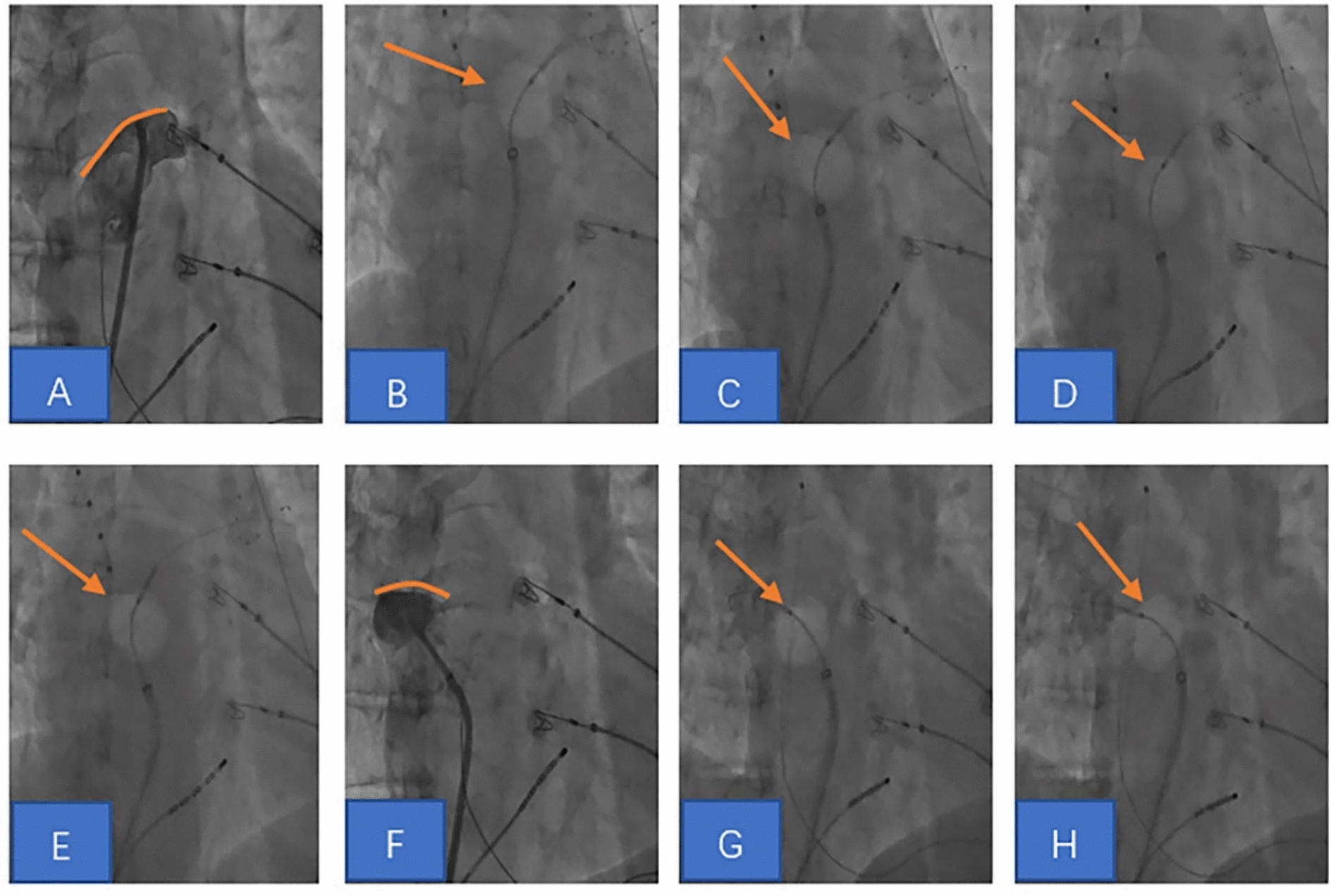
Cryoballoon ablation of the left atrial roof. A Confirmed the left atrial roof by the imaging. B-E Cyroballoon sequential ablation of left atrial roof from left to right. F Confirmed the right-side of the left atrial roof by the imaging. G-H Cyroballoon sequential ablation in the right-side segment of left atrial roof
Procedure endpoints
The procedure endpoints were intact PVI and intact left atrial roof line block zone. Verification of left atrial roof electrical isolation: the quadripolar electrode was located in the left atrial appendage for pacing, and the sequence of excitation was recorded from the posterior wall of the left atrium from bottom to top, confirmed left atrial roof electrical isolation (see Fig. 3 for details). After procedure, those who acquired sinus rhythm underwent burst pacing (frequency 200–300 ms) without inducing AF immediately and at least 30 min after ablation. AF did not occur after receiving synchronized electrical cardioversion, and the observation continued at least 30 min after ablation.
Fig. 3.
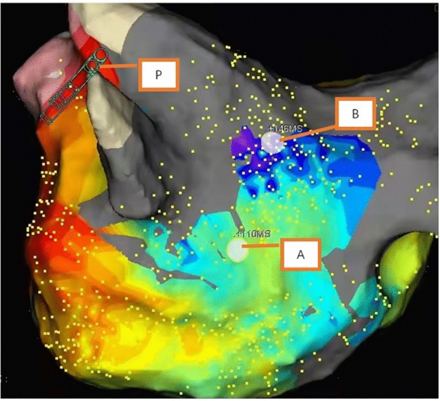
Left atrial roof electrical isolation verification. The left auricular pacing point (P) to the left atrial posterior wall point A excitation time of 110 ms and to the left atrial posterior wall point B excitation time of 146 ms, confirming that the sequence of left atrial posterior wall excitation is from bottom to top
Construction of three-dimensional electroanatomical-CT modeling
Under sinus rhythm, a 3D electroanatomical labeling model of the left atrial pulmonary vein was performed by Ensite Navx. Achieve electrodes were used to construct a voltage map, and the area with a voltage of < 0.2 mV was defined as a low-voltage area [9]. The 3D electroanatomical labeling model was fused with the CTA model of the left atrial pulmonary vein to build a 3D electroanatomical-CT model (3D-CT model). See Fig. 4 for details.
Fig. 4.
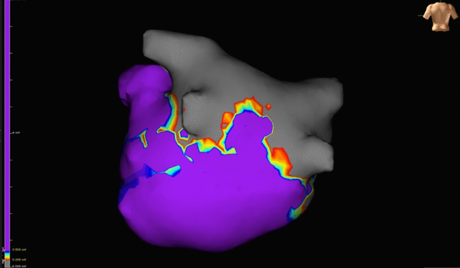
Three-dimensional electroanatomical-CT model; voltage < 0.2 mV in the gray area and > 0.5 mV in the purple area
Quantification of left atrial isolation surface area (ISA)
Define the pulmonary vein opening as the point of maximum inflection between the pulmonary vein wall and the left atrial wall [10]. See Fig. 5 for details.
Fig. 5.
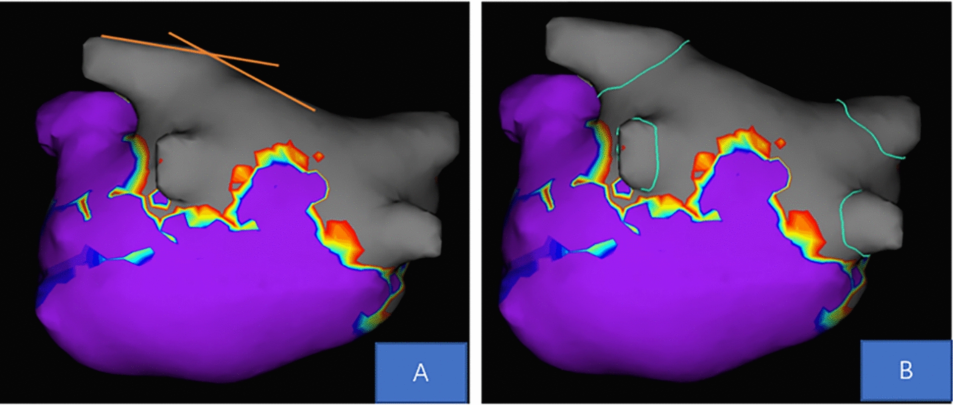
Three-dimensional electroanatomical-CT model. A The pulmonary vein opening is defined as the point of maximum inflection of the pulmonary vein wall with the left atrial wall. B Identification of the pulmonary vein ostium
According to the measurement tool of the Ensite Navx system, the left atrial surface area (LASA), left atrial isolated surface area (ISA), left atrial posterior wall surface area (PWSA), and posterior wall unablated surface area (residual-PWSA) were measured, respectively. LASA was defined as the surface area of the entire left atrium, excluding the surface area of the pulmonary vein, left atrial appendage and the mitral annulus. ISA was defined as the area between the opening of the pulmonary vein and the edge of the scar area with a voltage < 0.2 mV. ISA/LASA was defined as the surface area of the left atrial isolation area/the surface area of the left atrium × 100%. See Figs. 6, 7 for details.
Fig. 6.
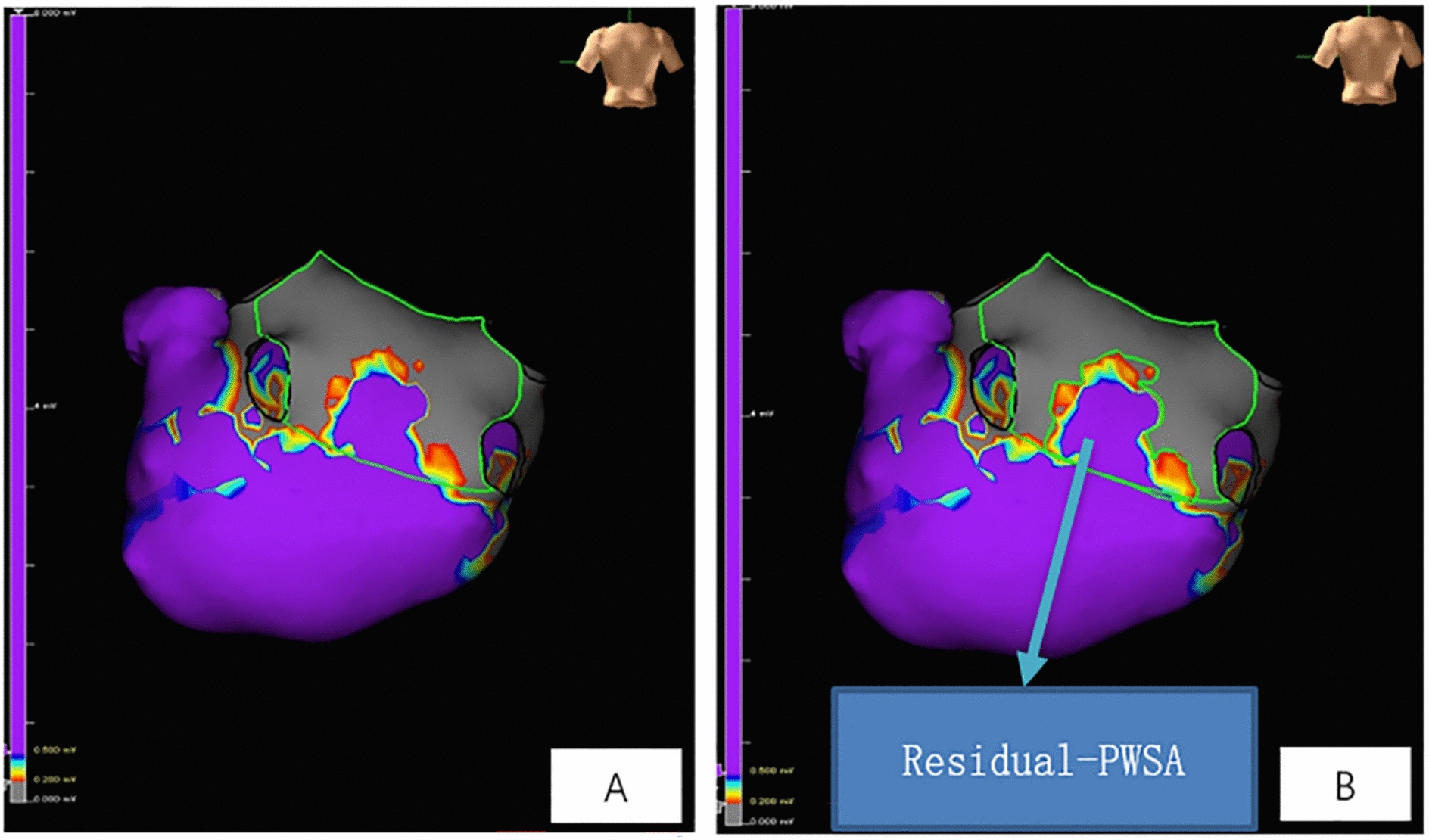
3D electroanatomical-CT model. A Green line area represents the surface area of the posterior wall of the left atrium; B in PWSA. The purple area represents the unablated area of the posterior wall of the left atrium
Fig. 7.
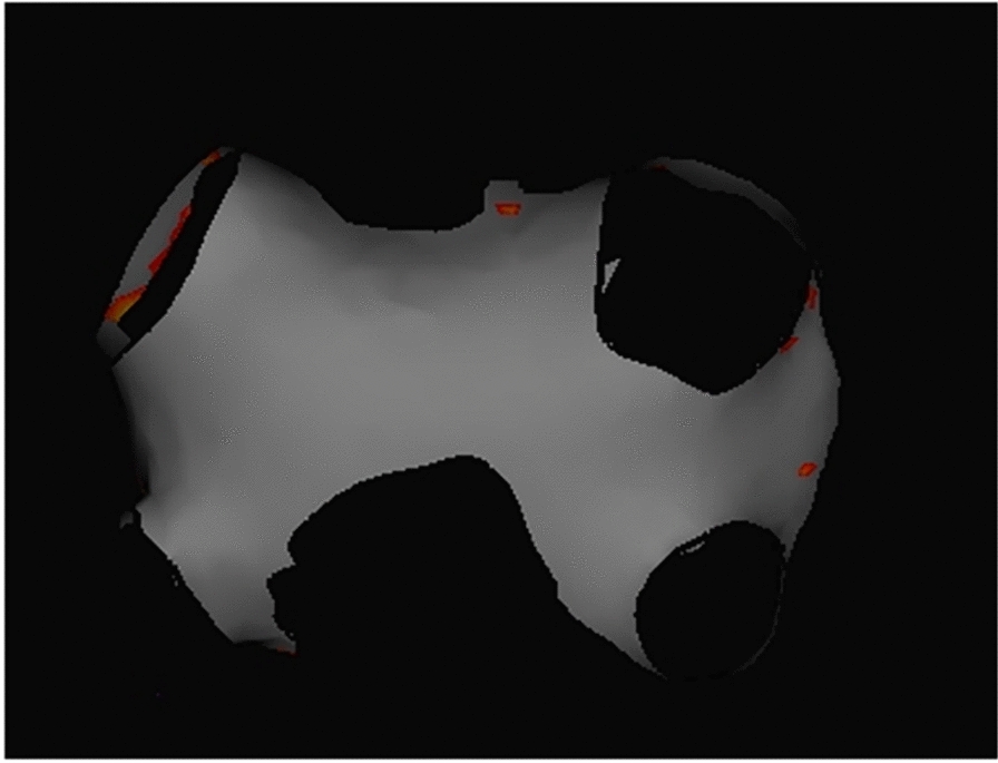
Three-dimensional electroanatomic-CT model. Left atrial isolation area surface area (ISA)
Postoperative management
Routine postoperative cardiac monitoring was performed. Low molecular heparin 5000 IU Q12H anticoagulation was given 6 h post-operation for 2–3 days and then changed to rivaroxaban or warfarin anticoagulation for 3 months, and the decision of whether to anticoagulated or not was made according to CHA2DS2-Vasc score after 3 months.
Postoperative follow-up and early recurrence definition
All patients were followed up at 3, 6, 9, and 12 months after procedure through the outpatient clinic specializing in atrial fibrillation, 12-lead electrocardiograms and 24-h ambulatory electrocardiograms were used whether or not they presented with palpitations, chest tightness, or other discomforts during the period. The patients were divided into the recurrence group and the non-recurrence group according to whether the postoperative follow-up was recurrent or not. Early postoperative recurrence was defined as atrial arrhythmias such as atrial tachycardia, atrial flutter, or atrial fibrillation lasting more than 30 s recorded on ECG or 24-h ambulatory electrocardiogram (EKG) from 3 to 12 months after procedure.
Statistical analysis
SPSS 26.0 software was applied for statistical analysis. Measured data conforming to normal distribution were statistically described using mean ± standard deviation for statistical description, and comparisons between groups were made using independent samples t-test; data not conforming to normal distribution were statistically described using interquartile spacing M (P25, P75), and comparisons between groups was done using rank sum test. Count data were statistically described using component ratios, and comparisons between groups were made using the Chi-square test, corrected Chi-square test, and Fisher’s exact probability method. Univariate and multifactorial Cox regression was used to analyze the risk factors for recurrence after ablation. The predictive power of ISA/LASA for early recurrence after ablation of persistent AF was described using subject operating characteristic curves (ROC curves), and the optimal cut-off value was selected.
Results
Baseline data
93 patients with persistent atrial fibrillation were included in this study, with a mean age of 59.2 ± 9.3 years, of whom 69 (74.2%) were male, with a mean LAD of 41.4 ± 6.4 mm, and 16 (19.4%) with combined heart failure. After the 1-year follow-up, there were 23 patients (24.7%) in the recurrence group and 70 patients (75.3%) in the non-recurrence group. The difference between the recurrent group and the non-recurrent group was statistically significant (P < 0.05) in terms of whether or not there was a combined left common trunk pulmonary vein, and the difference in the remaining baseline data was not statistically significant. For details, see Table 1.
Table 1.
Characteristics of baseline information
| Total (n = 93) | Non-recurrence group (n = 70) | Recurrence group (n = 23) | P | |
|---|---|---|---|---|
| Sex, male: cases (%) | 69 (74.2%) | 55 (78.6%) | 14 (60.9%) | 0.092 |
| Age (years) | 59.2 ± 9.3 | 58.7 ± 2.1 | 59.3 ± 9.1 | 0.788 |
| BMI (kg/m2) | 24.6 ± 3.0 | 24.7 ± 2.9 | 24.5 ± 3.5 | 0.851 |
| Duration of illness (months) | 12 (6, 48) | 12 (6, 48) | 24 (5, 72) | 0.768 |
| LAD (mm) | 41.4 ± 6.4 | 40.7 ± 6.0 | 43.7 ± 6.9 | 0.051 |
| Left ventricle (mm) | 46.6 ± 5.1 | 46.7 ± 4.5 | 46.2 ± 6.8 | 0.720 |
| CHA2DS2-Vasc score | 1 (0, 3) | 1 (0, 3) | 1 (1, 3) | 0.342 |
| HAS-BLED score | 1 (0, 1) | 0 (0, 1) | 1 (0, 1) | 0.628 |
| Complication | ||||
| Heart failure | 16 (17.2%) | 9 (12.9%) | 7 (30.4%) | 0.105 |
| Cerebral hemorrhage | 8 (8.6%) | 5 (7.1%) | 3 (13%) | 0.665 |
| Coronary heart disease | 5 (5.4%) | 4 (4.3%) | 2 (8.7%) | 0.779 |
| Hypertension | 41 (44.1%) | 30 (42.9) | 11 (47.8%) | 0.677 |
| Diabetes | 15 (16.1%) | 13 (18.6%) | 2 (8.7%) | 0.429 |
| Left common trunk pulmonary vein | 6 (6.5%) | 2 (2.9%) | 4 (17.4%) | 0.049 |
BMI body mass index, LAD left atrial internal diameter
Surface area analysis of the left atrial isolation area
Among the 93 patients included, the mean LASA was 126.1 ± 18.7 cm2, the mean PWSA was 21.8 ± 3.3 cm2, and the mean residual-PWSA was 6.2 ± 1.8 cm2, and none of the differences between the recurrent group and the non-recurrent group were statistically significant (P > 0.05). The mean ISA was 20 ± 3.4 cm2, and the ISA of the recurrent group was smaller than that of the non-recurrent group (18.7 ± 4.1 cm2 vs 20.4 ± 3.0 cm2), and the difference was statistically significant (P = 0.026). Pearson correlation analysis showed that there was a weak positive correlation between the ISA and the LASA (r = 0.271, P = 0.009), and therefore, the ISA/LASA was used. LASA was further analyzed, and the results showed that the ISA/LASA in the reoccurrence group was smaller than that in the non-relapse group (14.6 ± 2.6% vs 16.6 ± 3.2%), and the difference was statistically significant (P = 0.009). For details, see Table 2.
Table 2.
Comparison of the surface area of the left atrial segregation/isolation area between the recurrent and non-recurrent groups
| Total (n = 93) | Non-recurrence group (n = 70) | Recurrence group (n = 23) | P | |
|---|---|---|---|---|
| LASA (cm2) | 126.1 ± 18.7 | 125.5 ± 19.2 | 127.7 ± 17.1 | 0.636 |
| ISA (cm2) | 20.0 ± 3.4 | 20.4 ± 3.0 | 18.7 ± 4.1 | 0.026 |
| PWSA (cm2) | 21.8 ± 3.3 | 21.4 ± 3.1 | 22.8 ± 3.8 | 0.078 |
| Residual-PWSA (cm2) | 6.2 ± 1.8 | 6.0 ± 1.6 | 6.7 ± 2.3 | 0.111 |
| ISA/LASA (%) | 16.1 ± 3.2 | 16.6 ± 3.2 | 14.6 ± 2.6 | 0.009 |
LASA left atrial surface area, ISA left atrial segregation area surface area, PWSA posterior wall surface area of the left atrium, residual-PWSA unablated surface area of the posterior wall, ISA/LASA percentage of left atrial segregation area surface area
Predictors of early postoperative recurrence
Univariate Cox regression analysis showed that LAD, left common trunk pulmonary vein, ISA, and ISA/LASA were risk factors for early postoperative recurrence. Multifactorial cox regression analysis showed that the presence of combined left common trunk pulmonary vein (HR 7.754, 95% CI 2.256–26.651, P = 0.001) and ISA/LASA (HR 0, 95% CI 0–0.005, P = 0.008) were risk factors for early postoperative recurrence. See Table 3 for details. The ROC curve showed that an ISA/LASA of 15.2% was the best cut-off value for predicting early postoperative recurrence, at which point the sensitivity was 70% and the specificity was 70% (AUC 0.683; 95% CI 0.560–0.806; P = 0.009). See Fig. 8 for details.
Table 3.
Univariate and multivariate Cox regression analysis of predictors of early postoperative recurrence after CBA
| Single factor Cox | Multi-factor Cox | |||
|---|---|---|---|---|
| HR (95% CI) | P | HR (95% CI) | P | |
| Male | 0.540 (0.233–1.248) | 0.149 | ||
| Age (years) | 0.990 (0.947–1.036) | 0.679 | ||
| Duration of illness (months) | 1.002 (0.993–1.011) | 0.710 | ||
| LAD (mm) | 1.070 (1.001–1.143) | 0.046 | 0.956 (0.946–1.111) | 0.556 |
| Heart failure | 2.339 (0.962–5.689) | 0.061 | 1.870 (0.697–5.017) | 0.214 |
| Hypertension | 1.205 (0.532–2.731) | 0.655 | ||
| Diabetes | 2.187 (0.513–9.330) | 0.290 | ||
| Coronary heart disease | 1.626 (0.381–6.939) | 0.511 | ||
| Left common trunk pulmonary vein | 5.611 (1.877–16.770) | 0.002 | 7.754 (2.256–26.651) | 0.001 |
| LASA (cm2) | 1.005 (0.983–1.028) | 0.645 | ||
| ISA (cm2) | 0.850 (0.738–0.980) | 0.025 | ||
| PWSA (cm2) | 1.127 (0.994–1.277) | 0.061 | 1.107 (0.833–1.471) | 0.484 |
| R-PWSA (cm2) | 2.339 (0.962–5.689) | 0.078 | 1.167 (0.915–1.488) | 0.213 |
| ISA/LASA (%) | 1.205 (0.532–2.731) | 0.012 | 0 (0–0.005) | 0.008 |
LAD left atrial internal diameter, LASA left atrial surface area, ISA left atrial isolation area surface area, PWSA left atrial posterior wall surface area, R-PWSA left atrial posterior wall unablated surface area, ISA/LASA percentage of left atrial isolation area surface area
Fig. 8.
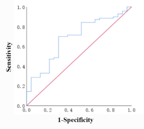
ROC curve of the predictive value of ISA/LASA for early recurrence after cryoablation of persistent AF
Complications
Three cases (3.2%) of related complications occurred in the 93 patients including, two cases from the recurrence group and one case in the non-recurrence group, and the difference between the two groups was not statistically significant [2/70 (2.9%) vs 1/23 (4.3%), P = 1.000]. One patient in the non-recurrent group developed severe sinus bradycardia during intraoperative conversion to sinus rhythm, was treated with a temporary pacemaker, and returned to normal sinus rhythm 2 days after returning to the ward, and the difference between the two groups was not statistically significant [1/70 (1.4%) vs 0/23 (0%), P = 1.000]. One patient in each group developed phrenic nerve injury paralysis during right upper pulmonary vein ablation, which recovered at 1 and 3 months after the procedure, respectively, and the difference between the two groups was not statistically significant [1/70 (1.4%) vs 1/23 (4.3%), P = 0.993]. No other procedure-related complications were seen.
Discussion
In this study, we found that 1-year AF-free rate after application of cryoballoon for antrum expansion combined with left atrial roof ablation in patients with persistent atrial fibrillation was 75.3%. ISA/LASA and left common trunk pulmonary vein are risk factors for early recurrence after cryoablation of persistent AF and ISA/LASA ≥ 15.2% may reduce early recurrence after persistent AF cryoablation.
Success rate of cryoablation for persistent AF
Previous literature has reported a 1-year success rate of approximately 60% after cryoablation for PVI in patients with persistent AF [1, 11], and the 1-year success rate in this study was 75.3%, which is an improvement in the success rate. This may be related to the greater elimination of the potentially arrhythmogenic areas of the left atrial pulmonary vein antrum, roof, and posterior wall, in which AF is developed and maintained.
The pulmonary vein antrum plays an important role in the development of atrial fibrillation and is the main trigger focus for the development of atrial fibrillation [12]. The pulmonary vein antrum has both myocardial and pulmonary vein tissues, and the left atrial myocardium extends into the pulmonary veins to form a myocardial sleeve, and the muscle fibers of the pulmonary vein antrum are unevenly arranged and intertwined and interlaced with each other, which constitutes the anatomical basis for the development of atrial fibrillation [13, 14]. The presence of P cells, Purkinje cells, and migratory cells in the pulmonary vein antrum, the autoregulation of P cells, and the conductivity of Purkinje cells and migratory cells constitute the histologic basis for the development of atrial fibrillation [15]. Pacing labeling techniques have confirmed that the pulmonary vein antrum is the main triggering foci of atrial fibrillation, and that in patients with atrial fibrillation, the pulmonary vein antrum potentials have significantly shorter effective refractory periods and prolonged conduction times, and that these specific potential alterations, which are conducive to the formation of microfracture, which constitute the electrophysiological basis for the occurrence of atrial fibrillation [12, 16–19]. In addition, the cardiac autonomic ganglionic plexus (GP) was found near the pulmonary vein antrum, and extremely fragmented atrial potentials could be recorded in the area where it was located. Tests have shown that highly activated GP can induce AF, and GP ablation can reduce AF recurrence, suggesting that GP in the pulmonary vein antrum is involved in the development and maintenance of AF [20–23]. Strips of ablation zones formed by cryoablation of the pulmonary vein antrum can accompany the ablation of GP [24].
In addition, the top of the left atrium was able to record fragmentation potentials, which may be associated with localized refractoriness and agitation, favoring the onset, development and maintenance of AF [25]. On the basis of PVI, combined top of the left atrium ablation can improve the clinical outcome, confirming the involvement of the top of the left atrium in the onset of AF [26]. However, it has been found that if left atrial roof ablation fails to create a complete electrical conduction block, the incidence of postprocedural macrofibrillatory atrial arrhythmias will be elevated [27]. Compared with radiofrequency ablation, cryoablation results in the formation of a striped block zone in the roof of the left atrium, which reduces the risk of incomplete ablation or incomplete conduction block lines [28–31]. In addition, the left atrial posterior wall and pulmonary veins share a common embryonic origin, and have more pronounced atrial remodeling, with cardiomyocytes displaying greater sodium currents, calcium content, and delay after depolarization, as well as more pronounced conduction abnormalities [32, 33]. The combination of PVI with ablation of the left atrial posterior wall facilitates the maintenance of sinus rhythm in the post-procedure period, and confirms the involvement of the left atrial posterior wall in the development and maintenance of atrial fibrillation [5]. In this study, we performed pulmonary vein antrum expansion combined with left atrial parietal cryoablation, applied three-dimensional voltage labeling and agonistic sequence labeling techniques, and intraoperatively confirmed the formation of a complete block zone and electrical isolation of the left atrial parietal, effectively intervening in the potential arrhythmogenic regions of the pulmonary vein antrum, left atrial parietal, and posterior wall, which may be an important factor in improving the postoperative success rate.
Safety of pulmonary vein antrum enlargement combined with atrial roof cryoablation
Kunniss et al. [3] showed that PVI combined with roof ablation of the left atrium did not increase overall complications compared to PVI alone (2.3% vs 2.3%) and no associated complications were seen during left atrium roof ablation. Ciconte et al. [19] reported that the overall post-operation complication rate for PVI combined with roof ablation with the cryoballoon is 6% with a phrenic nerve injury incidence of 4%. In this study, the overall complication rate of 93 patients with persistent atrial fibrillation who underwent antrum enlargement combined with left atrial roof cryoablation was 3.2% (3/93), of which the incidence of phrenic nerve injury paralysis was about 2.2% (2/93), all of which occurred during the ablation of the right upper pulmonary vein, and recovered at 1 month and 3 months after the procedure, respectively. The rates of associated complications were similar to those reported. Therefore, it is suggested that pulmonary vein antrum enlargement combined with roof ablation in patients with persistent atrial fibrillation can improve the success rate of cryoablation and does not increase the overall complication rate of cryoablation or the incidence of phrenic nerve injury.
In our study, one case of severe sinus bradycardia occurred during the ablation process, and was treated with a temporary pacemaker and returned to normal sinus rhythm 2 days after returning to the ward, which was considered to be related to vagal reaction. Vagal reaction occurs mostly in the left pulmonary vein, especially in the left upper pulmonary vein, and may be related to atrial tissue damage in the pulmonary vein antrum during ablation [34]. The incidence of vagal reactions may be reduced by performing right-sided pulmonary vein ablation before left-sided pulmonary vein ablation or by prophylactic use of atropine during cryoablation [35, 36]. Procedure-related complications such as atrioesophageal fistula, cardiac tamponade, femoral artery injury, and thrombus/air embolism were not seen in this study.
ISA/LASA and early recurrence after cryoablation of persistent atrial fibrillation
We considered the left atrial pulmonary vein antrum, roof and posterior wall as potential arrhythmogenic regions, and quantitatively evaluated the relationship between the ablation surface area of the potential arrhythmogenic regions of the left atrium and postoperative recurrence by fusing three-dimensional electroanatomical labeling with CTA of the left atrial pulmonary vein in this study. The results showed that ISA/LASA was a risk factor for early recurrence after cryoablation of persistent atrial fibrillation. An ISA/LASA ≥ 15.2% may be able to reduce early postoperative recurrence.
Circum-pulmonary vein antrum electrical isolation ablation is superior to segmental ablation of the pulmonary veins and can intervene in more arrhythmogenic regions of the left atrium, effectively reducing the recurrence of postoperative atrial arrhythmias [17, 37, 38]. The larger the surface area of pulmonary vein antrum isolation/surface area of the posterior wall of the left atrium, the lower the postoperative recurrence rate. When the surface area of pulmonary vein antrum isolation/surface area of the posterior wall of the left atrium is ≥ 55%, the recurrence of postoperative atrial arrhythmias can be effectively reduced [10]. However, the above study was conducted in patients with paroxysmal atrial fibrillation with radiofrequency ablation.
Studies have shown that the application of cryoballoon ablation in patients with persistent AF, with antrum enlargement and left atrial parietal ablation on the basis of PVI, can reduce the recurrence of postoperative atrial arrhythmias [2–4, 39]. However, up to date, few studies have quantitatively analyzed the relationship between the surface area of the left atrial isolation zone and postoperative recurrence.
According to previous studies, the voltage of healthy myocardia was ≥ 0.5 mV, and that of scarred myocardia was < 0.2 mV [40]. The other study defined the left atrial low-voltage region as < 0.5 mV [41]. Based on recently published research, low voltage in our study was defined as < 0.2 mV [9]. We fused the 3-dimensional electroanatomical left atrial model specimen with the CTA left atrial model, with the maximum inflection point between the pulmonary vein wall and the left atrial wall defined as the pulmonary vein opening, and the low-voltage area with a specimen voltage of < 0.2 mV under sinus rhythm defined as the ISA, and the results showed that a larger ISA and ISA/LASA were favorable to the maintenance of sinus rhythm after procedure. The ROC curve analysis found that an ISA/LASA of ≥ 15.2% might reduce early recurrence after cryoablation of persistent AF.
However, further expanding the ISA to eliminate more potentially arrhythmogenic regions of the left atrium for better clinical outcomes deserves further investigation.
Left common trunk pulmonary vein as a risk factor for early recurrence after cryoballoon ablation
In this study, we also found that left common trunk pulmonary vein was a risk factor for early recurrence after cryoballoon ablation, which is consistent with a previous finding reported by Shigeta et al. [42]. Several previous clinical studies on whether left common trunk pulmonary vein affects ablation efficacy have come to different conclusions [43]. Xu et al. [44] found that left common trunk pulmonary vein was associated with re-conduction after pulmonary vein electrical isolation. If the left common trunk pulmonary vein had a very large ostial diameter of 28 mm or more, antrum segmental ablation is mostly performed. Cryoablation would be executed in superior section, inferior sections, left lateral wall and right lateral wall of ostial region. The area between the bulbs may be under-ablated when segmental ablation is performed, which may also be a factor in early recurrence.
About freezing time
According to the Chinese expert consensus on atrial fibrillation ablation via cryoballoon catheter and experience of our center, we used 2 min of freezing time in the process of cryoablation. But Miyazaki et al. [45] reported that the success rate of LA roofline block creation was 77.2% with only 2 min of freezing, while Kunis et al. [3] reported that the freezing time was 3 min during LA roofline ablation, and the success rate of LA roofline block creation was 92%. Nishimura et al. [46] also reported that the success rate of LA roofline ablation using a cryoballoon was 92% with a freezing time of 3 to 4 min, while Shigeta [42] reported that the durability of LA roofline with the 3-min protocol was 75% durability rate with. Moreover, Miyazaki also reported that the durability of LA roofline using the 2-min protocol was 25%. In general, the longer the freezing time, the greater the extent of damage. In fact, in this study, the first acute success rate was about 50% when moving every cryoablation by the step of one-third balloon. The rate went up to 90% by the step of one-fourth balloon in the continuous roofline ablation, due to much more overlap areas were cryoablated for more than 2 min. Additionally, the areas with residual signals would accept much longer time of cryoablation to achieve completed isolation of roofline. So, the average time of each ablated site was more than 2 min in the roofline. The success rate of LA roofline block with different freezing times and their effect on the recurrence of atrial fibrillation will be further studied in the future, in which large and multi-center clinical studies are needed.
Limitations
Our study has some limitations. Firstly, this study is a single-center, non-randomized, retrospective study with a small sample size, which needs to be further supported by multi-center and large samples. Secondly, this study only measured the surface area of the isolation zone in the acute postoperative period, and the changes in the surface area of the isolation zone in the chronic period are unknown. Also, postoperative asymptomatic AF recurrence may be overlooked. Lastly, this study only analyzed the clinical outcomes of recurrence at 1 year after procedure, and longer follow-up is needed to observe long-term clinical outcomes.
Conclusion
In this study, we find that a greater ISA/LASA reduces early recurrence after cryoablation in patients with PeAF. An ISA/LASA of 15.2% may be the best cut-off value for predicting early recurrence after cryoablation for PeAF.
Author contributions
Conceptualization: JZ; data curation: QC, XL, YP; formal analysis: JH, ZY; funding acquisition: JZ, JC: supervision: JH, LJ; visualization: JH, LJ; writing—original draft: QC, JH, Panashe M.; writing—review and editing: QC, LJ, Panashe M., performing the experiments: MW, ZY, YP, JC; methodology: JZ, JC. All authors have read and approved the final manuscript.
Funding
This work was supported by grants obtained from the National Natural Science Foundation of China [No. 82070341], the Natural Science Foundation of Fujian Province [Nos: 2015J01370, 2019J01189, 2020J011074, 2023J011189], and the Fujian Province Science and Technology Innovation Joint Fund Project [No. 2023Y9350].
Data availability
No datasets were generated or analysed during the current study.
Declarations
Ethics approval and consent to participate
The study protocol was approved by the ethics committee of Fujian Provincial Hospital.
Competing interests
The authors declare no competing interests.
Footnotes
Publisher's Note
Springer Nature remains neutral with regard to jurisdictional claims in published maps and institutional affiliations.
Qian Chen, Jin-Jin Huang and Ling Jiang contributed equally to this work.
Contributor Information
Jian-Quan Chen, Email: ccjqemie@163.com.
Jian-Cheng Zhang, Email: fjzhangjiancheng@126.com.
References
- 1.Ciconte G, Ottaviano L, de Asmundis C, et al. Pulmonary vein isolation as index procedure for persistent atrial fibrillation: one-year clinical outcome after ablation using the second-generation cryoballoon. Heart Rhythm. 2015;12(1):60–6. [DOI] [PubMed] [Google Scholar]
- 2.Nanbu T, Yotsukura A, Sano F, et al. A relation between ablation area and outcome of ablation using 28-mm cryoballoon ablation: importance of carina region. J Cardiovasc Electrophysiol. 2018;29(9):1221–9. [DOI] [PubMed] [Google Scholar]
- 3.Kuniss M, Akkaya E, Berkowitsch A, et al. Left atrial roof ablation in patients with persistent atrial fibrillation using the second-generation cryoballoon: benefit or wasted time? Clin Res Cardiol. 2020;109(6):714–24. [DOI] [PubMed] [Google Scholar]
- 4.Nanbu T, Yotsukura A, Suzuki G, et al. Important factors in left atrial posterior wall isolation using 28-mm cryoballoon ablation for persistent atrial fibrillation-block line or isolation area? J Cardiovasc Electrophysiol. 2020;31(1):119–27. [DOI] [PubMed] [Google Scholar]
- 5.Aryana A, Baker JH, Espinosa Ginic MA, et al. Posterior wall isolation using the cryoballoon in conjunction with pulmonary vein ablation is superior to pulmonary vein isolation alone in patients with persistent atrial fibrillation: a multicenter experience. Heart Rhythm. 2018;15(8):1121–9. [DOI] [PubMed] [Google Scholar]
- 6.Chen L, Chen JQ, Zou T, et al. Efficacy of extended antrum ablation based on substrate mapping plus pulmonary vein isolation in the treatment of atrial fibrillation. Rev Port Cardiol. 2022;41(1):17–26. [DOI] [PubMed] [Google Scholar]
- 7.Lin YZ, Peng YM, Lian LH, et al. An evaluation of the clinical efficacy of the application of 28-mm cryoballoon for linear ablation of left atrial apex combined with enlarged pulmonary vein vestibule ablation for persistent atrial fibrillation. Hell J Cardiol. 2023;72:15–23. [DOI] [PubMed] [Google Scholar]
- 8.CSPE. Chinese expert consensus on atrial fibrillation ablation via cryoballoon catheter. Chin J Card Pacing Electrophysiol. 2020;34(2):95–109 (in Chinese). [Google Scholar]
- 9.Wakamatsu Y, Nakahara S, Nagashima K, et al. Hot balloon versus cryoballoon ablation for persistent atrial fibrillation: lesion area, efficacy, and safety. J Cardiovasc Electrophysiol. 2020;31(9):2310–8. [DOI] [PubMed] [Google Scholar]
- 10.Kiuchi K, Kircher S, Watanabe N, et al. Quantitative analysis of isolation area and rhythm outcome in patients with paroxysmal atrial fibrillation after circumferential pulmonary vein antrum isolation using the pace-and-ablate technique. Circ Arrhythm Electrophysiol. 2012;5(4):667–75. [DOI] [PubMed] [Google Scholar]
- 11.Su WW, Reddy VY, Bhasin K, et al. Cryoballoon ablation of pulmonary veins for persistent atrial fibrillation: results from the multicenter STOP persistent AF trial. Heart Rhythm. 2020;17(11):1841–7. [DOI] [PubMed] [Google Scholar]
- 12.Valles E, Fan R, Roux JF, et al. Localization of atrial fibrillation triggers in patients undergoing pulmonary vein isolation: importance of the carina region. J Am Coll Cardiol. 2008;52(17):1413–20. [DOI] [PubMed] [Google Scholar]
- 13.Saito T, Waki K, Becker AE. Left atrial myocardial extension onto pulmonary veins in humans: anatomic observations relevant for atrial arrhythmias. J Cardiovasc Electrophysiol. 2000;11(8):888–94. [DOI] [PubMed] [Google Scholar]
- 14.Nathan H, Eliakim M. The junction between the left atrium and the pulmonary veins. An anatomic study of human hearts. Circulation. 1966;34(3):412–22. [DOI] [PubMed] [Google Scholar]
- 15.Perez-Lugones A, McMahon JT, Ratliff NB, et al. Evidence of specialized conduction cells in human pulmonary veins of patients with atrial fibrillation. J Cardiovasc Electrophysiol. 2003;14(8):803–9. [DOI] [PubMed] [Google Scholar]
- 16.Marrouche NF, Dresing T, Cole C, et al. Circular mapping and ablation of the pulmonary vein for treatment of atrial fibrillation: impact of different catheter technologies. J Am Coll Cardiol. 2002;40(3):464–74. [DOI] [PubMed] [Google Scholar]
- 17.Chen SA, Hsieh MH, Tai CT, et al. Initiation of atrial fibrillation by ectopic beats originating from the pulmonary veins: electrophysiological characteristics, pharmacological responses, and effects of radiofrequency ablation. Circulation. 1999;100(18):1879–86. [DOI] [PubMed] [Google Scholar]
- 18.Jais P, Hocini M, Macle L, et al. Distinctive electrophysiological properties of pulmonary veins in patients with atrial fibrillation. Circulation. 2002;106(19):2479–85. [DOI] [PubMed] [Google Scholar]
- 19.Lee G, Spence S, Teh A, et al. High-density epicardial mapping of the pulmonary vein–left atrial junction in humans: insights into mechanisms of pulmonary vein arrhythmogenesis. Heart Rhythm. 2012;9(2):258–64. [DOI] [PubMed] [Google Scholar]
- 20.Nakagawa H, Scherlag BJ, Patterson E, et al. Pathophysiologic basis of autonomic ganglionated plexus ablation in patients with atrial fibrillation. Heart Rhythm. 2009;6(12 Suppl):S26-34. [DOI] [PubMed] [Google Scholar]
- 21.Zhou J, Scherlag BJ, Edwards J, et al. Gradients of atrial refractoriness and inducibility of atrial fibrillation due to stimulation of ganglionated plexi. J Cardiovasc Electrophysiol. 2007;18(1):83–90. [DOI] [PubMed] [Google Scholar]
- 22.Katritsis DG, Giazitzoglou E, Zografos T, et al. Rapid pulmonary vein isolation combined with autonomic ganglia modification: a randomized study. Heart Rhythm. 2011;8(5):672–8. [DOI] [PubMed] [Google Scholar]
- 23.Katritsis DG, Pokushalov E, Romanov A, et al. Autonomic denervation added to pulmonary vein isolation for paroxysmal atrial fibrillation: a randomized clinical trial. J Am Coll Cardiol. 2013;62(24):2318–25. [DOI] [PubMed] [Google Scholar]
- 24.Yorgun H, Aytemir K, Canpolat U, et al. Additional benefit of cryoballoon-based atrial fibrillation ablation beyond pulmonary vein isolation: modification of ganglionated plexi. Europace. 2014;16(5):645–51. [DOI] [PubMed] [Google Scholar]
- 25.Nademanee K, McKenzie J, Kosar E, et al. A new approach for catheter ablation of atrial fibrillation: mapping of the electrophysiologic substrate. J Am Coll Cardiol. 2004;43(11):2044–53. [DOI] [PubMed] [Google Scholar]
- 26.Hocini M, Jais P, Sanders P, et al. Techniques, evaluation, and consequences of linear block at the left atrial roof in paroxysmal atrial fibrillation: a prospective randomized study. Circulation. 2005;112(24):3688–96. [DOI] [PubMed] [Google Scholar]
- 27.Knecht S, Hocini M, Wright M, et al. Left atrial linear lesions are required for successful treatment of persistent atrial fibrillation. Eur Heart J. 2008;29(19):2359–66. [DOI] [PubMed] [Google Scholar]
- 28.Furnkranz A, Bordignon S, Schmidt B, et al. Improved procedural efficacy of pulmonary vein isolation using the novel second-generation cryoballoon. J Cardiovasc Electrophysiol. 2013;24(5):492–7. [DOI] [PubMed] [Google Scholar]
- 29.Giovanni GD, Wauters K, Chierchia GB, et al. One-year follow-up after single procedure cryoballoon ablation: a comparison between the first and second generation balloon. J Cardiovasc Electrophysiol. 2014;25(8):834–9. [DOI] [PubMed] [Google Scholar]
- 30.Okumura Y, Watanabe I, Iso K, et al. Mechanistic insights into durable pulmonary vein isolation achieved by second-generation cryoballoon ablation. J Atr Fibrillation. 2017;9(6):1538. [DOI] [PMC free article] [PubMed] [Google Scholar]
- 31.Sawhney N, Anousheh R, Chen W, et al. Circumferential pulmonary vein ablation with additional linear ablation results in an increased incidence of left atrial flutter compared with segmental pulmonary vein isolation as an initial approach to ablation of paroxysmal atrial fibrillation. Circ Arrhythm Electrophysiol. 2010;3(3):243–8. [DOI] [PubMed] [Google Scholar]
- 32.Aryana A, Pujara DK, Allen SL, et al. Left atrial posterior wall isolation in conjunction with pulmonary vein isolation using cryoballoon for treatment of persistent atrial fibrillation (PIVoTAL): study rationale and design. J Interv Card Electrophysiol. 2021;62(1):187–98. [DOI] [PMC free article] [PubMed] [Google Scholar]
- 33.Lim HS, Hocini M, Dubois R, et al. Complexity and distribution of drivers in relation to duration of persistent atrial fibrillation. J Am Coll Cardiol. 2017;69(10):1257–69. [DOI] [PubMed] [Google Scholar]
- 34.Zhou G, Ling TY, Jin Q, et al. Prediction of vagal reaction during cryoballoon ablation for the effect in patients with paroxysmal atrial fibrillation. Chin J Card Pacing Electrophysiol. 2015;20(06):515–8 (in Chinese). [Google Scholar]
- 35.Sun L, Dong JZ, Du X, et al. Prophylactic atropine administration prevents vasovagal response induced by cryoballoon ablation in patients with atrial fibrillation. Pacing Clin Electrophysiol. 2017;40(5):551–8. [DOI] [PubMed] [Google Scholar]
- 36.Miyazaki S, Nakamura H, Taniguchi H, et al. Impact of the order of the targeted pulmonary vein on the vagal response during second-generation cryoballoon ablation. Heart Rhythm. 2016;13(5):1010–7. [DOI] [PubMed] [Google Scholar]
- 37.Proietti R, Santangeli P, Di Biase L, et al. Comparative effectiveness of wide antral versus ostial pulmonary vein isolation: a systematic review and meta-analysis. Circ Arrhythm Electrophysiol. 2014;7(1):39–45. [DOI] [PubMed] [Google Scholar]
- 38.Arentz T, Weber R, Burkle G, et al. Small or large isolation areas around the pulmonary veins for the treatment of atrial fibrillation? Results from a prospective randomized study. Circulation. 2007;115(24):3057–63. [DOI] [PubMed] [Google Scholar]
- 39.Kuniss M, Greiss H, Pajitnev D, et al. Cryoballoon ablation of persistent atrial fibrillation: feasibility and safety of left atrial roof ablation with generation of conduction block in addition to antral pulmonary vein isolation. Europace. 2017;19(7):1109–15. [DOI] [PubMed] [Google Scholar]
- 40.Rolf S, Kircher S, Arya A, et al. Tailored atrial substrate modification based on low-voltage areas in catheter ablation of atrial fibrillation. Circ Arrhythm Electrophysiol. 2014;7(5):825–33. [DOI] [PubMed] [Google Scholar]
- 41.Jadidi AS, Lehrmann H, Keyl C, et al. Ablation of persistent atrial fibrillation targeting low-voltage areas with selective activation characteristics. Circ Arrhythm Electrophysiol. 2016;9(3): e002962. [DOI] [PubMed] [Google Scholar]
- 42.Shigeta T, Okishige K, Yamauchi Y, et al. Clinical assessment of cryoballoon ablation in cases with atrial fibrillation and a left common pulmonary vein. J Cardiovasc Electrophysiol. 2017;28(9):1021–7. [DOI] [PubMed] [Google Scholar]
- 43.Yorgun H, Canpolat U, Gumeler E, et al. Immediate and long-term outcomes of cryoballoon catheter ablation in patients with atrial fibrillation and left common pulmonary vein anatomy. J Interv Card Electrophysiol. 2020;59(1):57–65. [DOI] [PubMed] [Google Scholar]
- 44.Xu B, Xing Y, Xu C, et al. A left common pulmonary vein: anatomical variant predicting good outcomes of repeat catheter ablation for atrial fibrillation. J Cardiovasc Electrophysiol. 2019;30(5):717–26. [DOI] [PubMed] [Google Scholar]
- 45.Miyazaki S, Sekihara T, Hasegawa K, et al. The feasibility and safety of substrate modification on the left atrial roof area using a cryoballoon in atrial fibrillation ablation. Int J Cardiol. 2022;350:41–7. [DOI] [PubMed] [Google Scholar]
- 46.Nishimura T, Yamauchi Y, Aoyagi H, et al. The clinical impact of the left atrial posterior wall lesion formation by the cryoballoon application for persistent atrial fibrillation: feasibility and clinical implications. J Cardiovasc Electrophysiol. 2019;30(6):805–14. [DOI] [PubMed] [Google Scholar]
Associated Data
This section collects any data citations, data availability statements, or supplementary materials included in this article.
Data Availability Statement
No datasets were generated or analysed during the current study.


