Abstract
Introduction
Pyroptosis, inflammatory necrosis of cells, is a programmed cell death involved in the pathological process of diseases. Endoplasmic reticulum stress (ERS), as a protective stress response of cell, decreases the unfold protein concentration to inhibit the unfold protein agglutination. Whereas the relationship between endoplasmic reticulum stress and pyroptosis in pulmonary hypertension (PH) remain unknown. Previous evident indicated that circular RNA (circRNA) can participate in several biological process, including cell pyroptosis. However, the mechanism of circRNA regulate pyroptosis of pulmonary artery smooth muscle cells through endoplasmic reticulum stress still unclear. Here, we proved that circSSR1 was down-regulate expression during hypoxia in pulmonary artery smooth muscle cells, and over-expression of circSSR1 inhibit pyroptosis both in vitro and in vivo under hypoxic. Our experiments have indicated that circSSR1 could promote host gene SSR1 translation via m6A to activate ERS leading to pulmonary artery smooth muscle cell pyroptosis. In addition, our results showed that G3BP1 as upstream regulator mediate the expression of circSSR1 under hypoxia. These results highlight a new regulatory mechanism for pyroptosis and provide a potential therapy target for pulmonary hypertension.
Methods
RNA-FISH and qRT-PCR were showed the location of circSSR1 and expression change. RNA pull-down and RIP verify the circSSR1 combine with YTHDF1. Western blotting, PI staining and LDH release were used to explore the role of circSSR1 in PASMCs pyroptosis.
Results
CircSSR1 was markedly downregulated in hypoxic PASMCs. Knockdown CircSSR1 inhibited hypoxia induced PASMCs pyroptosis in vivo and in vitro. Mechanistically, circSSR1 combine with YTHDF1 to promote SSR1 protein translation rely on m6A, activating pyroptosis via endoplasmic reticulum stress. Furthermore, G3BP1 induce circSSR1 degradation under hypoxic.
Conclusion
Our findings clarify the role of circSSR1 up-regulated parental protein SSR1 expression mediate endoplasmic reticulum stress leading to pyroptosis in PASMCs, ultimately promoting the development of pulmonary hypertension.
Supplementary Information
The online version contains supplementary material available at 10.1186/s12931-024-02986-w.
Keywords: CircRNA, Endoplasmic reticulum stress, m6A, Pyroptosis, Pulmonary hypertension
Introduction
Pulmonary hypertension (PH) is a pathophysiological state of abnormally elevated blood pressure caused by several known or unknown reasons [1–3]. The pathological changes are extremely complicated in this process, among them, pulmonary vascular remodeling (PVR) is the major cause of PH [4]. Growing evidence has indicate that informed of PVR also including proliferation, autophagy, ferroptosis and apoptosis [5–8]. Undeniably, the discovery of novel pathological alterations holds pivotal importance in the quest for effective treatment modalities to pulmonary hypertension (PH).
Pyroptosis, also known as cell inflammatory necrosis, is a type of programmed cell death in which cells continue to be swelling until the cell membrane ruptures, resulting in the release of cell contents that activate a strong inflammatory response [9, 10]. Pyroptosis mainly relies on inflammatosomes to activate some proteins of the caspase family, causing them to cut and active Gasdermin protein. The activated Gasdermin protein is translocated to the membrane, forming pores, and finally leading to pyroptosis [11, 12]. Furthermore, it has been reported that pyroptosis participate in the occurrence and progression of infectious disease, nervous system disease, chronic liver disease and cancer [12–14]. Our previous studies provide evidence that pyroptosis is involved in the inflammatory process of pulmonary artery smooth muscle cells (PASMCs) and pulmonary artery endothelial cells (PAECs) in the model of hypoxia pulmonary hypertension [15, 16]. However, the regulatory factors and the regulatory mechanisms of pyroptosis in PH are not fully understand.
The endoplasmic reticulum stress (ERS) is a protective response of cell, the cell therefore reduces the concentration of unfold protein to prevent aggregating [17–19]. There are three membrane-related proteins in the endoplasmic reticulum of cells: inositolr equiring enzyme 1 (IRE1), PRKR-like endoplasmic reticulum kinase (PERK) and activating transcription factor 6 (ATF6). Under critical and enduring ER stress could actives cells apoptosis, manifest the nuclear rupture and cell membrane invaginate but not leading to release of inflammatory factors [20]. In infectious diseases, it is reported that IRE1 actuates the production of NO and release of IL-1β [21]. Our previous study has proved that BCAT1 combine with RNA binding protein ZNF423 to active autophagy through IRE1-XBP-1-RIDD axis in pulmonary hypertension [22]. In addition to this it is reported endoplasmic reticulum stress pathway is activated in pyroptosis. Li et al. showed endoplasmic reticulum stress contribute pyroptosis through NF-κB/NLRP3 pathway in diabetic nephropathy [23]. However, the mechanism of regulating pyroptosis through ER stress signaling pathway has been not further explored in pulmonary hypertension.
Circular RNAs (CircRNAs) are a special type of non-coding RNAs via back-splicing of linear RNA and do not have a 5’ terminal cap and 3’ terminal poly(A) tail [24]. In comparison to linear RNAs, it is not easy to degrade by exonucleases and more stable [25]. It had been reported that circRNA involved in the regulation of pyroptosis in many diseases. For instance, circRNA_0075723 act as sponge of miR-155-5p to promote SHIP1 expression directly inhibit macrophage pyroptosis in pneumonia-induced sepsis [26]. CircUSP9X interacts with binding protein EIF4A3 in the cytoplasm to regulate endothelial cell pyroptosis in atherosclerosis [27]. Our lab has demonstrated that circLrch3 with host gene form a R-Loop to regulate pyroptosis of pulmonary arterial smooth muscle cells caused by hypoxic [28]. All the data suggested that circRNAs are involved in the regulation of pyroptosis in multiple diseases with different mechanism.
In this study, we first proposed that circSSR1 regulates pyroptosis by activating endoplasmic reticulum stress via host protein SSR1 during hypoxia. Mechanically, circSSR1 regulate host gene expression by combined with m6A reader YTHDF1 under hypoxia. Our results highlight a new regulatory mechanism for pyroptosis and provide a potential therapy target for pulmonary hypertension.
Results
CircSSR1 was down-regulate expression during hypoxia in pulmonary artery smooth muscle cells
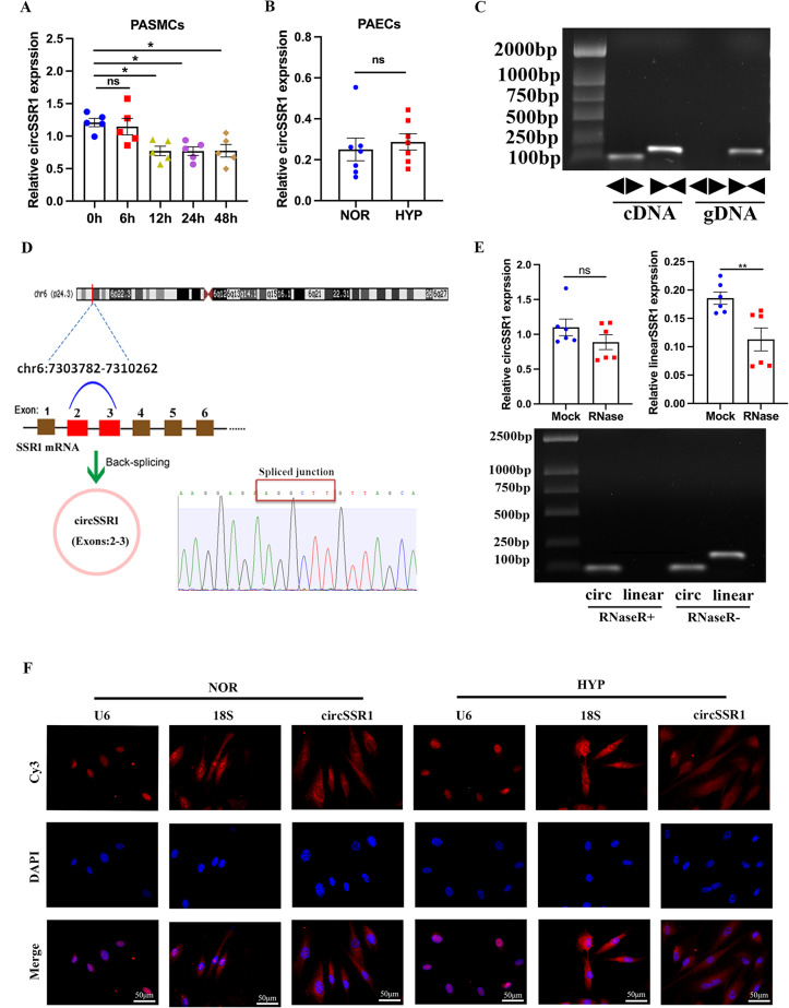
First, we detected the significance of circSSR1 in pulmonary artery smooth muscle cells, which were exposed to hypoxia in different period. Our results had proved circSSR1 is down-regulate under hypoxia PASMCs (Fig. 1A). Besides, to investigate the expression of circSSR1 in hypoxia PAECs, the results of quantitative PCR showed it was no significance compared with hypoxia (Fig. 1B). We extracted gDNA and cDNA to reveal the stability of RNA (Fig. 1C). The upstream and downstream primers were designed for cyclization sequence, to verify the cyclization of circSSR1 by quantitative polymerase chain reaction (qPCR), the results of sanger sequence also proved that circSSR1 formed by 2 and 3 exons, circSSR1 was located on human chromosome 6:7303782–7,310,262 (Fig. 1D). Using RNase R to treat with reverse-transcribed cDNA, the consequence of qPCR and agarose gel electrophoresis had showed circSSR1 was almost no change. These assays further approved the possible of cyclization (Fig. 1E). Next, we aimed to clarify the distribution of circSSR1 and circSSR1 was mainly distribute in cytoplasm showed by Fluorescence in Situ Hybridization (RNA FISH) (Fig. 1F).
Fig. 1.
CircSSR1 is down-regulate in PASMCs under hypoxia (HYP). A-B, The expression of circSSR1 in human pulmonary artery smooth muscle cells (PASMCs) and endothelial cells (n = 5–7). C, The product of qRT-PCR was detected by agarose gel electrophoresis (AGE) (cDNA, gDNA [genomic DNA]; n = 3). D, Sanger sequence was verified the correctness of circSSR1 cyclization site. E, CircSSR1 could resistance exonuclease (RNase R) (n = 6). F, PASMCs were cultured for 24 h in HYP conditions, and fluorescence in situ hybridization (FISH) was used to detected circSSR1 distribution,18 S and U6 were controls for localization for cytoplasm and nucleus. Scale bars, 50 μm. All values are presented as the mean ± SEM. *p<0.05, **p<0.01. Chr6 indicates chromosome 6; DAPI indicates 4ʹ, 6-diamidino-2-phenylindole; NOR, normoxia and HYP, hypoxia. NC: negative control
Over-expression of circSSR1 could inhibit endoplasm reticulum stress and pyroptosis under hypoxia
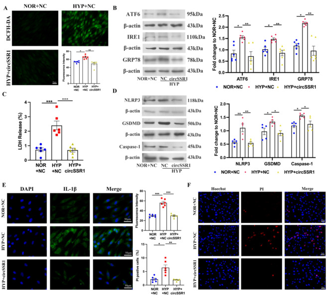
The PASMCs were transfected with SSR1 over-expressed plasmid and NC mimics were cultured in normal condition and we detected the over-expression efficiency by qPCR (Figure S1A). Besides, we detected the reactive oxygen species (ROS) standard, we find circSSR1 over-expressed under hypoxia can reverse the increased caused by hypoxia (Fig. 2A). Western blotting was proved that over-expression of circSSR1 could inhibit IRE1, ATF6 and GRP78 expression (Fig. 2B). We used LDH Cytotoxicity Assay Kit to detect lactate dehydrogenase to further confirm pyroptosis of PASMC when over-expression of circSSR1 (Fig. 2C). The results of western blot (wb) were used to illustrate the expression of pyroptosis-related indicators, we found the over-expression of circSSR1 could inhibit the proteins increased, such as Caspase-1, NLRP3 and Gasdermin D (GSDMD) (Fig. 2D). The immunofluorescence of IL-1β was also used to testify this phenomenon (Fig. 2E). The PI staining was used to demonstrate over-expression of circSSR1 could decrease the rate of positive cells caused by hypoxia (Fig. 2F). The above results indicate that over-expression of circSSR1 can prevent PASMCs pyroptosis and ERS in HYP.
Fig. 2.
Over-expression of circSSR1 inhibited vascular smooth muscle cell pyroptosis. A, Over-expression of circSSR1 reduce reactive oxygen species probes (DCFH-DA, green) staining in various groups (n = 6). B, Endoplasmic reticulum stress (ERS) related protein ATF6, IRE1 and GRP78 expression. C, LDH cytotoxicity assay kit (LDH) was used to detect LDH release (n = 6). D, Western blot to demonstrate pyroptosis related protein NLRP3, GSDMD and Caspase-1 expression (n = 5). E, Fluorescence staining of IL-1β in PASMCs under hypoxia (n = 6). F, Over-expression of circSSR1 plasmid decreased PI positive cells (n = 5). Scale bars, 50 μm. All values are presented as the mean ± SEM. *p<0.05, **p<0.01, ***p<0.001. NOR, normoxia and HYP, hypoxia. NC: negative control
CircSSR1 can inhibit pyroptosis via endoplasmic reticulum stress in hypoxia
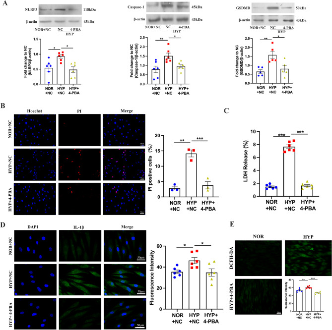
To identify circSSR1 regulate pyroptosis through endoplasmic reticulum stress, western blot analysis showed that hypoxia increased the expression of NLRP3, GSDMD and Caspase-1; however, the expression of them decreased after added the ERS inhibitor 4-phenylbutyric acid (4-PBA) (Fig. 3A). PI staining and LDH release assay were performed positive incidence of cells (Fig. 3B and C). The consistency of the findings was further validated by immunofluorescence analysis of the IL-1β (Fig. 3D). Moreover, we performed ROS assay to validate whether 4-PBA could restrain release of ROS in PASMCs (Fig. 3E). Recovery experiments were used to prove circSSR1 regulate pyroptosis through endoplasmic reticulum stress. We observed that the efficacy of si-1, in terms of its ability to interfere with circSSR1 expression, surpassed that of si-2, as evidenced by the quantitative PCR results (Figure S1B). Therefore, si-1 was used in subsequently experiments. The PASMCs were transfected with circSSR1 siRNA and added the ERS inhibitor in NOR for 24 h. The LDH release assay and immunofluorescence of IL-1β experiments demonstrated that the addition of 4-PBA reversed the effects of only interfering with circSSR1 in NOR (Figure S1C-S1D). Meantime, silencing of circSSR1 promoted vascular smooth cell pyroptosis in the normoxic, however, under identical conditions added ERS inhibitor could prevent the expression of pyroptosis related proteins expression including Caspase-1, NLRP3 and GSDMD (Figure S1E). Interference with circSSR1 lead to an increase in the number of positive cells detected by PI staining in NOR, whereas the addition of 4-PBA alleviated this effect (Figure S1F).
Fig. 3.
Added endoplasmic reticulum stress (ERS) 4-PBA can restrain pyroptosis caused by hypoxia. A, Western blot indicated adding 4-PBA can inhibit NLRP3, GSDMD and Caspase 1 expression during hypoxia (n = 5–6). B, The positive cells were demonstrated by PI staining (n = 3). C, LDH release assay detected the content of lactate dehydrogenase (n = 6). D, Immunofluorescence showed inhibitor of ERS reduce IL-1β expression in the hypoxia (n = 6). E, Fluorescent probe DCFH-DA detected ROS release of cells treated with 4-PBA (n = 6). Scale bars, 50 μm. All values are presented as the mean ± SEM. *p<0.05, **p<0.01, ***p<0.001. NOR, normoxia and HYP, hypoxia. NC: negative control
CircSSR1 regulates pyroptosis by activating endoplasmic reticulum stress via host protein SSR1 during hypoxic
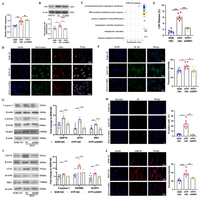
CircSSR1 was transcriptional by-product and subsequently we confirmed whether change of circSSR1 would affect the expression of linear mRNA or protein. The qPCR and western blot indicate that over-expression of circSSR1 was no change on mRNA level, however, it could decrease the protein of SSR1 (Fig. 4A and B). We used website to predict the function of SSR1, it is related with ERS and located on membrane (Fig. 4C). To elucidate the role of SSR1 on circSSR1 in regulating pyroptosis and ERS of pulmonary artery smooth cells, we construct a small RNA to interfere with SSR1 and the interference efficiency was detected by western blot. Western blot results reveal SSR1 protein expression level significantly down-regulated by si-3 rather than si-1 or si-2 (Figure S2A). Therefore, we chose the si-3 for the following experiments. Immunofluorescence co-localization analysis was used to examine the SSR1 and endoplasmic reticulum markers protein (Fig. 4D). The siRNA was transfected into PASMCs, and subsequent experiments measuring LDH release and fluorescence intensity of IL-1β were conducted to verify the induction of pyroptosis in the PASMCs (Fig. 4E and F). Moreover, the increased expression of the proteins NLRP3, Caspase-1, GSDMD and increased positivity of PI staining also get the same results (Fig. 4G and H). When transfect with SSR1 siRNA into PASMCs, ERS-related proteins encompassing IRE1, ATF6, GRP78 were down-regulate in hypoxia (Fig. 4I). And fluorescence intensity of GRP78 was increased exposure to hypoxia and reversed by silencing the SSR1 gene (Fig. 4J). Furthermore, we transfect SSR1 plasmid in PASMCs 4–6 h and then cultured in incubator overnight. The efficiency of over-expression was detected by western blot (Figure S2B). The PI staining and LDH release assay were showed that only expression of circSSR1 can reduce cell positive rate and LDH release caused by hypoxia, conversely, the combined over-expression of circSSR1 and SSR1 reversed this effect (Figure S2C-S2D). Meantime, immunofluorescence assay had indicated that co-transfected circSSR1 and SSR1 up-regulate expression of IL-1β and GRP78 (Figure S2E-S2F). These results showed circSSR1 regulate pyroptosis of PASMCs through parental protein SSR1.
Fig. 4.
CircSSR1 regulated pyroptosis and endoplasmic stress through acting on SSR1 expression under hypoxia. A and B, qRT-PCR and western blot showed over-expression of circSSR1 had no significance on mRNA (n = 6) but affect SSR1 translation (n = 4). C, DAVID website demonstrated Gene Ontology (GO) analysis showed SSR1 was related with unfold protein response. D, Endoplasmic reticulum specific probe (Green) and SSR1 (Red) can localization proved by ER-Tracker. E, Interfere with SSR1 under hypoxia can prevent hypoxia-induced LDH release (n = 6). F, Transfection siRNA decrease immunofluorescence intensity of IL-1β in HYP. G, Pyroptosis markable proteins expression after transfection siSSR1 (n = 5). H, PI staining showed the positive cells after transfect siRNA (n = 6). I, Western blot showed ER stress marker proteins expression after transfected with siSSR1 (n = 5–6). J, Immunofluorescence intensity of GRP78 decreased after transfect siRNA (n = 6). Scale bars, 50 μm. All values are presented as the mean ± SEM. *p<0.05, **p<0.01, ***p<0.001. NOR, normoxia and HYP, hypoxia. NC: negative control
CircSSR1 combined with YTHDF1 to regulate host gene expression
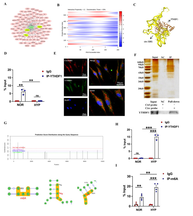
To explore the mechanism of circSSR1 regulate PASMC pyroptosis, we predicted the potential proteins by catRAPID and finally choose YTHDF1 protein as a target gene which was related with tranlation (Fig. 5A and B). The website was used to intuitive representative confirm the binding interaction between circSSR1 with YTHDF1 (Fig. 5C). RNA pull-down assay and RIP-qPCR assay were also performed to validate circSSR1 bound to YTHDF1 in PASMCs, western blot and qPCR analysis manifest the relationship between circSSR1 and YTHDF1 (Fig. 5D and F). Besides, fluorescence assay was visual observed that circSSR1 and YTHDF1 was colocalization in the cytoplasm of PASMCs (Fig. 5E). Previous studies had indicated YTHDF1 can facilitate the target gene translation. And simultaneously, YTHDF1 is one of a reader of m6A, we attempted to explore how m6A affects the SSR1 transcript under hypoxia conditions. First, we used website to be predicted whether m6A site on SSR1 mRNA (Fig. 5G). Then we used RIP assay to prove exist m6A site of SSR1 mRNA and YTHDF1 could recognize m6A of SSR1 mRNA to promote SSR1 translation. Above results indicated that YTHDF1 played an important role in promoting SSR1 translation (Fig. 5H and I). In addition to this, we detected the mRNA and protein level of YTHDF1 contract to over-expression of circSSR1 and found that it had no influence on mRNA expression (Figure S3A-S3B). Subsequently, we suppose circSSR1 could promote YTHDF1 degrade, to verify this hypothesis, co-ip assay was used to indicate YTHDF1 combine with ubiquitination protein to accelerate YTHDF1 degradation (Figure S3C). Likewise, the first one was detected the over-expression efficiency of YTHDF1 (Figure S4A). The results showed over-expression circSSR1 alone can restrain cell positive rate and LDH release increased by hypoxia; nevertheless, over-expression of YTHDF1 in the same condition can reverse this phenomenon (Figure S4B-S4C). We also used immunofluorescence to prove that over-expression of circSSR1 and YTHDF1 can increased over-expression of GRP78 and IL-1β under hypoxia (Figure S4D-S4E).
Fig. 5.
CircSSR1 regulate expression of SSR1 by bonding to YTHDF1. A, catRAPID website was used to predict YTHDF1 bound to circSSR1 and cytoscape software drawing interaction network diagram. B, The heatmap demonstrated that circSSR1 can interact with YTHDF1 by catRAPID. C, HNADOCK Server predicted and visualized the circSSR1 and YTHDF1. D, RNA immunoprecipitation (RIP) experiments showed that YTHDF1 can combine with circSSR1 rather than SSR1 mRNA (n = 3). E, CircSSR1 and YTHDF1 can co-localization in the cytoplasm of PASMCs. F, Fast silver stain kit and RNA pull-down kit were used to indicated circSSR1 bound to YTHDF1. G, SRAMP website predicted there was three m6A site on SSR1 mRNA. H-I, RIP experiment showed m6A is located on SSR1 mRNA and YTHDF1 can recognize m6A site of SSR1 to promote SSR1 translation (n = 3–4). Scale bars, 50 μm. All values are presented as the mean ± SEM. **p<0.01, ***p<0.001. NOR, normoxia and HYP, hypoxia. NC: negative control
G3BP1 regulate the expression of circSSR1 under hypoxia
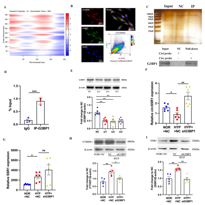
Recent studies have suggested the degradation of circRNAs rely on G3BP1 and UPF1 recognize the stem-loop structure of RNA. However, G3BP1 plays a significant role in degradation. We used website to predict interaction between circRNAs and G3BP1 (Fig. 6A). Fluorescence co-localization experiment, RNA pull-down and RIP assay were demonstrated that circSSR1 combine to G3BP1 in the cytoplasm (Fig. 6B–D). We transfected G3BP1 siRNA into PASMCs to silence G3BP1, three siRNA had the most obvious effects (Fig. 6E). To clarify the effect of G3BP1 on circSSR1 expression under hypoxia, PASMCs were transfected with G3BP1 siRNA, qRT-PCR results showed transfect G3BP1 siRNA could up-regulated circSSR1 expression (Fig. 6F). However, silence G3BP1 in PASMCs under hypoxia have no significance on SSR1 mRNA (Fig. 6G). In addition, we found YTHDF1 and SSR1 expression were evaluated using western blot (Fig. 6H and I). Based on the above findings, we have conclusively demonstrated that G3BP1 is responsible for the down regulation of circSSR1 under hypoxic conditions. This suggests a crucial role of G3BP1 in modulating the expression of circSSR1 under hypoxia.
Fig. 6.
G3BP1 can degraded circSSR1 during hypoxia. A, catRAPID predicted heatmap between circSSR1 and G3BP1. B, Immunofluorescence showed G3BP1 can co-localization with circSSR1. C, Fast silver stain kit and RNA pull-down kit were used to verify circSSR1-protein complex pulled down by the circSSR1 probe. D, G3BP1 can combine with circSSR1 proved by RIP experiment (n = 4). E, The small interference RNA (siRNA) efficiency of G3BP1 confirmed by western blot (n = 6). F, qRT-PCR detected the circSSR1 expression interfere with G3BP1 (n = 6). G, qRT-PCR showed G3BP1 not effect on linear SSR1 (n = 6). H-I, Western blot showed that the expression of YTHDF1 and SSR1 protein decreased after interfering with G3BP1 (n = 5). Scale bars, 50 μm. All values are presented as the mean ± SEM. *p<0.05, **p<0.01, ***p<0.001. NOR, normoxia and HYP, hypoxia. NC: negative control
Over-expression of circSSR1 reversed hypoxia-induced PH in vivo
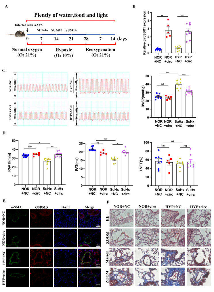
Although circSSR1 have no homology with mice and rat, we structure AAV5 virus carry humanized circSSR1 inject into mice (Fig. 7A). We use DNAMAN software to contrast the sequence of YTHDF1 between human and mice, it has higher homology (Figure S5A). RNA pull-down assay has proved that homolyzed circSSR1 can combine with YTHDF1 in mPASMCs (Figure S5B). Because of AAV5 could target lung tissue, qRT-PCR was used to detected expression of circSSR1 in mice lung tissue (Fig. 7B). We characterized the mice in detail, including right ventricular systolic pressure (RVSP), RV/left ventricular (LV) + Septum weight ratio, hemodynamics, cardiac function, and vascular remodeling (Fig. 7C and S5D). Echocardiographic analysis showed that the pulmonary artery acceleration time (PAAT) and pulmonary arterial velocity time integral (PAVTI) were significantly decreased, while the effect was prevented by circSSR1 over-expression. Meanwhile, our results show the left ventricular ejection fraction (LVEF) hardly any change (Fig. 7D). Immunofluorescent staining of GSDMD and YTHDF1 mouse lung tissue section indicate that the fluorescence activity in circSSR1 overexpressing mouse lung tissues was decreased compared with that in NC-treated mouse lung tissues after hypoxia exposure (Fig. 7E and S5C). Moreover, we found that circSSR1 over-expression reversed hypoxia-induced pulmonary vascular remodeling by HE is staining and Masson (Fig. 7F). These results confirmed that the conserved circSSR1 could regulate the pathology of PH through pyroptosis in vivo.
Fig. 7.
Over-expression of circSSR1 in vivo inhibit the symptoms of pulmonary hypertension. A, Construction of hypoxia model mouse. B, Specificity over-expression circSSR1 in the mouse lung tissue (n = 5–8). C, Right ventricular (RV) systolic pressure (RVSP), RV/left ventricular (LV) + S weight ratio and heart rate in the HYP-PH mice models. D, Pulmonary arterial velocity time integral (PAVTI) and Pulmonary artery acceleration time (PAT), left ventricular ejection fraction (LVEF) in the NOR + AAV5–negative control (NC), NOR + AAV5, HYP + AAV5–negative control (NC), and HYP + AAV5 groups are shown (n = 5–10). E, Immunofluorescence of GSDMD in mouse lung tissue section. Scale bars, 50 μm. F, HE and Masson staining showed circSSR1 over-expression prevented and reversed wall thickening. Scale bars, 100 μm. All values are presented as the mean ± SEM. *p<0.05, **p<0.01, ***p<0.001. NOR, normoxia and HYP, hypoxia. NC: negative control
Discussion
In this study, we provide compelling evidence that the decrease of circSSR1 in PASMCs, a novel circRNA, participate in progression of PASMCs pyroptosis in PH. Our research proves the following new concepts in vitro and in vivo. The first one is hypoxia induced PASMCs pyroptosis through circSSR1, a novel circRNA. Second, over-expression of circSSR1 reverse PASMC cells pyroptosis caused by hypoxia via ERS through combine with YTHDF1 to inhibit parental gene SSR1 translation. Third, G3BP1 as upstream to regulate the expression of circSSR1 under hypoxia. Our results provide potential target for nucleic acid therapy.
Pyroptosis is a form of programmed cell death, involved in several disease, such as gastric cancer, obstructive nephropathy and breast cancer. CircRNA plays a key role in cell pyroptosis [29]. It has been reported that circPUM1 serve as a scaffold for the interaction between UQCRC1 and UQCRC2 to regulate pyroptosis in esophageal squamous cell carcinoma [30]. CircPIBF1 could promoted the occurrence of lung adenocarcinoma (LUAD) though binding to nuclear factor erythroid 2-related factor 2 (Nrf2) [31]. Beyond that circ-calm4 acts as a sponge of miR-124-3p to promote pulmonary artery smooth muscle cells pyroptosis through PDCD6 in hypoxia [16]. In our experiments, we provide a solid evident that circSSR1 regulate pyroptosis through inhibit parental gene SSR1 translation. Our study proposed a new regulatory mechanism by which circRNA regulates pyroptosis, and a novel target molecule for the development of therapeutic approaches in pulmonary hypertension.
Endoplasmic reticulum stress (ERS) mainly includes the unfolded proteins in lumen and the accumulation of misfolded [32]. In the physiological state, the membrane-related proteins such as PERK, IRE1, and ATF6 partial bind to immunoglobulin heavy chain binding protein (BIP) [33]. These proteins are not active currently. When stress occurs, a large number of unfold proteins accumulate in the endoplasmic reticulum lumen, and BIP are released aim to combine with unfold protein, resulting in the activation of IRE1, PERK and ATF6, and the activation of stress pathways [34]. Although a few studies have reported data on the role of ER stress in PASMCs, the specific molecular regulatory mechanism is not completely understood. In this article, we have proved that SSR1 protein increased by over-expression of circSSR1 in hypoxia. The accumulate of SSR1 protein, which was located on ER membrane, activated ER stress. And even more important, we find that over-expression of circSSR1 in hypoxia can restrain mitochondrial reactive oxygen species (ROS) release, and the endoplasmic reticulum stress inhibitor affect the generation of ROS. In a word, circSSR1 down-expression induced by hypoxia regulate ER stress via host gene protein expression and finally facilitate the happen of pyroptosis.
Ras GTPase-activating protein-binding protein 1 (G3BP1), as upstream of circSSR1, regulate signal transduction stimulated by Ras protein [35]. A few research have indicated that circNUP50 could as a sponge of miR-197-3p and upregulate the expression of G3BP1 to mediate p53 ubiquitination resulting to OC platinum resistance [36]. Besides, it has been raised G3BP1 and UPF1 could degrade structure-mediated of circRNAs [37–39]. Some research show that G3BP1 is crucial to formulate stress granules, as a ribonucleoprotein (RNP) granule respond to various stress and produce aggregation in eukaryotic cells, which is participate in liquid-liquid phase separation (LLPS) [40–42]. At present, there is no report about G3BP1 as the circRNA upstream to promote the circRNA degrade in pulmonary hypertension now. Our study first propose G3BP1 could combine with circSSR1 and promote circSSR1 down-regulate in hypoxia. However, the specific mechanism of circSSR1 degradation by G3BP1 whether mediated by LLPS is unclear.
According to analysis the sequence of circSSR1 between human and mice, we find that the circSSR1 unlike other ncRNAs demonstrate highly homology. Just like Wang et al. research that homo-LncRNA lnc-TSI over-expression in mouses to inhibit renal fibrogenesis through negative regulation of the TGF-β/Smad pathway [13]. Therefore, we construct virus of AAV5 in vitro to over-expression humanized circSSR1 in mice through by the way of nose drop. Our study has proved that human circSSR1 can specific over-expression in lung tissue. This is a novel method to construct model of animal of PH. It is doubt reveal the circSSR1 as a key point in pulmonary hypertension.
In summary, our experiments have verified circSSR1 play a crucial role in regulation of pyroptosis in hypoxia PASMCs for the first put forward. As a circSSR1 binding protein, the m6A reader, YTHDF1 recognize m6A of SSR1 mRNA to promote translation to active ER stress. This is a original discovery to reveal mechanism of circSSR1 regulate pyroptosis through regulate the expression of host protein SSR1. Along with G3BP1 as upstream of circSSR1 promote degradation under hypoxia. And it may provide a novel in diagnosis and clinical treatment in pulmonary hypertension.
Materials and methods
Animals
To exclude the possible effects of estrogen on the chronic hypoxic and SuHx induced PH, adult C57BL/6 male mice weighing approximate 25 g were provided by Experimental Animal Center of Harbin Medical University. All experimental procedures were performed in accordance with the ethical standards in the 1964 Declaration of Helsinki and its later amendments and approved by the Ethics Committees of Harbin Medical University. As previously described by SuHx protocol, in brief, the mice were injected once a week with SU5416 at 20 mg/kg body weight per dose exposed in 10% oxygen for 3 weeks and re-exposure to normoxic 2 weeks. Construction of serotype 5 adenovirus-associated virus (AAV5) and the hypoxic animal model. To confirm the functional of circSSR1 in PH, the corresponding target RNA cloning construction and serotype 5 adenovirus-associated virus (AAV5) was packaged by Genechem (Shanghai, China). Mice were randomly divided into four groups and infected with the AAV5 vector at 1011 genome equivalents in 20 to 30 µL Hank balanced salt solution after isoflurane anesthesia, followed by nasal drops. The groups were as follow: normoxic environment plus normal control vector group (NOR + NC, n = 10), normoxic environment plus AAV5 group (NOR + AAV5, n = 10), hypoxic environment plus normal control vector group (HYP + NC, n = 15), hypoxic environment plus AAV5 group (HYP + AAV5, n = 15). The mice of HYP + NC and HYP + AAV5 group were exposed in hypoxic chambers (HUAYUE P110; Guangzhou, China, 10% O2) for 21 days. The control group mice were kept in the normoxic environment for 5 weeks. Lung tissues were taken for follow-up experiments after anesthesia, and the right ventricular (RV) hypertrophy index (ratio of RV free wall weight over the sum of septum plus left ventricular (LV) free wall weight), calculating RV/ (LV + S). The study protocol was approved by the Ethics Committees of Harbin Medical University (HMUDQ 20231120001).
Statistical analysis
Statistical analysis was performed with GraphPad Prism 8 software. Data were checked for normal distribution and equal variance (F test) before statistical. Students t test (unpaired) was used for 2-group analysis with equal variance, and Welch correction test was used for 2-group analysis with unequal variance. One-way ANOVA with Tukey post hoc test was used to compare multiple groups with equal variance, and Brown Forsythe and Welch ANOVA with Tamhane T2 post hoc test was used to compare multiple groups with unequal variance. Nonparametric analyses, including the Mann-Whitney U test for 2 groups or Kruskal-Wall is test followed by Dunn posttest for > 2 groups, were performed for nonnormally distributed data. Data are presented as the means ± SEM, and P < 0.05 was considered statistically significant.
Hematoxylin and eosin (HE) staining and Masson
Lung tissues of mice were immersed in 4% paraformaldehyde and then embedding in paraffin wax, cutting into 5-µm-thick sections and stained with hematoxylin and eosin and Masson. In situ hybridization was performed with kits following the manufacturer’s instructions (Boster, Wuhan, China).
Cell culture
PAECs and PASMCs in the experiment provided by Procell Life Science & Technology (Wuhan, China) were preserved in smooth muscle cell medium, which consists of ten milliliters fetal bovine serum (FBS), 5 mL smooth muscle cell growth supplement, and 5 mL penicillin/streptomycin solution. The system in the state of 37℃, 5% CO2, and 100% relative humidity. PAECs were incubated in endothelial cell medium (ScienCell) containing 25 mL FBS, 5 mL endothelial cell growth factor, and 5 mL penicillin/streptomycin solution at 37 °C in a 5% CO2 humidified incubator. The hypoxia cells were cultured in a Tri-Gas Incubator (Thermo Fisher, MA) for 24 h. The water saturated atmosphere included 3% O2, 5% CO2 and 91% N2 for 24 h.
Cell transfection
PASMCs were hungry culture one night in the incubator before transfection. The over-expression of circSSR1 plasmid, SSR1 plasmid and YTHDF1 plasmid were provided by GeneChem (Shanghai, China) and vector was used to as negative control (NC). The transfection reagent was Lipofectamine 2000 Reagent (11668019; Thermo Fisher, MA). Small interfere RNA for circSSR1, SSR1, YTHDF1 and G3BP1 were provided by GenePharma (Suzhou, China). The transfection reagent was X-tremeGene. Six hours after transfection, the cells were cultured in 5% culture medium and exposed to hypoxic conditions or normoxic conditions for 24 h.
Immunofluorescence staining
The cells were treated with hypoxic or transfection and washed with 1xPBS for 2–3 times, added the stationary buffer (Beyotime Biotechnology) for 10 min at room temperature. Removed the stationary buffer and washed with 1x PBS for 2–3 times, then added the transparent buffer (Beyotime Biotechnology) for 10 min at room temperature. Removed the transparent buffer and washed with 1xPBS for 2–3 times, then added confining buffer (Beyotime Biotechnology) for 10 min at room temperature. Added the prepared the antibody which was diluted by 1xPBS overnight at 4 ℃. The samples were washed with 1xPBS for 2–3 times and added fluorescent second antibody for 2 h at 37 ℃. All samples were stained DAPI for 10–15 min and washed with 1xPBS. Ultimately, took pictures by fluorescence microscope or laser confocal microscope.
PI staining/Hoechst 33,342
According to the illustrated of manual, the cells were stained with 6 µL Hoechst 33,342 solution and 6 µL PI at 4 °C avoiding the light for 20 min by using Apoptosis and Necrosis Assay Kit (Beyotime Biotechnology) and pyroptosis was observation with a fluorescence microscope or laser confocal microscope (AF6000; Leica, Germany).
Endoplasmic reticulum (ER)-tracker red staining
ER-Tracker Red, an endoplasmic reticulum-specific red fluorescent probe that can be used to stain the ER in living cells, was purchased from Beyotime Biotechnology (C1041). In brief, the cells were cleaning with 1xPBS, then prepared the ER-Tracker Red working solution. One microliter of ER-Tracker Red was added to 1 ml of dilute ER-Tracker Red and needed incubation at 37 °C before use. Removed the washing solution, and added the working solution which was incubated to the cells in the 371 ℃ for 15–30 min. Removed the ER-Tracker Red working solution and washing the cells by 1–2 times with cell culture. Ultimately, the samples were detected by fluorescence microscope or laser confocal microscope.
Western blot and antibodies
PASMCs were washed three times with cold 1xPBS and extracted the protein using lysis buffer (Beyotime Biotechnology), and the RIPA buffer was centrifugal at 4℃ for 20 min. The 5xloading buffer was added into the supernatant and denatured at 100℃ for 5 min. The protein samples were separated by SDS-PAGE. Antibodies were used as follows: anti-YTHDF1 (Proteintech, 1:500 dilution), anti-SSR1 (Boster, 1:500 dilution), anti-GRP78 (Boster, 1:500 dilution), anti-IRE1 (Boster, 1:500 dilution), anti-ATF6 (Boster, 1:500 dilution), anti-Caspase-1 (Abcam, 1:1000 dilution), anti-GSDMD-N (ABclonal, 1:500 dilution), anti-NLRP3 (Boster, 1:500 dilution), anti-actin (Boster, 1:4000 dilution), anti-m6A (Epigentek Group Inc, Farmingdale, NY, 1:1000 dilution).
LDH release assay
LDH release, an important indicator of cell membranes integrity, can be used to detect cytotoxicity and the LDH Cytotoxicity Assay Kit was purchase from Beyotime Biotechnology (C0017). In briefly, the cells were treated with hypoxia and the culture were centrifuge at 400 g for 5 min. Take supernatant added prepared LDH working solution and avoided the light incubation for 30 min at 25℃. Ultimately, the absorbance was detected by microplate at a wavelength of 490 nm.
Reactive oxygen species detection
Purchase reactive oxygen species (ROS) assay from Beyotime, no serum culture media containing 10 µM 2ʹ,7ʹ-dichlorodihydroflfluorescein diacetate probes (S0033S, Beyotime, Shanghai, China) was treated for cells in a 37 °C incubator for 20 min. Green fluo- rescence was captured (488 nm excitation/525 nm emission) from ≥ 3 optical fields.
RNA isolation and qRT-PCR
The total RNA was extracted from pulmonary arterial smooth muscle cells or tissues using Trizol (Beyotime). The trichloromethane was added, the lysis buffer was vortexed and centrifuged. The supernatant was transferred to a new centrifugal tube and added isopropyl alcohol stewing for a while. The samples were centrifuged and washed with prepared 75% ethanol. The sediment was air-dried and dissolved with no enzyme water. The total RNA was reversed transcription using an S1000 Thermal Cycler with random hexamer primers (Thermofisher, USA) or oligo (dt) and random primers (Haigene, China). A Roche Light Cycler 480II instrument was used for real-time quantitative fluorescence analysis with a two-step method. PCR was performed with 40 cycles at an annealing temperature of 53 °C. The RNA expression levels were normalized to β-actin.
RNA immunoprecipitation (RIP) assay
RNA immunoprecipitation Kit was used to research the relationship between protein and RNA and purchased by BersionBio (Guang zhou, China). In brief, the cells were collected in the room temperature, used the lysis buffer into the cytosol and added relevant antibody to combine with antigen overnight at 4 ℃. Extracted the RNA from immunoprecipitation and reverse transcription into cDNA and subjected to qRT-PCR assays.
RNA pull-down assay
RNA pulldown, a technology of researching the relationship between RNA and protein, was purchase from BersinBio (Guang zhou, China). In brief, the cells were collected and washed with PBS. The reagent was added into the cell pellet, vortexed blending and putted in -80 for 10 min. The lysate was centrifuged for 15 min at 4 ℃ after melted and transfer the supernatant to a new centrifuge tube. Then added reagents incubated for 1 h at 25 ℃ and rotated softy at 4℃ according to the specification to avoid nonspecific adsorption. The cell extract solution was added prepared probe-magnetic bead complex and added reagent incubated at 37℃ for 2 h. The sample was washed by solution and eluted at 37℃ for 2 h. Proteins were detected by western blot.
RNase R treatment
Total RNA (5 µg) was incubated with or without 3 U/µg of RNase R (Epicenter Biotechnologies, Madison, WI, USA) for 15 min at 37 °C according to the manufacturer’s protocol and used for subsequent experiments.
RNA-FISH assay
RNA Fluorescence in Situ Hybridization, an important technology of non-radioactive hybridization, was used to detect RNA location. The RNA FISH Kit and probe was purchased from GenePharma (Suzhou, China). In brief, the cells were inoculated in culture plate and incubated overnight at 37 ℃. Removed the culture and washed with 1x PBS before added 4% paraformaldehyde for 15 min at room temperature. Removed the paraformaldehyde, added 0.1% Buffer A (RNA FISH Kit) and washed with PBS before added with 2xBuffer C (RNA FISH Kit) for 30 min at 37℃. The Buffer E (RNA FISH Kit) and probe were treated with according to the description. The sample was avoided the light incubation with probe mixture at 37℃ overnight. The 0.1%Buffer F (RNA FISH Kit) and 2xBuffer C (RNA FISH Kit) were used to wash the sample in turn. Finally, the diluent DAPI working solution was incubated with sample and took picture with fluorescence microscope or laser confocal microscope before washed by PBS.
Electronic supplementary material
Below is the link to the electronic supplementary material.
Author contributions
XY G, HX D, XY W, and DL Z designed and supervised this study; XY G, XR Z, SY H, JN B, SS W and HY L performed the cell experiments; XR Z, H Y and KY W performed the animal experiments; XY G, XY W, LX Z and C M performed the statistical analysis; XY G, HX D, H Y and DL Z wrote the manuscript; All authors contributed to manuscript revision, and read and approved the submitted version.
Funding
This study was supported by National Natural Science Foundation of China [31820103007, 31971057 and 31771276 to DZ, 82170059 to CM, 32271171 to YH]; Heilongjiang Touyan Innovation Team Program; the Key Project of Natural Science Foundation of Heilongjiang Province [ZD2023H003 to CM; ZD2021H002 to YH]; the Fundamental Research Funds for the Provincial Universities [JFMSPY202101 to YH].
Data availability
Data that support the findings of this study have been deposited in the Harbin Medical University Ethical Review Report code HMUDQ20231120001.
Declarations
Ethical approval
Ethics approval and consent to participate.
Competing interests
The authors declare no competing interests.
Footnotes
Publisher’s note
Springer Nature remains neutral with regard to jurisdictional claims in published maps and institutional affiliations.
Xiaoyu Guan and Hongxia Du contributed equally to this work.
References
- 1.Wilkins MR. Pulmonary hypertension: the science behind the disease spectrum. Eur Respir Rev. 2012;21(123):19–26. 10.1183/09059180.00008411. [DOI] [PMC free article] [PubMed] [Google Scholar]
- 2.Montani D, Chaumais MC, Guignabert C, et al. Targeted therapies in pulmonary arterial hypertension. Pharmacol Ther. 2014;141(2):172–91. 10.1016/j.pharmthera.2013.10.002. [DOI] [PubMed] [Google Scholar]
- 3.McGlothlin D. Classification of pulmonary hypertension. Heart Fail Clin. 2012;8(3):301–17. 10.1016/j.hfc.2012.04.013. [DOI] [PubMed] [Google Scholar]
- 4.Leopold JA, Maron BA. Molecular mechanisms of Pulmonary Vascular Remodeling in Pulmonary arterial hypertension. Int J Mol Sci. 2016;17(5). 10.3390/ijms17050761. [DOI] [PMC free article] [PubMed]
- 5.Ren H, Dai R, Nik Nabil WN, Xi Z, Wang F, Xu H. Unveiling the dual role of autophagy in vascular remodelling and its related diseases. Biomed Pharmacother. 2023;168:115643. 10.1016/j.biopha.2023.115643. [DOI] [PubMed] [Google Scholar]
- 6.Mu M, Huang CX, Qu C, et al. Targeting Ferroptosis-Elicited Inflammation Suppresses Hepatocellular Carcinoma Metastasis and enhances Sorafenib Efficacy. Cancer Res. 2024;84(6):841–54. 10.1158/0008-5472.CAN-23-1796. [DOI] [PubMed] [Google Scholar]
- 7.Qu H, Khalil RA. Vascular mechanisms and molecular targets in hypertensive pregnancy and preeclampsia. Am J Physiol Heart Circ Physiol. 2020;319(3):H661–81. 10.1152/ajpheart.00202.2020. [DOI] [PMC free article] [PubMed] [Google Scholar]
- 8.Zhang Z, Tang J, Song J, et al. Elabela alleviates ferroptosis, myocardial remodeling, fibrosis and heart dysfunction in hypertensive mice by modulating the IL-6/STAT3/GPX4 signaling. Free Radic Biol Med. 2022;181:130–42. 10.1016/j.freeradbiomed.2022.01.020. [DOI] [PubMed] [Google Scholar]
- 9.Ji N, Qi Z, Wang Y, et al. Pyroptosis: a new regulating mechanism in Cardiovascular Disease. J Inflamm Res. 2021;14:2647–66. 10.2147/JIR.S308177. [DOI] [PMC free article] [PubMed] [Google Scholar]
- 10.Hu Y, Wang B, Li S, Yang S. Pyroptosis, and its role in Central Nervous System Disease. J Mol Biol. 2022;434(4):167379. 10.1016/j.jmb.2021.167379. [DOI] [PubMed] [Google Scholar]
- 11.Li Y, Yuan Y, Huang ZX, et al. GSDME-mediated pyroptosis promotes inflammation and fibrosis in obstructive nephropathy. Cell Death Differ. 2021;28(8):2333–50. 10.1038/s41418-021-00755-6. [DOI] [PMC free article] [PubMed] [Google Scholar]
- 12.Rao Z, Zhu Y, Yang P, et al. Pyroptosis in inflammatory diseases and cancer. Theranostics. 2022;12(9):4310–29. 10.7150/thno.71086. [DOI] [PMC free article] [PubMed] [Google Scholar]
- 13.Wang P, Luo ML, Song E, et al. Long noncoding RNA lnc-TSI inhibits renal fibrogenesis by negatively regulating the TGF-beta/Smad3 pathway. Sci Transl Med. 2018;10(462). 10.1126/scitranslmed.aat2039. [DOI] [PubMed]
- 14.Ozcan L. Endoplasmic reticulum stress in cardiometabolic disorders. Curr Atheroscler Rep. 2012;14(5):469–75. 10.1007/s11883-012-0270-z. [DOI] [PubMed] [Google Scholar]
- 15.Wang X, Li Q, He S, et al. LncRNA FENDRR with m6A RNA methylation regulates hypoxia-induced pulmonary artery endothelial cell pyroptosis by mediating DRP1 DNA methylation. Mol Med. 2022;28(1):126. 10.1186/s10020-022-00551-z. [DOI] [PMC free article] [PubMed] [Google Scholar]
- 16.Jiang Y, Liu H, Yu H, et al. Circular RNA Calm4 regulates Hypoxia-Induced Pulmonary arterial smooth muscle cells pyroptosis via the Circ-Calm4/miR-124-3p/PDCD6 Axis. Arterioscler Thromb Vasc Biol. 2021;41(5):1675–93. 10.1161/ATVBAHA.120.315525. [DOI] [PMC free article] [PubMed] [Google Scholar]
- 17.Han D, Kim H, Kim S, et al. Sestrin2 protects against cholestatic liver injury by inhibiting endoplasmic reticulum stress and NLRP3 inflammasome-mediated pyroptosis. Exp Mol Med. 2022;54(3):239–51. 10.1038/s12276-022-00737-9. [DOI] [PMC free article] [PubMed] [Google Scholar]
- 18.Ni Y, Zhang J, Zhu W, Duan Y, Bai H, Luan C. Echinacoside inhibited cardiomyocyte pyroptosis and improved heart function of HF rats induced by isoproterenol via suppressing NADPH/ROS/ER stress. J Cell Mol Med. 2022;26(21):5414–25. 10.1111/jcmm.17564. [DOI] [PMC free article] [PubMed] [Google Scholar]
- 19.Zhang J, Guo J, Yang N, Huang Y, Hu T, Rao C. Endoplasmic reticulum stress-mediated cell death in liver injury. Cell Death Dis. 2022;13(12):1051. 10.1038/s41419-022-05444-x. [DOI] [PMC free article] [PubMed] [Google Scholar]
- 20.Cao SS, Luo KL, Shi L. Endoplasmic reticulum stress interacts with inflammation in Human diseases. J Cell Physiol. 2016;231(2):288–94. 10.1002/jcp.25098. [DOI] [PMC free article] [PubMed] [Google Scholar]
- 21.Guimaraes ES, Gomes MTR, Sanches RCO, Matteucci KC, Marinho FV, Oliveira SC. The endoplasmic reticulum stress sensor IRE1alpha modulates macrophage metabolic function during Brucella abortus infection. Front Immunol. 2022;13:1063221. 10.3389/fimmu.2022.1063221. [DOI] [PMC free article] [PubMed] [Google Scholar]
- 22.Xin W, Zhang M, Yu Y, et al. Correction: BCAT1 binds the RNA-binding protein ZNF423 to activate autophagy via the IRE1-XBP-1-RIDD axis in hypoxic PASMCs. Cell Death Dis. 2021;13(1):27. 10.1038/s41419-021-04466-1. [DOI] [PMC free article] [PubMed] [Google Scholar]
- 23.Li Q, Zhang K, Hou L, et al. Endoplasmic reticulum stress contributes to pyroptosis through NF-kappaB/NLRP3 pathway in diabetic nephropathy. Life Sci. 2023;322:121656. 10.1016/j.lfs.2023.121656. [DOI] [PubMed] [Google Scholar]
- 24.Chen L, Wang C, Sun H, et al. The bioinformatics toolbox for circRNA discovery and analysis. Brief Bioinform. 2021;22(2):1706–28. 10.1093/bib/bbaa001. [DOI] [PMC free article] [PubMed] [Google Scholar]
- 25.Zhang W, He Y, Zhang Y. CircRNA in ocular neovascular diseases: fundamental mechanism and clinical potential. Pharmacol Res. 2023;197:106946. 10.1016/j.phrs.2023.106946. [DOI] [PubMed] [Google Scholar]
- 26.Yang D, Zhao D, Ji J, et al. CircRNA_0075723 protects against pneumonia-induced sepsis through inhibiting macrophage pyroptosis by sponging mir-155-5p and regulating SHIP1 expression. Front Immunol. 2023;14:1095457. 10.3389/fimmu.2023.1095457. [DOI] [PMC free article] [PubMed] [Google Scholar]
- 27.Xu S, Ge Y, Wang X, et al. Circ-USP9X interacts with EIF4A3 to promote endothelial cell pyroptosis by regulating GSDMD stability in atherosclerosis. Clin Exp Hypertens. 2023;45(1):2186319. 10.1080/10641963.2023.2186319. [DOI] [PubMed] [Google Scholar]
- 28.Liu H, Jiang Y, Shi R, et al. Super enhancer-associated circRNA-circLrch3 regulates hypoxia-induced pulmonary arterial smooth muscle cells pyroptosis by formation of R-loop with host gene. Int J Biol Macromol. 2024;130853. 10.1016/j.ijbiomac.2024.130853. [DOI] [PubMed]
- 29.Gao L, Jiang Z, Han Y, Li Y, Yang X. Regulation of pyroptosis by ncRNA: a Novel Research Direction. Front Cell Dev Biol. 2022;10:840576. 10.3389/fcell.2022.840576. [DOI] [PMC free article] [PubMed] [Google Scholar]
- 30.Gong W, Xu J, Wang Y, et al. Nuclear genome-derived circular RNA circPUM1 localizes in mitochondria and regulates oxidative phosphorylation in esophageal squamous cell carcinoma. Signal Transduct Target Ther. 2022;7(1):40. 10.1038/s41392-021-00865-0. [DOI] [PMC free article] [PubMed] [Google Scholar]
- 31.Zhang Z, Chen Q, Huang C, et al. Transcription factor Nrf2 binds to circRNAPIBF1 to regulate SOD2 in lung adenocarcinoma progression. Mol Carcinog. 2022;61(12):1161–76. 10.1002/mc.23468. [DOI] [PubMed] [Google Scholar]
- 32.Marciniak SJ, Chambers JE, Ron D. Pharmacological targeting of endoplasmic reticulum stress in disease. Nat Rev Drug Discov. 2022;21(2):115–40. 10.1038/s41573-021-00320-3. [DOI] [PubMed] [Google Scholar]
- 33.Oakes SA, Papa FR. The role of endoplasmic reticulum stress in human pathology. Annu Rev Pathol. 2015;10:173–94. 10.1146/annurev-pathol-012513-104649. [DOI] [PMC free article] [PubMed] [Google Scholar]
- 34.Kim G, Lee J, Ha J, Kang I, Choe W. Endoplasmic reticulum stress and its impact on adipogenesis: molecular mechanisms implicated. Nutrients. 2023;15(24). 10.3390/nu15245082. [DOI] [PMC free article] [PubMed]
- 35.Oi N, Yuan J, Malakhova M, et al. Resveratrol induces apoptosis by directly targeting Ras-GTPase-activating protein SH3 domain-binding protein 1. Oncogene. 2015;34(20):2660–71. 10.1038/onc.2014.194. [DOI] [PMC free article] [PubMed] [Google Scholar]
- 36.Zhu Y, Liang L, Zhao Y, et al. CircNUP50 is a novel therapeutic target that promotes cisplatin resistance in ovarian cancer by modulating p53 ubiquitination. J Nanobiotechnol. 2024;22(1):35. 10.1186/s12951-024-02295-w. [DOI] [PMC free article] [PubMed] [Google Scholar]
- 37.Fischer JW, Busa VF, Shao Y, Leung AKL, Structure-Mediated RNA. Decay by UPF1 and G3BP1. Mol Cell. 2020;78(1):70–e846. 10.1016/j.molcel.2020.01.021. [DOI] [PMC free article] [PubMed] [Google Scholar]
- 38.Liu S, Chen L, Chen H, Xu K, Peng X, Zhang M. Circ_0119872 promotes uveal melanoma development by regulating the miR-622/G3BP1 axis and downstream signalling pathways. J Exp Clin Cancer Res. 2021;40(1):66. 10.1186/s13046-021-01833-w. [DOI] [PMC free article] [PubMed] [Google Scholar]
- 39.Guo Y, Wei X, Peng Y. Structure-mediated degradation of CircRNAs. Trends Cell Biol. 2020;30(7):501–3. 10.1016/j.tcb.2020.04.001. [DOI] [PubMed] [Google Scholar]
- 40.Yang C, Wang Z, Kang Y, et al. Stress granule homeostasis is modulated by TRIM21-mediated ubiquitination of G3BP1 and autophagy-dependent elimination of stress granules. Autophagy. 2023;19(7):1934–51. 10.1080/15548627.2022.2164427. [DOI] [PMC free article] [PubMed] [Google Scholar]
- 41.Cai S, Zhang C, Zhuang Z, et al. Phase-separated nucleocapsid protein of SARS-CoV-2 suppresses cGAS-DNA recognition by disrupting cGAS-G3BP1 complex. Signal Transduct Target Ther. 2023;8(1):170. 10.1038/s41392-023-01420-9. [DOI] [PMC free article] [PubMed] [Google Scholar]
- 42.Ali N, Prasad K, AlAsmari AF, Alharbi M, Rashid S, Kumar V. Genomics-guided targeting of stress granule proteins G3BP1/2 to inhibit SARS-CoV-2 propagation. Int J Biol Macromol. 2021;190:636–48. 10.1016/j.ijbiomac.2021.09.018. [DOI] [PMC free article] [PubMed] [Google Scholar]
Associated Data
This section collects any data citations, data availability statements, or supplementary materials included in this article.
Supplementary Materials
Data Availability Statement
Data that support the findings of this study have been deposited in the Harbin Medical University Ethical Review Report code HMUDQ20231120001.


