Abstract
In order to identify the viral gene related to the pathogenicity of rabies virus, we tried to establish a reverse genetics system of the attenuated RC-HL strain, which causes nonlethal infection in adult mice after intracerebral inoculation. A full-length genome plasmid encoding the complete antigenomic cDNA of the RC-HL strain and helper plasmids containing cDNAs of the complete open reading frame of the N, P, and L genes, respectively, were constructed. After transfection of these plasmids into BHK-21 cells infected with the T7 RNA polymerase-expressing vaccinia virus, infectious rabies virus with almost the same biological properties as those of the wild-type RC-HL strain was rescued. Using this reverse genetics system of the RC-HL strain, we generated a chimeric virus with the open reading frame of the glycoprotein gene from the parent Nishigahara strain, which kills adult mice after intracerebral inoculation, in the background of the RC-HL genome. Since the chimeric virus killed adult mice following intracerebral inoculation, it became evident that the open reading frame of the glycoprotein gene is related to the pathogenicity of the Nishigahara strain for adult mice.
Rabies virus, which is a member of the genus Lyssavirus of the family Rhabdoviridae, causes a severe neurological disease and death in almost all kinds of mammals, including humans. The genome is an unsegmented negative-sense RNA of about 12 kb, encoding five structural proteins: nucleoprotein (N), phosphoprotein (P), matrix protein (M), glycoprotein (G), and large protein (L). The N, P, and L proteins form ribonucleoprotein together with the viral genomic or antigenomic RNA. The N protein is responsible for encapsidation of these viral RNAs, while the L protein, in cooperation with the P protein, functions as an RNA-dependent RNA polymerase. In contrast, the G and M proteins are located in the viral envelope. The G protein, in particular, participates in binding to receptors on host cells and induction of neutralizing antibody (31).
Furthermore, it has been reported that the G protein plays an important role in viral pathogenicity. Some previous studies (4, 25, 29) showed that an amino acid at position 333 on the G protein is a determinant of the virulence of fixed virus for adult mice. Strains that have arginine or lysine at position 333 on the G protein kill adult mice following intracerebral (i.c.) inoculation, whereas mutants with other amino acids at this site cause a nonlethal infection. This phenomenon can apply to all strains of representative fixed viruses, such as the CVS, ERA, PV, SAD B19, and HEP-Flury strains (1, 3, 14, 20, 27). However, we previously showed that the RC-HL strain, a fixed virus used for the production of animal vaccine in Japan, possesses arginine at position 333 on the G protein, even though this strain causes a nonlethal infection in adult mice (11). This indicates that mutation in another region is related to attenuation of the RC-HL strain and suggests the existence of a novel mechanism for pathogenicity of rabies virus.
The RC-HL strain was established from the Nishigahara strain, which had been maintained by rabbit brain passages, after 294 passages in chicken embryos, 8 passages in chicken embryo fibroblast cells, 5 passages in Vero cells, and 23 passages in hamster lung cells (10). In contrast to the RC-HL strain, which causes a mild disease with symptoms such as piloerection and body weight reduction in adult mice, the Nishigahara strain kills adult mice following i.c. inoculation. Recently we compared the complete genome sequences of the RC-HL strain and the Nishigahara strain (12). We found that the homology of the G gene was lower than those of the N, P, M, and L genes at both nucleotide and amino acid levels and that the percentage of radical amino acid substitution in the G protein was highest among these proteins. These findings suggested that the structure of the G protein is the most variable, and we therefore decided to try to determine whether the G gene is related to the difference between the pathogenicity of the RC-HL strain and that of the Nishigahara strain for adult mice, in accordance with the importance of the G protein in pathogenicity in other strains reported previously (4, 25, 29). Hence, we tried to produce a chimeric virus that has an open reading frame of the G gene (G-ORF) from the Nishigahara strain in the background of the RC-HL genome and to examine whether this chimeric virus kills adult mice or not. For this purpose, manipulation of the viral genome using the reverse genetics system was necessary.
The reverse genetics system of rabies virus has been already established in the SAD B19 strain (24). Afterward, various viruses belonging to the order Mononegavirales, including vesicular stomatitis virus (15, 30), measles virus (21), human respiratory syncytial virus (2) and Sendai virus (6, 13), were recovered from cloned cDNA using almost the same principle as that used for rabies virus. However, a second rescue of rabies virus from cDNA has not been reported.
In this study, we established a reverse genetics system of the RC-HL strain and compared the biological properties of the virus rescued from cloned cDNA to those of the wild-type RC-HL strain. Furthermore, a chimeric virus having the G-ORF of the Nishigahara strain in the background of the RC-HL genome was produced by this system. Since the chimeric virus, in contrast to the attenuated RC-HL strain, killed adult mice after i.c. inoculation, it became evident that G-ORF is related to pathogenicity of the Nishigahara strain for adult mice.
MATERIALS AND METHODS
Cells and viruses.
Baby hamster kidney (BHK-21) cells were maintained in Eagle's minimal essential medium (MEM) supplemented with 10% tryptose phosphate broth (TPB) and 5% calf serum. Mouse neuroblastoma (NA) cells were grown in Eagle's MEM containing 10% fetal calf serum. Virus stocks of the RC-HL and Nishigahara strains were prepared in BHK-21 cells and in a 4-week-old mouse brain, respectively. Recombinant vaccinia virus, vTF7-3 (5) (kindly provided by B. Moss), which expresses T7 RNA polymerase, was propagated in Vero cells grown in Eagle's MEM containing 10% TPB and 10% calf serum.
Reverse transcription (RT) and PCR.
Genomic RNA from the virus stocks was extracted using Isogen (Nippon Gene, Tokyo, Japan). Subsequently, single-stranded cDNAs were synthesized by RT, using Ready-To-Go You-Prime First-Strand Beads (Amersham Pharmacia Biotech, Little Chalfont, England). PCRs were performed with TaKaRa Ex-Taq (Takara Shuzo, Shiga, Japan) using the RT product or a plasmid as template.
Cloning and sequencing of cDNA fragments amplified by RT-PCR and PCR.
The amplified cDNA fragments were ligated with a pT7Blue T-vector (Novagen, Madison, Wis.) following agarose gel purification with UltraClean 15 (MO BIO Laboratories, Solana Beach, Calif.). The ligation products were used to transform competent Escherichia coli (DH5α or JM109) cells. Sequencing was carried out using an AutoCycle Sequencing Kit and an ALF DNA Sequencer (Amersham Pharmacia Biotech).
Construction of the helper and full-length genome plasmids of the RC-HL strain.
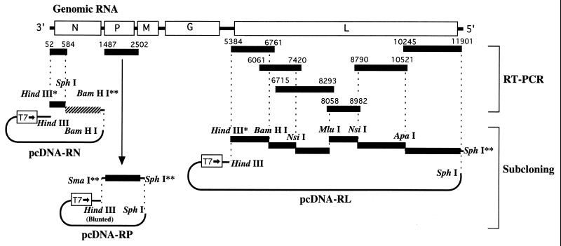
Helper plasmids carrying the N, P, and L genes of the RC-HL strain were constructed as shown in Fig. 1. All of the cDNA fragments, except for an SphI-BamHI fragment of the N gene, which originated with pETGN10 (7), were amplified by RT-PCR. After cloning and sequencing, these cDNA fragments were subcloned into pcDNA1.1/Amp (Invitrogen, Groningen, The Netherlands). For construction of the L helper plasmid, a total of six fragments were assembled by stepwise subclonings. Consequently, complete ORFs of the N, P, and L genes were positioned downstream of the T7 promoter in pcDNA1.1/Amp. The resulting plasmids were designated as pcDNA-RN, -RP, and -RL, respectively.
FIG. 1.
Construction of the helper plasmids pcDNA-RN, -RP, and -RL. All of the cDNA fragments, except for the SphI-BamHI fragment (hatched) of the N gene, which was derived from pETGN10 (7), were amplified by RT-PCR and subcloned into pcDNA1.1/Amp (Invitrogen) after cloning and sequencing. The number above each amplified fragment indicates the genome nucleotide number of the RC-HL strain. Restriction enzyme sites with an asterisk and double asterisks originated from the primer sequence and the plasmid vector, respectively. For subcloning of the P gene, pcDNA1.1/Amp was digested with HindIII and blunt ended with the Klenow fragment, followed by digestion with SphI. For construction of the L helper plasmid, a total of six fragments were assembled by stepwise subclonings.
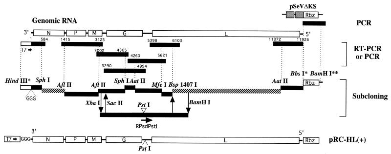
A full-length (11,926 nucleotides) cDNA of the RC-HL strain was constructed by stepwise assembly of a total of nine cDNA fragments (Fig. 2). Seven of the nine cDNA fragments were amplified by RT-PCR or PCR using the genomic RNA extracted from virion or viral cDNA-containing plasmids (12) as a template, respectively, followed by cloning and sequencing. The remaining two fragments, an SphI-AflII fragment of the N gene and a Bsp1407I-AatII fragment of the L gene, were produced from pETGN10 (7) and pcDNA-RL, respectively. The complete genomic cDNA was constructed on pUC19 and was located between a T7 promoter sequence and a cDNA copy of self-cleaving ribozyme (Rbz) from the hepatitis delta virus antigenome. The Rbz cDNA was amplified by PCR using pSeVΔKS (9) (kindly provided by A. Kato) as a template. This full-length genome plasmid directs synthesis of positive (antigenomic)-sense RNA under the control of T7 RNA polymerase. In order to distinguish the virus rescued from the full-length genome plasmid (r RC-HL strain) from the wild-type (wt) RC-HL strain, a recognition site for the restriction enzyme PstI was constructed as a genetic marker in the G-L noncoding region of the genome plasmid by changing an adenine residue at position 4925 (indicated as positive sense) to a cytosine residue using a U.S.E. mutagenesis kit (Amersham Pharmacia Biotech) with an RPsdPstI primer (5′-GCT TCA AGT TCT GCA GAT CAC CTT CCA TCT AAG TCT GG-3′) (Mutation to cytosine residue is underlined.). The resulting plasmid was designated pRC-HL(+).
FIG. 2.
Construction of the full-length genome plasmid pRC-HL(+). A total of nine cDNA fragments were assembled by stepwise subcloning. Of the nine fragments, two (SphI-AflII and Bsp1407I-AatII fragments) (hatched) originated from pETGN10 (7) and pcDNA-RL, respectively. Two fragments containing the 3′ and 5′ terminal regions were amplified by PCR using the terminal cDNA-carrying plasmids (12) as a template. The remaining five fragments were amplified by RT-PCR. The number above each amplified fragment indicates the genome nucleotide number of the RC-HL strain. The self-cleaving ribozyme (Rbz) cDNA of the hepatitis delta virus antigenome was also amplified by PCR using pSeVΔKS (9) as a template. These amplified cDNA fragments were used for subcloning after cloning and sequencing. Restriction enzyme sites with asterisks and double asterisks originated from the primer sequence and the plasmid vector, respectively. For construction of the recognition site for the restriction enzyme PstI, used as a genetic marker, site-directed mutagenesis was performed with a RPsdPstI mutagenesis primer.
Details of the construction of these plasmids and sequences of the primers are available from the authors on request.
Construction of full-length genome plasmid for the chimeric R(G) strain.
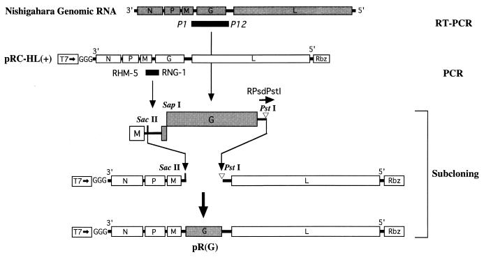
The full-length genome plasmid to produce a chimeric virus, the R(G) strain with the G-ORF of the Nishigahara strain in the background of the RC-HL genome, was constructed as shown in Fig. 3. A cDNA including the full-length G-ORF from the Nishigahara strain was amplified by RT-PCR using a set of primers, P1 and P12, designed for a previous study (12). Furthermore, a set of primers, positive-sense RHM-5 (5′-TTA AGA CAC AAA TGT CTG AAG AGG-3′) and negative-sense RNG-1 (5′-ATG GGT ACA AGC AGA AGA GCT TGC-3′) acting at positions from nucleotide 3053 to 3076 and from 3325 to 3348 (based on the nucleotide number of the RC-HL genome), respectively, were employed for PCR using pRC-HL(+) as a template. The RNG-1 primer was designed on the basis of the nucleotide sequence of the Nishigahara G gene and contained a complementary sequence for a SapI site. The cDNA fragment amplified with the RHM-5 and RNG-1 primers contains the M-G noncoding region of the RC-HL strain and a part of the G coding region of the Nishigahara strain. After cloning and sequencing of these cDNA fragments, the plasmid with the former cDNA fragment was mutated for construction of a PstI site in the G-L noncoding region, using mutagenesis with the RPsdPstI primer. Following connection of the two cDNA fragments at the SapI site, a SacII-PstI fragment was inserted into the same sites of pRC-HL(+). The resulting plasmid was designated pR(G).
FIG. 3.
Construction of the full-length genome plasmid for the chimeric R(G) strain. Two cDNA fragments containing the Nishigahara G-ORF and the RC-HL M-G noncoding region were amplified by RT-PCR and PCR, respectively. The P1 and P12 primers (italics) were designed for a previous study (12). For construction of a PstI site in the G-L noncoding region, site-directed mutagenesis was performed with a RPsdPstI mutagenesis primer. After cloning and sequencing, the two fragments were connected and subcloned into pRC-HL(+).
Rescue of rabies virus from cloned cDNA.
BHK-21 cells grown in six-well plates were infected with vTF7-3 at a multiplicity of infection (MOI) of 5. One hour after inoculation, the cells were washed twice with Dulbecco's MEM and transfected with 2.5 μg of pRC-HL(+) or pR(G) and 2.5, 2.5, and 0.5 μg of pcDNA-RN, -RP, and -RL per well, respectively, using 20 μl of Cellfectin (Life Technolologies, Gaithersburg, Md.) in a final volume of 1 ml. The transfection was allowed to proceed for 8 h or overnight. The transfecting medium was removed from the cells and replaced with Eagle's MEM supplemented with 10% TPB, 5% fetal calf serum, and 25 μg of cytosine arabinoside (Ara C)/ml. The transfected cells were incubated at 32°C for 3 days and were then harvested together with the medium by scraping with a rubber policeman. The cell suspension was cocultured with NA cells in a 6-cm dish at 32°C for 4 days in a medium containing 25 μg of Ara C/ml. The culture medium was then collected and centrifuged at 1,500 × g for 10 min. The supernatant was inoculated into NA cells grown in 24-well plates. After incubation for 2 h, the cells were cultured at 32°C with medium containing 25 μg of Ara C/ml. After 3 days, an indirect fluorescence antibody (IFA) test was performed for detection of rabies virus antigen using anti N protein monoclonal antibody (N-MAb) 8-1 (18), following harvest of the medium. The medium from rabies virus antigen-positive wells was inoculated into NA cells for a second passage. After 3 days, the medium was collected and centrifuged at 13,000 × g for 10 min at 4°C. Afterward, the supernatant was centrifuged again at 13,000 × g for 10 min at 4°C. To remove vaccinia virus completely, the final supernatant was filtered using a sterile Millex-GP 0.22-μm-pore-size filter unit (Millipore, Bedford, Mass.) and stored as the original stock virus at −80°C.
Confirmation of the virus rescue from cloned cDNA by RT-PCR and restriction enzyme digestion.
For detection of genomic RNA of the virus rescued from cloned cDNA, RT-PCR was performed with one set of primers, positive-sense RGP-14 (5′-GTT GTA GAA AAG TCG ATC GGC CAG-3′) and negative-sense RHL-27 (5′-GGA TCA ATG GGG TCA TCA TAG ACC-3′) primers acting at positions from nucleotide 4752 to 4775 and from 5427 to 5452 (based on the nucleotide number of the RC-HL genome), respectively, and another set of primers, the RHM-5 primer described above and the P6 primer designed for our previous study (12). The cDNA fragments amplified with the RGP-14 and RHL-27 primers were digested with the restriction enzyme PstI for detection of the genetic marker. The cDNA fragments amplified with the RHM-5 and P6 primers were digested with BglII or SapI to distinguish cDNAs of the G gene from the RC-HL and Nishigahara strains.
Titration of virus.
The virus was titrated by a focus assay on confluent monolayers of NA or BHK-21 cells in 24-well plates. For staining of viral foci, an IFA test was performed using N-MAb 8-1 (18). The relative tropism ratio of virus in NA and BHK-21 cells was determined by division of the titer of virus stock in NA cells by the titer of the identical stock in BHK-21 cells.
Virus growth.
Monolayer cultures of 3 × 10⁶ NA cells and 5 × 10⁶ BHK cells were infected with individual viruses at an MOI of 0.01 focus-forming units (FFU) that had been titrated in NA cells. After adsorption of virus for 1 h, the cells were washed three times with Hanks' balanced salt solution. Afterward, the cultures were replenished with fresh medium and incubated at 37°C. Samples of the culture medium were harvested at 1, 3, and 5 days postinoculation. The titer of virus in NA cells was examined by the focus assay as described above.
Inoculation of virus into mice.
Virulence of the virus for adult and suckling mice was measured in 4-week-old female and 2-day-old ddY mice, respectively. Groups of five adult or suckling mice were inoculated i.c. with 30 and 10 μl of serial 10-fold dilutions of each virus, respectively. The fifty percent lethal dose (LD50) of each virus was calculated by the method of Reed and Müench (22).
RESULTS
Rescue of the RC-HL strain from the full-length genome plasmid.
Helper plasmids carrying the N, P, and L genes of the RC-HL strain, respectively, were constructed in pcDNA1.1/Amp. After transfection of each plasmid into BHK-21 cells infected with vTF7-3, the expressed N, P, and L proteins were analyzed by Western blotting with the specific antibodies, namely N-MAb 12-2 (18), and anti-P (26) and anti-L (19) rabbit sera (kindly provided by A. Kawai), respectively. These expressed proteins were indistinguishable by molecular weight from the respective corresponding proteins produced in the cells infected with the RC-HL strain (data not shown).
In order to optimize transfection conditions for rescue of the RC-HL strain from cDNA, we constructed a minigenome plasmid encoding a negative-sense luciferase gene between the genomic sense 3′ and 5′ terminal sequences of the RC-HL strain, which contained the transcriptional signals. Based on the measured level of luciferase activity in the BHK-21 cells transfected with this minigenome plasmid and helper plasmids, pcDNA-RN, -RP, and -RL, the amounts of the helper plasmids to be transfected, incubation temperature (32°C), and concentration of Ara C after transfection were optimized (data not shown). After carrying out the processes described in Materials and Methods, N protein antigens of the rabies virus were detected by the IFA test in NA cells inoculated with the supernatants from coculturing of the transfected BHK-21 cells with NA cells. The N protein antigens observed in NA cells had the same granular form as those in cells infected with the rabies virus. Rabies virus antigen was also seen in NA cells inoculated with the culture medium from N protein antigen-positive cells. Therefore, it was clear that the supernatant contained infectious rabies virus. Infectious virus was generated in all wells of a six-well plate in which the transfected BHK-21 cells were cultured. Thus, based on the cell number per well, it was estimated that at least one infectious virus had emerged from 5 × 10⁵ BHK-21 cells.
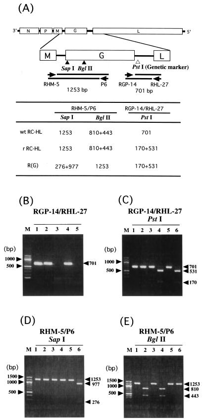
In order to exclude the possibility of contamination with the wt RC-HL strain in this experimental system, the presence of the genetic marker, PstI site, in the G-L noncoding region of the rescued virus (r RC-HL strain) was examined. For this purpose, a cDNA fragment including this region was amplified by RT-PCR using genomic RNA of the rescued virus as a template and was digested with a restriction enzyme, PstI (Fig. 4). PCR with a set of the primers RGP-14 and RHL-27 amplified a fragment with the expected size of 701 bp, the same as that in the case of the wt RC-HL strain (Fig. 4A and B). PCR using the same primers without an RT step failed to produce any products, indicating that the DNA band did not originate from the full-length genome plasmid used for transfection (Fig. 4B). The amplified cDNA fragment was digested with PstI, and 531-bp and 170-bp bands were observed, whereas cDNA from the wt RC-HL strain could not be digested (Fig. 4A and C).
FIG. 4.
Confirmation of rescue of the r RC-HL and chimeric R(G) strains from cloned cDNA by RT-PCR and restriction enzyme digestion. (A) Schematic diagram of the rabies virus genome showing the annealing positions of primers, the genetic marker PstI site in the G-L noncoding region, SapI and BglII sites in the G gene, and the predicted fragment size after digestion of the respective restriction enzyme. (B) RT-PCR products of wt RC-HL (lane 1), r RC-HL (lanes 2 and 3), and chimeric R(G) (lanes 4 and 5) strains amplified by the RGP-14 and RHL-27 primers with (lanes 1, 2, and 4) or without (lanes 3 and 5) the RT step. (C, D and E) Restriction enzyme digestions of the cDNA fragments amplified by RT-PCR. cDNA fragments of wt RC-HL (lanes 1 and 2), r RC-HL (lanes 3 and 4), and chimeric R(G) (lanes 5 and 6) strains amplified by RGP-14 and RHL-27 primers (C) or RHM-5 and P6 primers (D and E). These cDNA fragments were treated with (lanes 2, 4, and 6) or without (lanes 1, 3, and 5) a restriction enzyme, PstI (C), SapI (D), or BglII (E). M, molecular size marker.
Subsequently, eradication of vaccinia virus, vTF7-3, from the original stock of the r RC-HL strain was confirmed by IFA and PCR methods. The vaccinia antigen was not detected in BHK-21 cells inoculated with the undiluted original stock by IFA testing with an anti-vaccinia virus rabbit serum (data not shown). The vTF7-3 gene was also not amplified from an extract of these BHK-21 cells by the PCR method, which can detect the DNA from 20 PFU of vTF7-3/ml (data not shown). Thus, the results indicated that the method used in this study enabled complete removal of vTF7-3 from the original stock of the r RC-HL strain.
Comparison of several biological properties of the wt and r RC-HL strains.
In order to examine whether the r RC-HL strain had the same biological properties as the wt RC-HL strain, growth of the r RC-HL strain in cultured cells and its virulence for mice were determined and compared with those of the wt RC-HL strain.
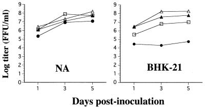
Infectivities of the wt and r RC-HL strains in both neuronal NA cells and nonneuronal BHK-21 cells were determined by the focus assay to examine their cell tropism (Table 1). The relative tropism ratios of the wt and r RC-HL strains were almost the same (1.4 and 1.6, respectively). Furthermore, multiple-step growth curves of the wt and r RC-HL strains were similar in NA and BHK-21 cells (Fig. 5).
TABLE 1.
Relative cell tropism of the wt RC-HL, r RC-HL, Nishigahara, and chimeric R(G) strains in NA and BHK cells
| Viral strain | Titer (FFU/ml) in cell line:
|
Relative tropism ratioa (NA/BHK) | |
|---|---|---|---|
| NA | BHK | ||
| wt RC-HL | 8.1 × 10⁸ | 5.7 × 10⁸ | 1.4 |
| r RC-HL | 2.5 × 10⁸ | 1.6 × 10⁸ | 1.6 |
| R(G) | 6.0 × 10⁷ | 2.7 × 10⁶ | 22 |
| Nishigahara | 2.2 × 10⁷ | 1.3 × 10³ | 17,000 |
The relative tropism ratio was determined by dividing the titer of virus stock in NA cells by the titer of the identical stock in BHK-21 cells.
FIG. 5.
Growth curves of wt RC-HL (▴), r RC-HL (▵), and Nishigahara (●) strains and the chimeric R(G) (□) strain in NA and BHK-21 cells. Each virus was inoculated with an MOI of 0.01. The virus in the culture fluid was harvested at 1, 3, and 5 days postinoculation and titrated in NA cells.
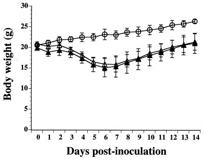
Next, the LD50s of the wt and r RC-HL strains were determined in suckling and adult mice by i.c. inoculation (Table 2). The r RC-HL strain as well as the wt RC-HL strain killed suckling mice, and the LD50s were comparable (1.8 × 10⁻¹ and 3.2 × 10⁻¹ FFU, respectively). However, i.c. inoculation of the wt or r RC-HL strain, even inoculation with 1.0 × 10⁶ FFU of the virus, did not kill any of the adult mice (Table 2). The adult mice developed a mild disease with symptoms such as piloerection and body weight reduction. Figure 6 shows body weight change in the adult mice inoculated with 1.0 × 10⁶ FFU of the wt and r RC-HL strains. In contrast to the continuous increase in body weights of mock-infected mice during the 14-day observation period, the body weights both of the mice infected with the wt RC-HL strain and of those infected with the r RC-HL strain decreased up to about 6 days postinoculation and then gradually increased. The body weight curves of the two groups of mice were very similar.
TABLE 2.
LD50s of the wt RC-HL, r RC-HL, Nishigahara, and chimeric R(G) strains in suckling and adult mice
| Mouse group | LD50 (FFU) of strain:
|
|||
|---|---|---|---|---|
| wt RC-HL | r RC-HL | R(G) | Nishigahara | |
| Suckling | 3.2 × 10⁻¹ | 1.8 × 10⁻¹ | NDa | ND |
| Adult | >1.0 × 10⁶ | >1.0 × 10⁶ | 1.0 × 100 | 6.3 × 10⁻² |
ND, not determined.
FIG. 6.
Body weight changes in adult mice inoculated with wt or r RC-HL strains. Five mice per group were inoculated i.c. with wt (▴) and r RC-HL (▵) strains of 106 FFU per mouse or were mock inoculated (○). The values in the graph are averages and standard deviations of body weight.
Rescue of the chimeric R(G) strain from cloned cDNA.
The chimeric R(G) strain, which has the G-ORF of the Nishigahara strain in the background of the RC-HL genome, was rescued from cloned cDNA in the same manner as the r RC-HL strain, as demonstrated by the fact that the genetic marker, the PstI site, constructed in the G-L noncoding region was detected by digestion of cDNA of the rescued virus with PstI. (Fig. 4A and C). In order to examine whether the viral genome had the G-ORF from the Nishigahara strain, a 1,253-bp cDNA fragment including the M-G noncoding region and the first half of the G-ORF of the R(G) strain was amplified by RT-PCR with RHM-5 and P6 primers (Fig. 4A). Afterward, the cDNA fragment was digested with the restriction enzyme SapI or BglII. SapI digests cDNA of the G-ORF from the Nishigahara strain but not from the RC-HL strain, and vice versa for BglII (Fig. 4A). The cDNA fragment from the R(G) strain was digested with SapI, generating 977-bp and 276-bp fragments, whereas those from the wt and r RC-HL strains were not digested (Fig. 4C). On the other hand, BglII digested cDNA fragments from the wt and r RC-HL strains, generating 810-bp and 443-bp fragments, but did not digest that from the R(G) strain (Fig. 4E). Furthermore, the amplified cDNA fragment from the R(G) strain was directly sequenced, and it was confirmed that the G-ORF and the M-G noncoding region were derived from the Nishigahara and RC-HL strains, respectively (data not shown).
Pathogenicity of the chimeric R(G) strain for adult mice.
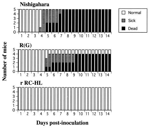
To examine the pathogenicity of the chimeric R(G) strain, the strain was inoculated i.c. into adult mice. The mice inoculated with the R(G) strain developed neurological symptoms, such as paralysis and fury, and died, as did the mice inoculated with the virulent Nishigahara strain. Rabies virus antigen, but not vaccinia virus antigen, was detected in tissue from one dead mouse brain by the IFA test. Furthermore, the rabies virus gene was amplified from the mouse brain by RT-PCR with RHM-5 and P6 primers. Moreover, the nucleotide sequence of the amplified cDNA fragment coincided with that of the corresponding gene region in the R(G) strain as expected (data not shown). The LD50 of the chimeric virus in adult mice was 1 FFU (Table 2). This value was about 16-fold higher than that of the Nishigahara strain, whereas the LD50 of the r RC-HL strain was more than 106 FFU. Figure 7 shows morbidity and mortality changes in mice inoculated i.c. with 10 FFU of the Nishigahara, r RC-HL, and R(G) strains. Although mice inoculated with the r RC-HL strain were apparently normal during the 14-day observation period, the R(G) strain caused disease and death in four of the five mice. Onset of disease in mice inoculated with the R(G) strain was delayed compared to that in mice inoculated with the Nishigahara strain.
FIG. 7.
Morbidity and mortality changes in adult mice inoculated i.c. with 10 FFU of the Nishigahara, r RC-HL, and chimeric R(G) strains.
Tropism and growth of the chimeric R(G) strain in NA and BHK-21 cells.
Table 1 shows the relative tropism ratios of the chimeric R(G) strain in NA and BHK-21 cells. The titer of the R(G) strain determined in NA cells was about 22-fold higher than that in BHK-21 cells, indicating that tropism of the R(G) strain to BHK-21 cells was less distinct than those of the wt and r RC-HL strains. However, tropism of the R(G) strain was approximately 800-fold higher than that of the Nishigahara strain.
The multiple-step growth curve of the R(G) strain in neuronal NA cells was similar to those of the wt and r RC-HL strains and the Nishigahara strain (Fig. 5). The rate of propagation of the R(G) strain in nonneuronal BHK-21 cells was lower than those of the wt and r RC-HL strains but greater than that of the Nishigahara strain, whose titer remained constant at about 104 FFU/ml, corresponding to the tropisms of these strains in NA and BHK-21 cells.
DISCUSSION
The infectious rabies virus that was recovered from BHK-21 cells transfected with full-length cDNA of the RC-HL strain had a genetic marker in the G-L noncoding region on its genome, and the virus had almost the same biological properties as the wt RC-HL strain. These findings indicated that the RC-HL strain was rescued from cloned cDNA. This is the second report on the establishment of a reverse genetics system of rabies virus and the first report on rescue of an attenuated strain that causes nonlethal infection in adult mice after i.c. inoculation.
We chose the RC-HL strain for establishment of the reverse genetics system for the following reasons. First, it was thought to be easier to rescue the RC-HL strain from plasmid-transfecting cultured cells, because the RC-HL strain is more highly adapted to cultured cells than the parental Nishigahara strain, which had been maintained by rabbit brain passages. Second, the attenuated RC-HL strain is suitable for identification of the gene of the Nishigahara strain related to the pathogenicity for adult mice, since the two strains have a close genetical relationship despite there being a great difference in their pathogenicities (12).
It has been estimated that one infectious virion can be rescued from 107 transfected cells in the reverse genetics system of the SAD B19 strain (23, 24). The system of the RC-HL strain established here generated more than one infectious virus from 5 × 10⁵ transfected cells, indicating that the rescue efficiency of the RC-HL strain is at least 20-fold higher than that of the SAD B19 strain. There are several possible reasons for this. First, the high transfection efficiency (about 40%) in BHK-21 cells (data not shown) might have increased the numbers of cells that were transfected with all of the full-length genome plasmid and the three helper plasmids. Second, conditions such as the amounts of transfected helper plasmids, incubation temperature, and supplementation of Ara C, optimized by the minigenome system (data not shown), may also have increased the efficiency of rescue of the virus. Third, coculturing of transfected BHK-21 cells with confluent NA cells may have enabled a more efficient recovery of the virus than the routine method of freeze-thawing of transfected cells (13, 15, 24). Finally, the RC-HL strain may have been more highly adapted to BHK-21 cells than the SAD B19 strain. This high efficiency of rescue of the virus will help the genome of the RC-HL strain to be easily manipulated.
We tried to produce a chimeric virus, the R(G) strain, that has the G-ORF of the Nishigahara strain in the background of the RC-HL genome. Using the same procedure as that used for the rescue of the RC-HL strain, infectious virus was rescued from a chimeric cDNA. Analyses of amplified cDNA fragments from the virus with restriction enzyme digestion and sequencing revealed that this virus had the G-ORF from the Nishigahara strain, indicating that the designed chimeric virus, the R(G) strain, was produced by the reverse genetics system.
This study showed that the chimeric R(G) strain acquired lethality for adult mice. Therefore, it became evident that the G-ORF is closely related to the pathogenicity of the Nishigahara strain for adult mice. This is consistent with previous reports of the G protein playing an important role in the pathogenicity of rabies virus for adult mice (4, 25, 29). However, it is obvious that an amino acid at position 333 in the G protein, which is known as a determinant of pathogenicity in representative fixed viruses (4, 25, 29), is not associated with the difference between the virulence of the RC-HL strain and that of the Nishigahara strain, because an amino acid change was not observed at this position between the two strains (11, 12). This clearly indicates that the Nishigahara G protein includes a novel determinant of pathogenicity other than the amino acid at position 333. It also suggests that the determinant of pathogenicity in the G protein is very complex. We previously reported that 14 amino acid substitutions in the G protein were found in a comparison between the RC-HL and Nishigahara strains and that 9 of these 14 substitutions were clustered in the region at amino acid positions 164 to 303 (12). Since five of the nine substitutions are radical and this region includes the putative binding domain (residues 189 to 214) for the nicotinic acetylcholine receptor, which is known to be a receptor for rabies virus (8, 17, 16), the cluster of substitutions is likely to be related to some functional changes in the G protein and consequently to the difference between the pathogenicities of the two strains.
Although the chimeric R(G) strain killed adult mice after i.c. inoculation, the LD50 of this strain was about 16-fold higher than that of the Nishigahara strain (Table 2). Furthermore, morbidity and mortality changes in the mice inoculated with 10 FFU of each virus also showed that the R(G) strain is less virulent than the Nishigahara strain (Fig. 7). These findings suggest that a genomic region other than the G-ORF may also be related to the difference between the pathogenicities of the two strains. The cluster of amino acid differences in the L protein between the two strains (positions 1157 to 1592) found in our previous study (12) might be associated with this minor difference between the pathogenicities of the R(G) and Nishigahara strains.
The RC-HL strain has been established from the rabbit brain-passaged Nishigahara strain, as a consequence of the adaptation to nonneuronal cells (10). Tuffereau et al. (28) postulated that adaptation of rabies virus to cells is partly due to the ability of the virus to use ubiquitous receptors that are present on every cell type. This postulation seems reasonable, since the chimeric R(G) strain has less nonneuronal-BHK-21-cell tropism than does the RC-HL strain (Table 1; Fig. 5). However, the great difference in BHK-21-cell tropism between the R(G) and Nishigahara strains indicates that a genomic region other than the G-ORF is more closely related to adaptation of the RC-HL strain to BHK-21 cells. Comparison of the RNA transcriptional activities of the RC-HL, Nishigahara, and R(G) strains might elucidate the distinct cell tropisms of these strains. Although it is possible that the adaptation of the RC-HL strain to nonneuronal cells is closely correlated with attenuation of the strain, the data presented here failed to clearly indicate the correlation. In order to make clear whether the distinct cell tropisms contribute to the differences among the pathogenicities of the RC-HL, Nishigahara, and R(G) strains, an immunohistochemical study using mouse brains infected with these strains is now in progress.
In this study, we obtained direct evidence that the G-ORF is closely related to the pathogenicity of the Nishigahara strain for adult mice. This finding supports the previously reported importance of the G gene as a determinant of the pathogenicity of a strain for adult mice (4, 25, 29). In order to elucidate the mechanism generating the difference between the pathogenicities of the RC-HL and Nishigahara strains, functional analyses of G proteins of the two strains are needed. Propagative kinetics of the R(G) strain in the mouse brain should also be compared with those of the RC-HL and Nishigahara strains. Furthermore, in order to determine the significance of the cluster of amino acid substitutions (at positions 164 to 303) in the G protein for viral pathogenicity, production and analysis of another chimeric virus that has the variable region of the G gene from the Nishigahara strain in the background of the RC-HL genome are needed.
ACKNOWLEDGMENTS
We are grateful to B. Moss (National Institutes of Health, Bethesda, Md.) for providing vTF7-3 and to A. Kato (National Institute of Infectious Diseases, Tokyo, Japan) for providing the plasmid pSeVΔKS and for valuable technical advice. We also thank A. Kawai (Kyoto University, Kyoto, Japan) for the gifts of anti-P and anti-L rabbit sera, K. K. Conzelmann (Max-von-Pettenkofer Institute, Munich, Germany) for technical support, and Sri Kantha (Faculty of Agriculture, Gifu University, Gifu, Japan) and Frank Roerink (Central Laboratories, Kyoritsu Shoji Corporation, Ibaraki, Japan) for advice in preparing the manuscript.
This study was supported in part by a Grant-in-Aid for Scientific Research from the Ministry of Education, Science, Sports and Culture, Japan (no. 10556072).
REFERENCES
- 1.Anilionis A, Wunner W H, Curtis P J. Structure of the glycoprotein gene in rabies virus. Nature. 1981;294:275–278. doi: 10.1038/294275a0. [DOI] [PubMed] [Google Scholar]
- 2.Collins P L, Hill M G, Camargo E, Grosfeld H, Chanock R M, Murphy B R. Production of infectious human respiratory syncytial virus from cloned cDNA confirms an essential role for the transcription elongation factor from the 5′ proximal open reading frame of the M2 mRNA in gene expression and provides a capability for vaccine development. Proc Natl Acad Sci USA. 1995;92:11563–11567. doi: 10.1073/pnas.92.25.11563. [DOI] [PMC free article] [PubMed] [Google Scholar]
- 3.Conzelmann K K, Cox J H, Schneider L G, Thiel H J. Molecular cloning and complete nucleotide sequence of the attenuated rabies virus SAD B19. Virology. 1990;175:485–499. doi: 10.1016/0042-6822(90)90433-r. [DOI] [PubMed] [Google Scholar]
- 4.Dietzschold B, Wunner W H, Wiktor T J, Lopes A D, Lafon M, Smith C L, Koprowski H. Characterization of an antigenic determinant of the glycoprotein that correlates with pathogenicity of rabies virus. Proc Natl Acad Sci USA. 1983;80:70–74. doi: 10.1073/pnas.80.1.70. [DOI] [PMC free article] [PubMed] [Google Scholar]
- 5.Fuerst T R, Niles E G, Studier F W, Moss B. Eukaryotic transient-expression system based on recombinant vaccinia virus that synthesizes bacteriophage T7 RNA polymerase. Proc Natl Acad Sci USA. 1986;83:8122–8126. doi: 10.1073/pnas.83.21.8122. [DOI] [PMC free article] [PubMed] [Google Scholar]
- 6.Garcin D, Pelet T, Calain P, Roux L, Curran J, Kolakofsky D. A highly recombinogenic system for the recovery of infectious Sendai paramyxovirus from cDNA: generation of a novel copy-back nondefective interfering virus. EMBO J. 1995;14:6087–6094. doi: 10.1002/j.1460-2075.1995.tb00299.x. [DOI] [PMC free article] [PubMed] [Google Scholar]
- 7.Goto H, Minamoto N, Ito H, Luo T R, Sugiyama M, Kinjo T, Kawai A. Expression of the nucleoprotein of rabies virus in Escherichia coli and mapping of antigenic sites. Arch Virol. 1995;140:1061–1074. doi: 10.1007/BF01315415. [DOI] [PubMed] [Google Scholar]
- 8.Hanham C A, Zhao F, Tignor G H. Evidence from the anti-idiotypic network that the acetylcholine receptor is a rabies virus receptor. J Virol. 1993;67:530–542. doi: 10.1128/jvi.67.1.530-542.1993. [DOI] [PMC free article] [PubMed] [Google Scholar]
- 9.Hasan M K, Kato A, Shioda T, Sakai Y, Yu D, Nagai Y. Creation of an infectious recombinant Sendai virus expressing the firefly luciferase gene from the 3′ proximal first locus. J Gen Virol. 1997;78:2813–2820. doi: 10.1099/0022-1317-78-11-2813. [DOI] [PubMed] [Google Scholar]
- 10.Ishikawa Y, Samejima T, Nunoya T, Motohashi T, Nomura Y. Biological properties of the cell culture-adapted RC-HL strain of rabies virus as a candidate strain for an inactivated vaccine. J Jpn Vet Med Assoc. 1989;42:637–643. [Google Scholar]
- 11.Ito H, Minamoto N, Watanabe T, Goto H, Rong L T, Sugiyama M, Kinjo T, Mannen K, Mifune K, Konobe T, Yoshida I, Takamizawa A. A unique mutation of glycoprotein gene of the attenuated RC-HL strain of rabies virus, a seed virus used for production of animal vaccine in Japan. Microbiol Immunol. 1994;38:479–482. doi: 10.1111/j.1348-0421.1994.tb01812.x. [DOI] [PubMed] [Google Scholar]
- 12.Ito N, Kakemizu M, Ito K A, Yamamoto A, Yoshida Y, Sugiyama M, Minamoto N. A comparison of complete genome sequences of the attenuated RC-HL strain of rabies virus used for production of animal vaccine in Japan, and the parental Nishigahara strain. Microbiol Immunol. 2001;45:51–58. doi: 10.1111/j.1348-0421.2001.tb01274.x. [DOI] [PubMed] [Google Scholar]
- 13.Kato A, Sakai Y, Shioda T, Kondo T, Nakanishi M, Nagai Y. Initiation of Sendai virus multiplication from transfected cDNA or RNA with negative or positive sense. Genes Cells. 1996;1:569–579. doi: 10.1046/j.1365-2443.1996.d01-261.x. [DOI] [PubMed] [Google Scholar]
- 14.Lai C Y, Dietzschold B. Amino acid composition and terminal sequence analysis of the rabies virus glycoprotein: identification of the reading frame on the cDNA sequence. Biochem Biophys Res Commun. 1981;103:536–542. doi: 10.1016/0006-291x(81)90485-x. [DOI] [PubMed] [Google Scholar]
- 15.Lawson N D, Stillman E A, Whitt M A, Rose J K. Recombinant vesicular stomatitis viruses from DNA. Proc Natl Acad Sci USA. 1995;92:4477–4481. doi: 10.1073/pnas.92.10.4477. [DOI] [PMC free article] [PubMed] [Google Scholar]
- 16.Lentz T L, Benson R J, Klimowicz D, Wilson P T, Hawrot E. Binding of rabies virus to purified Torpedo acetylcholine receptor. Brain Res. 1986;387:211–219. doi: 10.1016/0169-328x(86)90027-6. [DOI] [PubMed] [Google Scholar]
- 17.Lentz T L, Wilson P T, Hawrot E, Speicher D W. Amino acid sequence similarity between rabies virus glycoprotein and snake venom curaremimetic neurotoxins. Science. 1984;226:847–884. doi: 10.1126/science.6494916. [DOI] [PubMed] [Google Scholar]
- 18.Minamoto N, Tanaka H, Hishida M, Goto H, Ito H, Naruse S, Yamamoto K, Sugiyama M, Kinjo T, Mannen K, Mifune K. Linear and conformation-dependent antigenic sites on the nucleoprotein of rabies virus. Microbiol Immunol. 1994;38:449–455. doi: 10.1111/j.1348-0421.1994.tb01806.x. [DOI] [PubMed] [Google Scholar]
- 19.Morimoto K, Akamine T, Takamatsu F, Kawai A. Studies on rabies virus RNA polymerase: 1. cDNA cloning of the catalytic subunit (L protein) of avirulent HEP-flury strain and its expression in animal cells. Microbiol Immunol. 1998;42:485–496. doi: 10.1111/j.1348-0421.1998.tb02314.x. [DOI] [PubMed] [Google Scholar]
- 20.Morimoto K, Ohkubo A, Kawai A. Structure and transcription of the glycoprotein gene of attenuated HEP-Flury strain of rabies virus. Virology. 1989;173:465–477. doi: 10.1016/0042-6822(89)90559-x. [DOI] [PubMed] [Google Scholar]
- 21.Radecke F, Spielhofer P, Schneider H, Kaelin K, Huber M, Dotsch C, Christiansen G, Billeter M A. Rescue of measles viruses from cloned DNA. EMBO J. 1995;14:5773–5784. doi: 10.1002/j.1460-2075.1995.tb00266.x. [DOI] [PMC free article] [PubMed] [Google Scholar]
- 22.Reed L J, Müench H. A simple method of estimating fifty per cent endpoints. Am J Hyg. 1938;27:493–497. [Google Scholar]
- 23.Roberts A, Rose J K. Recovery of negative-strand RNA viruses from plasmid DNAs: a positive approach revitalizes a negative field. Virology. 1998;247:1–6. doi: 10.1006/viro.1998.9250. [DOI] [PubMed] [Google Scholar]
- 24.Schnell M J, Mebatsion T, Conzelmann K K. Infectious rabies viruses from cloned cDNA. EMBO J. 1994;13:4195–4203. doi: 10.1002/j.1460-2075.1994.tb06739.x. [DOI] [PMC free article] [PubMed] [Google Scholar]
- 25.Seif I, Coulon P, Rollin P E, Flamand A. Rabies virulence: effect on pathogenicity and sequence characterization of rabies virus mutations affecting antigenic site III of the glycoprotein. J Virol. 1985;53:926–934. doi: 10.1128/jvi.53.3.926-934.1985. [DOI] [PMC free article] [PubMed] [Google Scholar]
- 26.Takamatsu F, Asakawa N, Morimoto K, Takeuchi K, Eriguchi Y, Toriumi H, Kawai A. Studies on the rabies virus RNA polymerase: 2. Possible relationships between the two forms of the non-catalytic subunit (P protein) Microbiol Immunol. 1998;42:761–771. doi: 10.1111/j.1348-0421.1998.tb02350.x. [DOI] [PubMed] [Google Scholar]
- 27.Tordo N, Poch O, Ermine A, Keith G, Rougeon F. Walking along the rabies genome: is the large G-L intergenic region a remnant gene? Proc Natl Acad Sci USA. 1986;83:3914–3918. doi: 10.1073/pnas.83.11.3914. [DOI] [PMC free article] [PubMed] [Google Scholar]
- 28.Tuffereau C, Benejean J, Blondel D, Kieffer B, Flamand A. Low-affinity nerve-growth factor receptor (P75NTR) can serve as a receptor for rabies virus. EMBO J. 1998;17:7250–7259. doi: 10.1093/emboj/17.24.7250. [DOI] [PMC free article] [PubMed] [Google Scholar]
- 29.Tuffereau C, Leblois H, Benejean J, Coulon P, Lafay F, Flamand A. Arginine or lysine in position 333 of ERA and CVS glycoprotein is necessary for rabies virulence in adult mice. Virology. 1989;172:206–212. doi: 10.1016/0042-6822(89)90122-0. [DOI] [PubMed] [Google Scholar]
- 30.Whelan S P, Ball L A, Barr J N, Wertz G T. Efficient recovery of infectious vesicular stomatitis virus entirely from cDNA clones. Proc Natl Acad Sci USA. 1995;92:8388–8392. doi: 10.1073/pnas.92.18.8388. [DOI] [PMC free article] [PubMed] [Google Scholar]
- 31.Wiktor T J, Gyorgy E, Schlumberger D, Sokol F, Koprowski H. Antigenic properties of rabies virus components. J Immunol. 1973;110:269–276. [PubMed] [Google Scholar]


