



ABSTRACT
Aspergillus fumigatus is the predominant pathogen responsible for aspergillosis infections, with emerging drug-resistant strains complicating treatment strategies. The role of mitochondrial functionality in fungal resistance to antifungal agents is well-documented yet not fully understood. In this study, the mitochondrial protein Bcs1A, a homolog of yeast Bcs1, was found to regulate colony growth, ion homeostasis, and the response to antifungal drugs in A. fumigatus. Microscopic observations revealed substantial colocalization of Bcs1A-GFP fusion protein fluorescence with mitochondria. Bcs1A deletion compromised colony growth and the utilization of non-fermentable carbon sources, alongside causing abnormal mitochondrial membrane potential and reduced reactive oxygen species production. These findings underscore Bcs1A’s vital role in maintaining mitochondrial integrity. Phenotypic analysis and determinations of minimum inhibitory concentrations indicated that the Δbcs1A mutant was more resistant to various antifungal agents, such as azoles, terbinafine, and simvastatin, compared to wild-type strain. RNA sequencing and RT-qPCR analysis highlighted an upregulation of multiple efflux pumps in the Δbcs1A mutant. Furthermore, loss of the principal drug efflux pump, mdr1, decreased azole tolerance in the Δbcs1A mutant, suggesting that Bcs1A’s modulated of azoles response via efflux pump expression. Collectively, these results establish Bcs1A as essential for growth and antifungal drug responsiveness in A. fumigatus mediated through mitochondrial regulation.
IMPORTANCE
Drug resistance presents a formidable obstacle in the clinical management of aspergillosis. Mitochondria are integral to various biochemical pathways, including those involved in fungi drug response, making mitochondrial proteins promising therapeutic targets for drug therapy. This study confirms that Bcs1A, a mitochondrial respiratory chain protein, is indispensable for mitochondrial functionality and multidrug tolerance in Aspergillus fumigatus. Mutation of Bcs1A not only leads to a series of drug efflux pumps upregulated but also shows that loss of the primary efflux pump, mdr1, partial reduction in drug tolerance in the Bcs1A mutant, highlighting that Bcs1A’s significant influence on mitochondria-mediated drug resistance.
KEYWORDS: Aspergillus fumigatus, Bcs1A, mitochondria, drug resistance, vegetative growth
INTRODUCTION
Invasive aspergillosis represents a significant threat to human health, especially among immune-compromised individuals, leading to substantial high morbidity and mortality rates (1–3). Aspergillus fumigatus, a ubiquitous environmental saprotrophic fungus, is the principal causative agent of this disease (3–5). In 2022, the World Health Organization recognized A. fumigatus as a critical fungal pathogen affecting human health (6).
Clinically, aspergillosis is treated with various antifungal agents, including azoles, echinocandins, and polyenes (7–10). Azoles are often preferred due to their efficacy and lower side effects profile (11, 12). Nevertheless, the rise in azole-resistant strains, driven by their extensive use in both clinical and agricultural settings, poses a significant challenge in treating aspergillosis (13–17). The mechanisms of azole resistance have been extensively explored, focusing on mutations in the target protein, enhanced efflux pump expression, and activation of stress response pathways (18–22). Particularly, alterations in the cyp51/erg11 gene have been identified as a major resistance factor (20, 23). However, recent data indicate a growing prevalence of mechanisms that do not involve cyp51, underscoring the urgency of understanding these novel resistance pathways to develop more effective therapeutic strategies(24).
The mitochondrion plays a multifaceted role in fungal cells, impacting lipid metabolism, ion homeostasis, and cell wall integrity (25–27). Research has highlighted a potential link between mitochondrial function and antifungal resistance (28, 29). For instance, in yeast, mitochondrial DNA (mtDNA) deletions have been associated with increased azole resistance (30, 31). Moreover, fungal mitochondria undergo continuous fission and fusion to increase adaptability (32–34). In the pathogenic fungus Candida albicans, mutations in the Fzo1 gene lead to defective mitochondrial fusion and increase fungus susceptibility to azole (35). Conversely, the deletion of mitochondrial fission genes (Δfis1, Δdnm1, and Δmdv1) in the filamentous pathogenic fungus A. fumigatus can significantly increase resistance to azole (36). Furthermore, mutations in heme biosynthetic pathway genes (cox7, cox10, and cox15) lead to mitochondrial dysfunction that contributes to drug resistance in both yeast and A. fumigatus (37–39). Likewise, in Candida glabrata, impaired mitochondrial respiration resulted in reduced sensitivity to azoles (40). Overall, maintaining optimal mitochondrial function is critical for drug resistance in fungi, suggesting that targeting mitochondrial proteins could be a promising avenue for developing novel antifungal drugs.
Mitochondrial oxidative phosphorylation (OXPHOS) is crucial for ATP production within the cell, relying on the assembly of five multi-subunit complexes (I–V) within the inner mitochondrial membrane (41). The assembly of these complexes involves subunits encoded both by mtDNA and nuclear DNA (41–44). Among these, Bcs1, a conserved protein containing ATPases associated with diverse cellular activities (AAA) domain, is integral to the assembly of the mitochondrial respiratory chain cytochrome bc1 complex (45). Bcs1 facilitates the transport and insertion of the Rieske Fe/S protein, Rip1, into respiratory complex III (46–49). In yeast, an absence of Bcs1 results in an accumulation of Rip1 in the mitochondrial matrix and deficiencies in complex III (50, 51). Mutations in BCS1L, the human homolog of Bcs1, have been associated with several serious diseases (52). The Bcs1 is critical to hyphal growth and the maintenance of intracellular lipid homeostasis in Beauveria bassiana (53). Recent studies have confirmed the involvement of Bcs1 in the response to azole (54), although the precise underlying molecular mechanism is poorly understood.
In this study, we investigated the role of the mitochondrial protein Bcs1A in regulating various cellular processes such as growth, reactive oxygen species (ROS) production, maintenance of the mitochondrial membrane potential, ion homeostasis, and the multidrug response in A. fumigatus. Importantly, our findings demonstrated that the primary mechanism driving Bcs1A-mediated antifungal tolerance may be the upregulation of efflux pumps.
RESULTS
Bcs1A is the homolog of the yeast Bcs1 in A. fumigatus
Homologs of the yeast mitochondrial electron transfer chain complex III protein Bcs1 (bc1 synthesis) were explored by BLASTP searches in the Fungal Genome Database. The search identified a putative mitochondrial protein, Bcs1A (AFUA_3G13000), showing 46.95% identity and an E-value of 0.0, suggesting a significant similarity. Phylogenetic analyses of Bcs1 proteins from a diverse range of eukaryotes, from filamentous fungi to Homo sapiens, confirmed the presence of orthologs of Bcs1A. Notably, all selected Bcs1 homologs exhibited key domains including BCS1_N and AAA domains (Fig. 1A).
Fig 1.

Mitochondria-localized Bcs1A is required for hyphal growth in A. fumigatus. (A) Bioinformatic analysis of selected Bcs1 homologs from eukaryotes. The phylogenetic tree was constructed using MEGA 7 software. Domains were analyzed using SMART (http://smart.embl-heidelberg.de/). (B) Subcellular localization of green fluorescent protein (GFP)-tagged Bcs1A. MitoTracker was used to visualize mitochondria. Scale bar, 10 µm. Colony phenotypes (C) and diameters (D) of the wild-type (WT), Δbcs1A, and bcs1AC strains grown in minimal medium (MM) and yeast extract-glucose (YG) media at 37°C for 48 h. ns, not significant; ***, P < 0.001.
To elucidate the function of Bcs1A, a green fluorescent protein (GFP)-tagged Bcs1A strain was created by the incorporation of GFP at the C-terminus of Bcs1A under the control of its natural promoter (Fig. S1A). The resultant strain displayed similar colony phenotypes compared to the wild-type (WT) strain (Fig. S1B), confirming the functionality of the GFP-tagged Bcs1A. Fluorescence microscopy images showed mitochondria expression of Bcs1A-GFP (Fig. 1B), analogous to ScBcs1 in yeast (47). Mitochondrial localization was further validated using MitoTracker Red CMXRos, which showed an overlap of red fluorescence with the GFP signal, clearly indicating the mitochondrial residence of Bcs1A (Fig. 2B).
Fig 2.
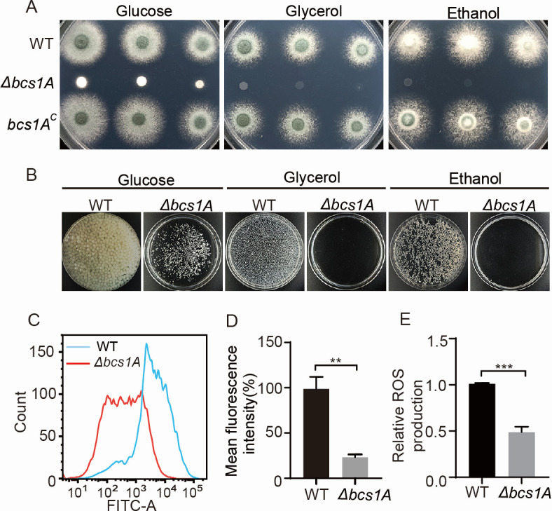
Bcs1A is essential for maintaining optimal mitochondrial function in A. fumigatus. (A) Colony morphologies of the WT, Δbcs1A, and bcs1AC strains on solid medium containing glucose, glycerol, and ethanol as carbon sources, respectively, at 37°C for 48 h. (B) The indicated strains were cultured in a liquid medium supply with glucose, glycerol, and ethanol as carbon sources, respectively, with shaking at 37°C, 220 rpm for 48 h. (C) The mitochondrial membrane potential was assessed using flow cytometry, with FITC-A on the x-axis indicating the relative fluorescence intensity value. (D) Quantitative and statistical analyses of the mean fluorescence intensity and (E) ROS generation in the WT and Δbcs1A mutant strains. **, P < 0.01; ***, P < 0.001.
To investigate the role of Bcs1A more deeply, a Δbcs1A mutant strain lacking the full-length bcs1A gene was constructed through homologous recombination, substituting with the nutritional marker pyrG in the WT background (Fig. S2A). This deletion was verified using diagnostic PCR (Fig. S2B). Comparative growth analysis demonstrated that the Δbcs1A mutant exhibited significant growth impairments on both minimal medium (MM) and yeast extract-glucose (YG) medium, in stark contrast to the WT strain (Fig. 1C and D). Conversely, the bcs1A complementation strain, where the Bcs1A gene was reintroduced, restored normal growth, paralleling the WT phenotype. These results underscore the crucial role of Bcs1A in supporting hyphal growth and overall vitality in A. fumigatus.
Bcs1A is required for proper mitochondrial function
Previous research indicates that Bcs1 is a mitochondrial protein essential for the biogenesis of the respiratory chain complex III in eukaryotic organisms (45–47). Based on this understanding, it was hypothesized that Bcs1A plays a similar critical role in mitochondrial function within A. fumigatus. Mitochondria are pivotal for the proper metabolism of intracellular carbon sources. Notably, the bcs1A mutant displayed profound growth impairments when cultured on both solid (Fig. 2A) and liquid minimal media (Fig. 2B) supplemented with non-fermentable carbon sources such as glycerol or ethanol, compared to the parental WT and complemented strains. To further explore the mitochondrial function in these strains, rhodamine 123 (Rh123), a marker for mitochondrial membrane potential (MMP), was employed. Flow cytometry analysis revealed a significant reduction in mean fluorescence intensity in the Δbcs1A mutant relative to the WT, indicating impaired mitochondrial function in the mutant (Fig. 2C and D). Additionally, the intracellular levels of mitochondria-derived ROS were assessed. The Δbcs1A mutant exhibited decreased ROS production compared to the WT, further supporting the compromised mitochondrial function (Fig. 2E). Previous studies have also highlighted the role of mitochondria, as osmosensing organelles capable of directly responding to osmotic stress in eukaryotes (55). To test the osmotic sensitivity of the Δbcs1A mutant, strains were grown on MM supplemented with osmotic stressors such as KCl, NaCl, or sorbitol. The results showed that the Δbcs1A mutant was more susceptible to osmotic stress compared to the WT strain (Fig. S3). Collectively, these findings underscore the essential role of Bcs1A in maintaining mitochondrial function in A. fumigatus, affecting not only energy metabolism and ROS production but also the organism’s ability to respond to environmental stress.
Bcs1A is required for the antifungal drug response in A. fumigatus
As the mitochondria play important roles in the fungal response to antifungal drugs, the optimal functioning of the organelles is crucial. This study examined the phenotypes of Bcs1A-related strains following exposure to media containing various fungicidal agents, including azoles, simvastatin, terbinafine, amphotericin B, and caspofungin. Notably, except for amphotericin B and caspofungin, the Δbcs1A mutant exhibited increased tolerance to voriconazole, itraconazole (ITC), bifonazole, simvastatin, and terbinafine compared with both the parental WT and bcs1A-complemented strains (Fig. 3A), suggesting the bcs1A plays a role in multidrug tolerance in A. fumigatus. To further quantify this tolerance, commercial E‐test strips were then used to measure the minimum inhibitory concentration (MIC) values of Δbcs1A mutant to the azole compounds, such as itraconazole and voriconazole. As depicted in Fig. 3B, the MIC of itraconazole for the Δbcs1A mutant strain was 6 µg/mL, significantly higher than the 2 µg/mL observed for the WT strain. Similarly, the MIC value of voriconazole for the Δbcs1A mutant strain (0.38 µg/mL) was markedly higher than that of the WT strain (0.125 µg/mL). These findings corroborate the critical role of bcs1A in modulating multidrug tolerance in A. fumigatus.
Fig 3.

Loss of bcs1A reduces susceptibility to antifungal drugs in A. fumigatus. (A) Colony morphology of the strains after cultivation on minimal medium at 37°C for 2 days, as well as after exposure to various concentrations of antifungal drugs (0.15 µg/mL itraconazole, 0.5 µg/mL voriconazole, 1 µg/mL bifonazole, 0.8 µg/mL terbinafine, 10 µg/mL simvastatin, 1 µg/mL amphotericin B, and 0.8 µg/mL caspofungin) at 37°C for 3 days. (B) MIC values of itraconazole and voriconazole were determined using E-test strips on plates containing a mixture of conidia (1 × 10⁵) of the WT or Δbcs1A strains in solid YG medium, followed by incubation at 37°C for 3 days.
In yeast, Bcs1 facilitates the assembly of respiratory complex III by transporting and inserting the Rip1 into the mitochondrial membrane. To further determine if the Δbcs1A’s drug-resistant phenotype is caused by the dysfunction of complex III in OXPHOS or it is specific to the loss of function of Bcs1A, the Δrip1 deletion mutant was constructed. As shown in Fig. S4, The Δrip1 mutant strain exhibited defects in colony growth and conidiation, as well as resistance to the antifungal drugs, consistent with the phenotypes observed in the ΔbcslA mutant strain. Therefore, we hypothesize that the drug-resistant phenotype of ΔbcslA is attributed to the dysfunction of complex III rather than solely to the loss of Bcs1A function.
Deletion of bcs1A upregulates numerous multidrug tolerance-associated transport genes, and loss of the drug efflux pump mdr1 decreases azole tolerance in A. fumigatus
To delve deeper into the molecular mechanisms underlying the involvement of Bcs1A in multidrug tolerance, the whole-genome transcriptome profiles of the WT and Δbcs1A strains were analyzed by RNA sequencing (RNA-seq). The total number of reads for each sample is presented in Table S1, with subsequent mapping to the genome database using HISAT2 software (http://ccb.jhu.edu/software/hisat2/index.shtml; Table S2). Normalization of expression levels was performed using fragments per kilobase per million fragments. Identification of differentially expressed genes (DEGs) showed that 1,349 genes were upregulated and 1,174 were downregulated in the Δbcs1A mutant in comparison with the WT strain (Fig. 4A and B; Data set 1), including a series of mitochondrial localization proteins (Data set 1). Furthermore, Gene Ontology (GO) enrichment analysis showed that the upregulated genes involved in multifaceted activities, including GO terms oxidoreductase activity, secondary metabolic processes, and transition metal ion binding, among others (Fig. 4C). Further investigation of pathways associated with the upregulated genes using the Kyoto Encyclopedia of Genes and Genomes (KEGG) database indicated the upregulated genes’ involvement in a variety of metabolic signaling pathways. Specifically, the bcs1A deletion mutant showed significant enrichment in pathways associated with ATP-binding cassette (ABC) transporters (Fig. 4D). These are a type of drug efflux pump and include tpo1, mdr2, atrB, atrA, abcE, abcA, and the widely recognized drug efflux pump mdr1 (Fig. 5A).
Fig 4.

RNA-sequencing analysis of the bcs1A mutant and WT strains. (A) Volcano plot showing DEGs in the Δbcs1A mutant compared to the WT strain. (B). Heatmap comparing the RNA-seq data of the Δbcs1A mutant relative to the WT strain. (C) GO analysis of the functional category and (D) KEGG analysis of pathway enrichment of DEGs (P < 0.05 and lg2FC > 1 or < −1).
Fig 5.

Loss of bcs1A leads to upregulating various multidrug resistance-associated transport genes. (A) Heatmap showing RNA-seq data of multidrug transport-related genes. Real-time quantitative PCR analysis of indicated genes in the WT and Δbcs1A strains without ITC treatment (B) and with ITC treatment for 1 h (C). ns, not significant; *, P < 0.05; ***, P < 0.001; **, P < 0.01; ***, P < 0.001. (D). Colony morphologies of the indicated strains cultured in MM containing different ITC concentrations (0, 0.15, 0.2, 0.3 µg/mL).
To confirm the RNA-seq findings, quantitative real-time PCR (qRT-PCR) was conducted using biological replicate samples from both the Δbcs1A mutant and the WT control. As depicted in Fig. 5B, the results indicated varying degrees of upregulation of the aforementioned transport genes in the Δbcs1A strain relative to the WT control. The expression of transport genes is critical for fungal adaptation to environments containing drugs; hence, the mRNA levels of the ABC transporters in the WT and Δbcs1A mutant strains were measured by qRT-PCR after the treatment with ITC. Likewise, compared to the WT, the selected genes showed significant upregulation in the Δbcs1A mutant (Fig. 5C), implying that Bcs1A may be involved in drug susceptibility by affecting the expression of drug transporters. To further verify whether the multidrug resistance of the Δbcs1A mutant is caused by the upregulation of efflux pumps, the gene mdr1, a key drug efflux pump in fungi, was deleted in both the WT and Δbcs1A mutant. As shown in Fig. 5D, the double bcs1A and mdr1 mutants exhibited increased susceptibility compared to the single bcs1A mutant when treated with the antifungal drug ITC. Taken together, these data suggest that the bcs1A may contribute to multidrug tolerance by affecting the transcriptional levels of efflux pumps.
Bcs1A regulates metal ion homeostasis in A. fumigatus
The RNA-seq analysis demonstrated that the transcriptional levels of genes associated with metal ion transport, including iron, calcium, magnesium, copper, and zinc transporters, were affected in the bcs1A deletion mutant compared to the WT control (Fig. 4C and 6A). To confirm these findings, qRT-PCR was performed on 11 selected genes, which demonstrated that seven genes (sidA, mirB, mirD, sidE, zrfA, zrfB, zrfC) were downregulated, while four genes (pmcC, mrs2, mac1, Afu3g07860) were upregulated (Fig. S4). These results suggest that Bcs1A plays an important role in maintaining metal ion homeostasis in A. fumigatus. Considering the well-documented correlation between intracellular metal ion homeostasis and the fungal reaction to antifungal medication (39). It was hypothesized that disrupted metal ion regulation might contribute to the observed multidrug resistance shown by the Δbcs1A mutant. The WT, Δbcs1A mutant, and bcs1A complementary strains were then cultured on media containing different metal ions with and without the addition of ITC. As shown in Fig. 6B, metal ion supplementation did not affect colony growth among the bcs1A-related strains under normal conditions. However, when cultured with ITC, the addition of metal ions could ameliorate the drug-induced growth defects in the WT and bcs1A complementary strains (Fig. 6C), suggesting that metal ion homeostasis is linked to antifungal drug resistance, potentially mediating the enhanced multidrug resistance phenotype seen in the Δbcs1A mutant.
Fig 6.

Addition of metal ions can increase azole tolerance of A. fumigatus. (A) Heatmap showing metal ion transport-related genes in the Δbcs1A mutant and its parental WT strain. (B) Colony morphologies of strains grown on MM in the presence of various metal ions (FeCl3 1 mM; CaCl2 50 mM; CuSO4 100 µM, MgCl2 50 mM; ZnCl2 1 mM) at 37°C for 2 days or (C) with 0.2 µg/mL ITC at 37°C for 2.5 days.
DISCUSSION
Mitochondria are commonly referred to as the powerhouses of the eukaryotic cell, underpinning numerous critical biological processes (34, 36, 43). Previous studies highlight the essential role of Bcs1 orthologs in mitochondrial function, primarily through facilitating the transport and integration of the folded Rip1 into developing complex III of the mitochondrial respiratory chain (46, 47, 49).
Bcs1 was the first identified chaperone in yeast (48, 50). Subsequently, six Bcs domain proteins (Bbbcs1a–f) were identified, while only Bbbcs1c is considered the ortholog of yeast bcs1 in the filamentous entomopathogenic fungus B. bassiana (53). The findings of the present study showed that Bcs1A is a homolog of the yeast Bcs1 and contributes to proper mitochondrial function in the filamentous fungal pathogen A. fumigatus (Fig. 1A and 2). Consistent with previous reports on the unicellular yeast and multicellular B. bassiana, analysis of subcellular localization showed that the Bcs1A was located in the mitochondria of A. fumigatus (Fig. 1B). Further bioinformatic analysis revealed that Bcs1 orthologs are widely distributed in eukaryotes, and the two domains (BCS1_N and AAA) are contained in all selected Bcs1 homologs, suggesting the strong conservation of Bcs1 as a mitochondrial protein from fungi to Homo sapiens (Fig. 1A). Phenotypic analysis showed serious impairment of the growth of the Δbcs1A strain compared with the WT and complemented strains (Fig. 1C and D), suggesting Bcs1A is the A. fumigatus homolog of the yeast Bcs1. Further functional analysis showed that a series of mitochondria-related functions, including carbon source metabolism, the generation of ROS and ATP, the MMP, and osmosensing, were abnormal in the Δbcs1A mutant strain, indicating that Bcs1A is critical for proper mitochondrial function in A. fumigatus (Fig. 2).
Numerous studies have reported that impaired vegetative growth caused by dysfunctional mitochondria is an adaptive strategy used by fungi to counteract the presence of harmful antifungal drugs (29, 39). The phenotypic analysis conducted in this study revealed that the Δbcs1A mutant strain displayed decreased susceptibility to antifungal drugs such as azoles, statin, and allylamine (Fig. 3A). The further results of the MIC test were another demonstration that Bcs1A is critical for the fungal response to azoles (Fig. 3B). The antifungal mechanisms of these drugs have been extensively studied. Azoles inhibit the synthesis of the cell membrane lipid ergosterol by targeting lanosterol demethylation (Cyp51/Erg11), resulting in the accumulation of toxic intermediate metabolites and ultimately cell death (19). Statins and allylamines have fungicidal activity by targeting 3-Hydroxy-3-methylglutaryl coenzyme A (HMG CoA) reductase and squalene epoxidase, two key enzymes in the ergosterol biosynthesis pathway (56, 57). Thus, ergosterol synthesis may underlie the Bcs1A-mediated response to drugs. However, there was no detective difference between the Δbcs1A and the WT strains when grown with amphotericin B that induces fungicidal effects by direct binding to ergosterol in the cell membrane (Fig. 3A), resulting in membrane permeabilization and cell death, suggesting the mechanism of Bcs1A-mediated drug tolerance is multifaceted.
In yeast, mitochondrial dysfunction can lead to azole resistance by activating the pleiotropic drug resistance pathway, a direct regulator of the expression of drug efflux pumps (29, 35). RNA-seq and qRT-PCR results revealed an upregulation of multiple efflux pumps in the Δbcs1A mutant (Fig. 4D, 5A through C). Subsequent confirmatory experiments involving the deletion of mdr1 showed a partial reduction in azole tolerance (Fig. 5D). Therefore, it is postulated that the increased expression of drug efflux pumps resulting from mitochondrial dysfunction may be the primary factor contributing to drug resistance in the Δbcs1A mutant. Recent studies have shown a close association between mitochondrial metal ion homeostasis and drug efflux pump expression (39, 58). For example, intracellular calcium disorder induced by mitochondrial dysfunction causes persistent activation of the zinc finger transcription factor CrzA, triggering a global overexpression of multidrug transport genes (39). Both RNA-seq and RT-qPCR showed the abnormal expression of metal ion-related genes in the Δbcs1A mutant (Fig. 4C and 6A). Subsequent phenotypic analysis showed that the WT strain exhibited increased tolerance to itraconazole in media supplemented with various metal ions (FeCl3, CaCl2, CuSO4, MgCl2, ZnCl2) (Fig. 6B and C), suggesting that metal ion homeostasis may be another reason for the increased azole tolerance in the Δbcs1A mutant. Besides, the activity of energy-dependent efflux pumps is crucial for drug tolerance in fungi (59). Hence, the abnormal ATP generation may contribute to drug tolerance by improving the activities of drug efflux pumps and increasing the efflux of intracellular drugs in the Δbcs1A mutant.
Numerous studies have demonstrated that the components of respiratory chain complex III, Bcs1 included, are excellent potential drug targets as they are easily targeted by many natural antibiotics (49, 60). Defective BCS1L (BCS1-like), the human homolog of the yeast Bcs1, may cause various complex III-related pathologies, such as neonatal proximal tubulopathy, hepatic involvement, and encephalopathy (52). In agreement with previous studies, the bioinformatic analysis showed conservation of Bcs1 mitochondrial proteins from fungi to humans (Fig. 1A). Hence, drugs that bind directly to Bcs1A may have side effects when used for the clinical treatment of aspergillosis. Of note, it is reported that the use of pentamidine and clarithromycin, two FDA-approved drugs, can partially rescue the bcs1 mutant in yeast (41). There is thus a possibility that the use of pentamidine or clarithromycin together with antifungal drugs may represent a novel treatment strategy for aspergillosis caused by Bcs1A-mediated (mitochondrial dysfunction) drug resistance.
Collectively, our research underscores the critical role of the mitochondrial protein Bcs1A in modulating antifungal drug responses through the regulation of drug efflux pumps. Nevertheless, the widespread conservation of Bcs1A among different species highlights its importance, which may hinder its potential as a viable drug target in humans due to the likelihood of adverse effects.
MATERIALS AND METHODS
Strains, media, and culture conditions
The A. fumigatus strains utilized in this study are detailed in Table 1. The culture media employed encompassed MM consisting of 50 mL/L 20 × salt solution, 1% glucose, 1 mL/L trace elements, and 2% agar, with a pH of 6.5, as well as YG medium containing 0.5% yeast extract, 2% glucose, 1 mL/L trace elements, and 2% agar (61). Agar was excluded from all liquid media utilized. The glycerol or ethanol medium mirrored the glucose medium, except for substituting glycerol or ethanol as the carbon source. Cultivation of all strains occurred at 37°C for the specified durations.
TABLE 1.
Strains used in this study
| Strain | Genotype | Source |
|---|---|---|
| A1160 | Δku80, pyrG | LL-lab |
| WT | Δku80, A1160::pyrG | LL-lab |
| Bcs1A-GFP | ∆ku80, pyrG, bcs1A::gfp::pyrG | This study |
| Δbcs1A | Δku80, pyrG, Δbcs1A::pyrG | This study |
| bcs1AC | Δku80, pyrG, Δbcs1A::pyrG, bcs1A::bcs1A::hph | This study |
| Δmdr1 | ∆ku80, pyrG, Δmdr1::hph | This study |
| Δmdr1Δbcs1A | Δku80, pyrG, Δbcs1A::pyrG, Δmdr1::hph | This study |
| Δrip1 | Δku80, pyrG, Δrip1::pyrG | This study |
Deletion and complementation of bcs1A
All primers used in this study are displayed in Table 2. For the construction of the bcs1A deletion strain, the open reading frame (ORF) of bcs1A was substituted with the selective marker pyrG. The pyrG marker was amplified from the plasmid pXDRFP4 using the primer pair PyrG-F/PyrG-R. The approximately 1.5 kb of the upstream and downstream flanking sequences was amplified by using the primer pairs bcs1A-P1/P3 as well as bcs1A-P4/P6, respectively. The abovementioned three PCR products were purified and then employed as templates to generate the bcs1A deletion cassette by using the primer pair bcs1A-P2/P5. The resulting fusion product was subsequently used to transform the recipient strain A1160 (62).
TABLE 2.
Primers used in this study
| Name | Sequence (5´ to 3´) |
|---|---|
| bcs1A-gfp-p1 | GGGTGTGTAAGACGCGTCTG |
| bcs1A-gfp-p3 | CCAGCGCCTGCACCAGCTCCTGGTCGCCCTCGGCTGAATCAT |
| bcs1A-gfp-p4 | CATCAGTGCCTCCTCTCAGACAGGTATGTACATTATCATGACGTG |
| bcs1A-gfp-p6 | AGGAATAGTGTACCTCAAG |
| bcs1A-gfp-p2 | TACATACTCACTTTGCACTGG |
| bcs1A-gfp-p5 | CTTTTGATGAGAGCCTACCTG |
| gfp-pyrG F | GGAGCTGGTGCAGGCGCTGG |
| gfp-pyrG R | CTGTCTGAGAGGAGGCACTGATG |
| bcs1A-p1 | CCGCACGGTATCCAGTATCTC |
| bcs1A-p3 | CCTCTAGATGCATGCTCGAGC CCCAGTCTACGGAGATGAGC |
| bcs1A-p4 | CATCAGTGCCTCCTCTCAGACAG TATGTACATTATCATGACGTGT |
| bcs1A-p6 | GAATAGTAACCTATCGGGATG |
| bcs1A-p2 | CATATTCTTAATTCCCGAGC |
| bcs1A-p5 | TTGAGGGTAGTACTGCTGTT |
| PyrG-F | GCCTCAAACAATGCTCTTCACC |
| PyrG-R | CTGTCTGAGAGGAGGCCTGATG |
| bcs1A-self-F | CAAGGACGATTCTTATCCAT |
| bcs1A-self-R | AGGAAAATGATACGCTCCTC |
| Com-bcs1A-F | ACTGCAGGCATGCAAGCTT AATTTCTTCGCAGGTTCGAG |
| Com-bcs1A-R | CGACGGCCAGTGCCAAGCTTCCAACATTGACATATACCCCTC |
| RT-tubA F | TTCCGTCCCGACAACTTCGT |
| RT-tubA R | TCACAGCCTTCAGCCTCACG |
| RT-tpo1 F | CTGCGCCCTATTCAAATGCTT |
| RT-tpo1 R | GAAAAGGTACAGGATTCCGTACACCA |
| RT-mdr1 F | GCTCTGCCTGAAGGTTACG |
| RT-mdr1 R | TGTGGGCTGTTTTGATTGT |
| RT-atrB F | CTGGCCTCGACGGTCAATCC |
| RT-atrB R | TTGGCCAACAGCAACAGGGT |
| RT-abcA F | CGGGCTTTTGGATTTTCATGTACC |
| RT-abcA R | TCAATATCTGAGCACTTGACGCTGG |
| RT-atrA F | GCATCCACGAGTCCAAGCGA |
| RT-atrA R | CCGCGCATATGCCAAGCATC |
| RT-abcE F | GCCACCGACCCAAAGCAGGT |
| RT-abcE R | TGTGCATGGTAAGGCGGCAA |
| RT-mdr2 F | AGATCCGATACGATTATTGTCC |
| RT-mdr2 R | TGCCACTCCATCAATTTGGT |
| RT-sidA F | AACACTTGAGACCTACGGGACA |
| RT-sidA R | TTACAACCTTGAAGCCAGATGC |
| RT-mirB F | AGCTTGATCTATTCGTCCCTCC |
| RT-mirB R | CCCTTGGTCTGAGTCATGTTTT |
| RT-mirD F | ACTACTACACATCCTACCTGC |
| RT-mirD R | AATAGCCAGACTCCCGAGA |
| RT-sidE F | AATGTTCCTGTGGACCGCTTC |
| RT-sidE R | GGCGCTTCTCAGATTGCTTC |
| RT-pmcC F | ACACCGTCATCTTCAACACC |
| RT-pmcC R | GCGTTGATCACCATGAACCA |
| RT-mrs2 F | ACCAAGAGCCGTCCACTCCT |
| RT-mrs2 R | GTAGCCGTGGCTCGTTCGTT |
| RT-mac1 F | TGACAACCCTGATGCCACT |
| RT-mac1 R | AGCTGCCATTTGGACT |
| RT-3g07690 F | TCTGTAAGACCGAGAAGTCC |
| RT-3g07690 R | AACCTTGAATCCGTATCCAG |
| RT-zrfC F | TGTCTTCCACCAGTTCTTCGAGG |
| RT-zrfC R | ATCACGGCAAAGGTCCC |
| RT-zrfB F | TTCATCCATCTTTTGGGACCG |
| RT-zrfB R | GAACAGCACAACAATGGTCA |
| RT-zrfA F | CGCATCTCCTCCATCTTCGT |
| RT-zrfA R | CAAAGTATCGCCCGAACAGGT |
| Rip1-P1 | CACCAGCAACAGCACAAACC |
| Rip1-P2 | CGTGTGAACCAGAGCTTTGTC |
| Rip1-P3 | CGATTAAGTTGGGTAACGCCAGGCTGGGGCCCACGACAATTC |
| Rip1-P4 | ATAAGTAGCCAGTTCCCGAAAGCCTCTTCGTAAGTCGCTAGATG |
| Rip1-P5 | CTGCTCTTACATCCACATCG |
| Rip1-P6 | GTCGCAAACGACTCGTGAAG |
| Rip1-up | GCTCTCTTCCCCCCTCAACT |
| Rip1-down | ACGGGCTTTCCACGCCACTT |
The transformants were grown on MM and verified by diagnostic PCR using primers bcs1A-P1/PyrG-R, bcs1A1-self-F/R, and PyrG-F/bcs1A-P6, respectively. For complementation of the Δbcs1A mutant, the bcs1A-complemented fragment was amplified using the primer pair Com-bcs1A-F/R with the wild-type genomic DNA as a template, which contains the native promoter sequence, the complete ORF, and the termination sequence of bcs1A. This fragment was then cloned into the Hind III site of the plasmid pAN7‐1 and then transformed into the Δbcs1A strain.
Construction of Bcs1A-GFP strain
To label Bcs1A with a GFP tag at the C terminus, we first amplified the upstream sequence (without the termination codon) and downstream sequence of bcs1A from A1160 strain gDNA using the primers bcs1A-gfp-p1/p3 and bcs1A-gfp-p4/p6, respectively. Then, a GFP‐pyrG fragment was amplified from the plasmid pFNO3 using primer pair gfp-pyrG F/R. The abovementioned three DNA fragments were then employed as templates to generate the Bcs1A-gfp cassette using the primers bcs1A-gfp-P2/p5. The fusion production was then transformed to A1160 strain and transformants were detected by diagnostic PCR using the primer pairs bcs1A-gfp-p1/gfp-pyrG R and gfp-pyrG F/bcs1A-gfp-p6.
Fluorescence microscopy
To visualize the subcellular distribution of Bcs1A, the GFP-labeled strain was incubated in 500 µL of liquid MM on glass coverslips for a duration of 12 h. Samples were fixed in 4% formaldehyde for 30 min and then incubated in the dark with the mitochondrial dye MitoTracker Red CMXRos (Beyotime) for 5 min used at a final concentration of 5 nM. Images were obtained by using an Olympus BX53 microscope (Tokyo, Japan).
Flow cytometry
The accumulation of Rh123 was carried out to determine mitochondrial membrane potential in A. fumigatus as described previously (63). Briefly, a total of 5 × 10⁶ spores of the related strains were incubated in 1.5 mL of liquid YG medium at 37°C until conidia began to germinate. The samples were then washed three times with phosphate-buffered saline (PBS) and incubated with Rh123 (Beyotime) at a final concentration of 10 µM for 30 min in the dark, and then washed three times with PBS to remove Rh123. Fluorescence intensity was measured by flow cytometry (BD FACSVerse, USA).
Detection of reactive oxygen species
To measure the production of ROS, 1 × 10⁷ spores of the related strains were inoculated into 100 mL of liquid MM at 37°C, 220 rpm, until conidia grew into mycelia. Then, 2´,7´-dichlorodihydrofluorescein diacetate (MedChemExpress) was added to the medium at a final concentration of 15 µM and incubated at 37°C for 1.5 h in the dark. The collected mycelia were pulverized with liquid nitrogen and subjected to incubation in PBS. The supernatant was next collected by centrifugation at 12,000 × g and 10 min at 4°C. Fluorescence intensity was measured with an excitation wavelength of 504 nm and an emission wavelength of 524 nm using PerkinElmer EnSight (USA). The relative production of ROS was normalized by the fluorescence intensity to the total protein concentration, which was measured by a Bicinchoninic Acid Assay (BCA) protein assay kit (Beyotime, China).
RNA-seq and qRT-PCR
For RNA-seq, 1×10⁸ conidia of relative stains were inoculated into liquid MM in a rotary shaker at 37°C, 220 rpm, 18 h. Then, the mycelium was collected and frozen with liquid nitrogen. Three biological replicates of the WT and Δbcs1A samples were subsequently submitted to Personal Biotechnology (Nanjing, China) for transcriptome analysis using the Illumina platform. The raw data have been submitted to SRA (https://www.ncbi.nlm.nih.gov/sra) at NCBI with six accession numbers SRR28521163–SRR19440565 (WT-1, WT-2, and WT-3, three biological replicate samples of WT) and SRR28521166–SRR19440568 (Δbcs1A-1, Δbcs1A -2, and Δbcs1A -3, three biological replicate samples of Δbcs1A).
For qRT-PCR, samples were prepared as aforementioned. Total RNA was isolated using a UNIQ-10 column total RNA purification kit (Shanghai Sangon Biotech, Shanghai, China) and cDNA was generated using a HiScript II Q RT SuperMix for qPCR kit (Vazyme) according to the manufacturer’s instructions. Quantitative PCR was performed with AceQ qPCR SYBR Green Master Mix (Vazyme) by LightCycler 480 (Roche). Results were normalized to tubA expression, and expression levels were calculated using the 2-∆∆CT method.
ACKNOWLEDGMENTS
This work was financially supported by the National Natural Science Foundation of China (grant no. 32100152), the Natural Science Foundation of Anhui Province (grant no. 2108085QC90), and the Scientific Research Foundation of the Higher Education Institutions of Anhui Province (grant no. KJ2021A0775) to X.Z. The Graduate Research and Innovation Projects of Bengbu Medical University (grant no. Byycxz23011) to W.S. The Undergraduate Research and Innovation Project of Anhui Province (grant no. S202110367007, S202310367042).
Contributor Information
Huaxue Wang, Email: huaxuew2010@163.com.
Xiaogang Zhou, Email: Zhouxg@bbmc.edu.cn.
Gustavo H. Goldman, Universidade de Sao Paulo, Ribeirao Preto, Sao Paulo, Brazil
SUPPLEMENTAL MATERIAL
The following material is available online at https://doi.org/10.1128/spectrum.01172-24.
Fig. S1-S5; Tables S1 and S2.
Different expression genes of RNA-Seq.
ASM does not own the copyrights to Supplemental Material that may be linked to, or accessed through, an article. The authors have granted ASM a non-exclusive, world-wide license to publish the Supplemental Material files. Please contact the corresponding author directly for reuse.
REFERENCES
- 1. Latgé J-P, Chamilos G. 2019. Aspergillus fumigatus and aspergillosis in 2019. Clin Microbiol Rev 33:e00140-18. doi: 10.1128/CMR.00140-18 [DOI] [PMC free article] [PubMed] [Google Scholar]
- 2. de Castro PA, Colabardini AC, Manfiolli AO, Chiaratto J, Silva LP, Mattos EC, Palmisano G, Almeida F, Persinoti GF, Ries LNA, Mellado L, Rocha MC, Bromley M, Silva RN, de Souza GS, Loures FV, Malavazi I, Brown NA, Goldman GH. 2019. Aspergillus fumigatus calcium-responsive transcription factors regulate cell wall architecture promoting stress tolerance, virulence and caspofungin resistance. PLoS Genet 15:e1008551. doi: 10.1371/journal.pgen.1008551 [DOI] [PMC free article] [PubMed] [Google Scholar]
- 3. Abad A, Fernández-Molina JV, Bikandi J, Ramírez A, Margareto J, Sendino J, Hernando FL, Pontón J, Garaizar J, Rementeria A. 2010. What makes Aspergillus fumigatus a successful pathogen? Genes and molecules involved in invasive aspergillosis. Rev Iberoam Micol 27:155–182. doi: 10.1016/j.riam.2010.10.003 [DOI] [PubMed] [Google Scholar]
- 4. van de Veerdonk FL, Gresnigt MS, Romani L, Netea MG, Latgé J-P. 2017. Aspergillus fumigatus morphology and dynamic host interactions. Nat Rev Microbiol 15:661–674. doi: 10.1038/nrmicro.2017.90 [DOI] [PubMed] [Google Scholar]
- 5. Earle K, Valero C, Conn DP, Vere G, Cook PC, Bromley MJ, Bowyer P, Gago S. 2023. Pathogenicity and virulence of Aspergillus fumigatus. Virulence 14:2172264. doi: 10.1080/21505594.2023.2172264 [DOI] [PMC free article] [PubMed] [Google Scholar]
- 6. World Health Organization . 2022. WHO fungal priority pathogens list to guide research, development and public health action. Available from: https://www.who.int/publications/i/item/9789240060241
- 7. Walsh TJ, Anaissie EJ, Denning DW, Herbrecht R, Kontoyiannis DP, Marr KA, Morrison VA, Segal BH, Steinbach WJ, Stevens DA, van Burik J-A, Wingard JR, Patterson TF, Infectious Diseases Society of America . 2008. Treatment of aspergillosis: clinical practice guidelines of the infectious diseases society of America. Clin Infect Dis 46:327–360. doi: 10.1086/525258 [DOI] [PubMed] [Google Scholar]
- 8. Nocua-Báez LC, Uribe-Jerez P, Tarazona-Guaranga L, Robles R, Cortés JA. 2020. [Azoles of then and now: a review]. Rev Chilena Infectol 37:219–230. doi: 10.4067/s0716-10182020000300219 [DOI] [PubMed] [Google Scholar]
- 9. Szymański M, Chmielewska S, Czyżewska U, Malinowska M, Tylicki A. 2022. Echinocandins - structure, mechanism of action and use in antifungal therapy. J Enzyme Inhib Med Chem 37:876–894. doi: 10.1080/14756366.2022.2050224 [DOI] [PMC free article] [PubMed] [Google Scholar]
- 10. Chandrasekar P. 2011. Management of invasive fungal infections: a role for polyenes. J Antimicrob Chemother 66:457–465. doi: 10.1093/jac/dkq479 [DOI] [PubMed] [Google Scholar]
- 11. Roemer T, Krysan DJ. 2014. Antifungal drug development: challenges, unmet clinical needs, and new approaches. Cold Spring Harb Perspect Med 4:a019703. doi: 10.1101/cshperspect.a019703 [DOI] [PMC free article] [PubMed] [Google Scholar]
- 12. Lass-Flörl C. 2011. Triazole antifungal agents in invasive fungal infections: a comparative review. Drugs 71:2405–2419. doi: 10.2165/11596540-000000000-00000 [DOI] [PubMed] [Google Scholar]
- 13. Lockhart SR, Chowdhary A, Gold JAW. 2023. The rapid emergence of antifungal-resistant human-pathogenic fungi. Nat Rev Microbiol 21:818–832. doi: 10.1038/s41579-023-00960-9 [DOI] [PMC free article] [PubMed] [Google Scholar]
- 14. Camps SMT, van der Linden JWM, Li Y, Kuijper EJ, van Dissel JT, Verweij PE, Melchers WJG. 2012. Rapid induction of multiple resistance mechanisms in Aspergillus fumigatus during azole therapy: a case study and review of the literature. Antimicrob Agents Chemother 56:10–16. doi: 10.1128/AAC.05088-11 [DOI] [PMC free article] [PubMed] [Google Scholar]
- 15. Chowdhary A, Kathuria S, Xu J, Meis JF. 2013. Emergence of azole-resistant Aspergillus fumigatus strains due to agricultural azole use creates an increasing threat to human health. PLoS Pathog 9:e1003633. doi: 10.1371/journal.ppat.1003633 [DOI] [PMC free article] [PubMed] [Google Scholar]
- 16. Faria-Ramos I, Farinha S, Neves-Maia J, Tavares PR, Miranda IM, Estevinho LM, Pina-Vaz C, Rodrigues AG. 2014. Development of cross-resistance by Aspergillus fumigatus to clinical azoles following exposure to prochloraz, an agricultural azole. BMC Microbiol 14:155. doi: 10.1186/1471-2180-14-155 [DOI] [PMC free article] [PubMed] [Google Scholar]
- 17. Chen S, Zhu G, Lin H, Guo J, Deng S, Wu W, Goldman GH, Lu L, Zhang Y. 2024. Variability in competitive fitness among environmental and clinical azole-resistant Aspergillus fumigatus isolates. mBio 15:e0026324. doi: 10.1128/mbio.00263-24 [DOI] [PMC free article] [PubMed] [Google Scholar]
- 18. Shapiro RS, Robbins N, Cowen LE. 2011. Regulatory circuitry governing fungal development, drug resistance, and disease. Microbiol Mol Biol Rev 75:213–267. doi: 10.1128/MMBR.00045-10 [DOI] [PMC free article] [PubMed] [Google Scholar]
- 19. Wei X, Zhang Y, Lu L. 2015. The molecular mechanism of azole resistance in Aspergillus fumigatus: from bedside to bench and back. J Microbiol 53:91–99. doi: 10.1007/s12275-015-5014-7 [DOI] [PubMed] [Google Scholar]
- 20. Lelièvre L, Groh M, Angebault C, Maherault A-C, Didier E, Bougnoux M-E. 2013. Azole resistant Aspergillus fumigatus: an emerging problem. Med Mal Infect 43:139–145. doi: 10.1016/j.medmal.2013.02.010 [DOI] [PubMed] [Google Scholar]
- 21. Lamping E, Baret PV, Holmes AR, Monk BC, Goffeau A, Cannon RD. 2010. Fungal PDR transporters: phylogeny, topology, motifs and function. Fungal Genet Biol 47:127–142. doi: 10.1016/j.fgb.2009.10.007 [DOI] [PMC free article] [PubMed] [Google Scholar]
- 22. Cowen LE. 2008. The evolution of fungal drug resistance: modulating the trajectory from genotype to phenotype. Nat Rev Microbiol 6:187–198. doi: 10.1038/nrmicro1835 [DOI] [PubMed] [Google Scholar]
- 23. Chowdhary A, Sharma C, Hagen F, Meis JF. 2014. Exploring azole antifungal drug resistance in Aspergillus fumigatus with special reference to resistance mechanisms. Future Microbiol 9:697–711. doi: 10.2217/fmb.14.27 [DOI] [PubMed] [Google Scholar]
- 24. Ener B, Ergin Ç, Gülmez D, Ağca H, Tikveşli M, Aksoy SA, Otkun M, Siğ AK, Öğünç D, Özhak B, et al. 2022. Frequency of azole resistance in clinical and environmental strains of Aspergillus fumigatus in Turkey: a multicentre study. J Antimicrob Chemother 77:1894–1898. doi: 10.1093/jac/dkac125 [DOI] [PubMed] [Google Scholar]
- 25. Horvath SE, Daum G. 2013. Lipids of mitochondria. Prog Lipid Res 52:590–614. doi: 10.1016/j.plipres.2013.07.002 [DOI] [PubMed] [Google Scholar]
- 26. Nam E, Han J, Suh JM, Yi Y, Lim MH. 2018. Link of impaired metal ion homeostasis to mitochondrial dysfunction in neurons. Curr Opin Chem Biol 43:8–14. doi: 10.1016/j.cbpa.2017.09.009 [DOI] [PubMed] [Google Scholar]
- 27. Borcherding N, Brestoff JR. 2023. The power and potential of mitochondria transfer. Nature 623:283–291. doi: 10.1038/s41586-023-06537-z [DOI] [PMC free article] [PubMed] [Google Scholar]
- 28. Shingu-Vazquez M, Traven A. 2011. Mitochondria and fungal pathogenesis: drug tolerance, virulence, and potential for antifungal therapy. Eukaryot Cell 10:1376–1383. doi: 10.1128/EC.05184-11 [DOI] [PMC free article] [PubMed] [Google Scholar]
- 29. Song J, Zhou J, Zhang L, Li R. 2020. Mitochondria-mediated azole drug resistance and fungal pathogenicity: opportunities for therapeutic development. Microorganisms 8:1574. doi: 10.3390/microorganisms8101574 [DOI] [PMC free article] [PubMed] [Google Scholar]
- 30. Zhang X, Moye-Rowley WS. 2001. Saccharomyces cerevisiae multidrug resistance gene expression inversely correlates with the status of the F(0) component of the mitochondrial ATPase. J Biol Chem 276:47844–47852. doi: 10.1074/jbc.M106285200 [DOI] [PubMed] [Google Scholar]
- 31. Gohil VM, Hayes P, Matsuyama S, Schägger H, Schlame M, Greenberg ML. 2004. Cardiolipin biosynthesis and mitochondrial respiratory chain function are interdependent. J Biol Chem 279:42612–42618. doi: 10.1074/jbc.M402545200 [DOI] [PubMed] [Google Scholar]
- 32. Koshiba T, Holman HA, Kubara K, Yasukawa K, Kawabata S, Okamoto K, MacFarlane J, Shaw JM. 2011. Structure-function analysis of the yeast mitochondrial Rho GTPase, Gem1p: implications for mitochondrial inheritance. J Biol Chem 286:354–362. doi: 10.1074/jbc.M110.180034 [DOI] [PMC free article] [PubMed] [Google Scholar]
- 33. Okamoto K, Shaw JM. 2005. Mitochondrial morphology and dynamics in yeast and multicellular eukaryotes. Annu Rev Genet 39:503–536. doi: 10.1146/annurev.genet.38.072902.093019 [DOI] [PubMed] [Google Scholar]
- 34. Youle RJ, van der Bliek AM. 2012. Mitochondrial fission, fusion, and stress. Science 337:1062–1065. doi: 10.1126/science.1219855 [DOI] [PMC free article] [PubMed] [Google Scholar]
- 35. Thomas E, Roman E, Claypool S, Manzoor N, Pla J, Panwar SL. 2013. Mitochondria influence CDR1 efflux pump activity, Hog1-mediated oxidative stress pathway, iron homeostasis, and ergosterol levels in Candida albicans. Antimicrob Agents Chemother 57:5580–5599. doi: 10.1128/AAC.00889-13 [DOI] [PMC free article] [PubMed] [Google Scholar]
- 36. Neubauer M, Zhu Z, Penka M, Helmschrott C, Wagener N, Wagener J. 2015. Mitochondrial dynamics in the pathogenic mold Aspergillus fumigatus: therapeutic and evolutionary implications. Mol Microbiol 98:930–945. doi: 10.1111/mmi.13167 [DOI] [PubMed] [Google Scholar]
- 37. Wei X, Chen P, Gao R, Li Y, Zhang A, Liu F, Lu L. 2017. Screening and characterization of a non-cyp51a mutation in an Aspergillus fumigatus cox10 strain conferring azole resistance. Antimicrob Agents Chemother 61:e02101-16. doi: 10.1128/AAC.02101-16 [DOI] [PMC free article] [PubMed] [Google Scholar]
- 38. Chen M, Zhong G, Wang S, Chen P, Li L. 2022. Deletion of cox7c results in pan-azole resistance in Aspergillus fumigatus. Antimicrob Agents Chemother 66:e0015122. doi: 10.1128/aac.00151-22 [DOI] [PMC free article] [PubMed] [Google Scholar]
- 39. Li Y, Zhang Y, Zhang C, Wang H, Wei X, Chen P, Lu L. 2020. Mitochondrial dysfunctions trigger the calcium signaling-dependent fungal multidrug resistance. Proc Natl Acad Sci U S A 117:1711–1721. doi: 10.1073/pnas.1911560116 [DOI] [PMC free article] [PubMed] [Google Scholar]
- 40. Peng Y, Dong D, Jiang C, Yu B, Wang X, Ji Y. 2012. Relationship between respiration deficiency and azole resistance in clinical Candida glabrata. FEMS Yeast Res 12:719–727. doi: 10.1111/j.1567-1364.2012.00821.x [DOI] [PubMed] [Google Scholar]
- 41. Panozzo C, Laleve A, Tribouillard-Tanvier D, Ostojić J, Sellem CH, Friocourt G, Bourand-Plantefol A, Burg A, Delahodde A, Blondel M, Dujardin G. 2017. Chemicals or mutations that target mitochondrial translation can rescue the respiratory deficiency of yeast bcs1 mutants. Biochim Biophys Acta Mol Cell Res 1864:2297–2307. doi: 10.1016/j.bbamcr.2017.09.003 [DOI] [PubMed] [Google Scholar]
- 42. Fox TD. 2012. Mitochondrial protein synthesis, import, and assembly. Genetics 192:1203–1234. doi: 10.1534/genetics.112.141267 [DOI] [PMC free article] [PubMed] [Google Scholar]
- 43. Tang JX, Thompson K, Taylor RW, Oláhová M. 2020. Mitochondrial OXPHOS biogenesis: co-regulation of protein synthesis, import, and assembly pathways. Int J Mol Sci 21:3820. doi: 10.3390/ijms21113820 [DOI] [PMC free article] [PubMed] [Google Scholar]
- 44. Ostojić J, Panozzo C, Lasserre J-P, Nouet C, Courtin F, Blancard C, di Rago J-P, Dujardin G. 2013. The energetic state of mitochondria modulates complex III biogenesis through the ATP-dependent activity of Bcs1. Cell Metab 18:567–577. doi: 10.1016/j.cmet.2013.08.017 [DOI] [PubMed] [Google Scholar]
- 45. Sawamura R, Ogura T, Esaki M. 2014. A conserved α helix of Bcs1, a mitochondrial AAA chaperone, is required for the Respiratory Complex III maturation. Biochem Biophys Res Commun 443:997–1002. doi: 10.1016/j.bbrc.2013.12.084 [DOI] [PubMed] [Google Scholar]
- 46. Wagener N, Ackermann M, Funes S, Neupert W. 2011. A pathway of protein translocation in mitochondria mediated by the AAA-ATPase Bcs1. Mol Cell 44:191–202. doi: 10.1016/j.molcel.2011.07.036 [DOI] [PubMed] [Google Scholar]
- 47. Wagener N, Neupert W. 2012. Bcs1, a AAA protein of the mitochondria with a role in the biogenesis of the respiratory chain. J Struct Biol 179:121–125. doi: 10.1016/j.jsb.2012.04.019 [DOI] [PubMed] [Google Scholar]
- 48. Cui TZ, Smith PM, Fox JL, Khalimonchuk O, Winge DR. 2012. Late-stage maturation of the Rieske Fe/S protein: Mzm1 stabilizes Rip1 but does not facilitate its translocation by the AAA ATPase Bcs1. Mol Cell Biol 32:4400–4409. doi: 10.1128/MCB.00441-12 [DOI] [PMC free article] [PubMed] [Google Scholar]
- 49. Tang WK, Borgnia MJ, Hsu AL, Esser L, Fox T, de Val N, Xia D. 2020. Structures of AAA protein translocase Bcs1 suggest translocation mechanism of a folded protein. Nat Struct Mol Biol 27:202–209. doi: 10.1038/s41594-020-0373-0 [DOI] [PMC free article] [PubMed] [Google Scholar]
- 50. Nouet C, Truan G, Mathieu L, Dujardin G. 2009. Functional analysis of yeast bcs1 mutants highlights the role of Bcs1p-specific amino acids in the AAA domain. J Mol Biol 388:252–261. doi: 10.1016/j.jmb.2009.03.018 [DOI] [PubMed] [Google Scholar]
- 51. Stan T, Brix J, Schneider-Mergener J, Pfanner N, Neupert W, Rapaport D. 2003. Mitochondrial protein import: recognition of internal import signals of BCS1 by the TOM complex. Mol Cell Biol 23:2239–2250. doi: 10.1128/MCB.23.7.2239-2250.2003 [DOI] [PMC free article] [PubMed] [Google Scholar]
- 52. de Lonlay P, Valnot I, Barrientos A, Gorbatyuk M, Tzagoloff A, Taanman JW, Benayoun E, Chrétien D, Kadhom N, Lombès A, de Baulny HO, Niaudet P, Munnich A, Rustin P, Rötig A. 2001. A mutant mitochondrial respiratory chain assembly protein causes complex III deficiency in patients with tubulopathy, encephalopathy and liver failure. Nat Genet 29:57–60. doi: 10.1038/ng706 [DOI] [PubMed] [Google Scholar]
- 53. Hou J, Ding JL, Peng YJ, Feng MG, Ying SH. 2023. Genome-wide identification of BCS1 domain-containing proteins reveals the mitochondrial bcs1 essential for growth, stress response, and virulence of the filamentous entomopathogenic fungus Beauveria bassiana. Microbiol Res 267:127262. doi: 10.1016/j.micres.2022.127262 [DOI] [PubMed] [Google Scholar]
- 54. Klugherz I, Basch M, Ng N, Zhu Z, Wagener N, Wagener J. 2023. Only one of three Bcs1 homologs in Aspergillus fumigatus confers respiratory growth. J Fungi (Basel) 9:1074. doi: 10.3390/jof9111074 [DOI] [PMC free article] [PubMed] [Google Scholar]
- 55. Ikizawa T, Ikeda K, Arita M, Kitajima S, Soga T, Ichijo H, Naguro I. 2023. Mitochondria directly sense osmotic stress to trigger rapid metabolic remodeling via regulation of pyruvate dehydrogenase phosphorylation. J Biol Chem 299:102837. doi: 10.1016/j.jbc.2022.102837 [DOI] [PMC free article] [PubMed] [Google Scholar]
- 56. Tavakkoli A, Johnston TP, Sahebkar A. 2020. Antifungal effects of statins. Pharmacol Ther 208:107483. doi: 10.1016/j.pharmthera.2020.107483 [DOI] [PubMed] [Google Scholar]
- 57. White TC, Marr KA, Bowden RA. 1998. Clinical, cellular, and molecular factors that contribute to antifungal drug resistance. Clin Microbiol Rev 11:382–402. doi: 10.1128/CMR.11.2.382 [DOI] [PMC free article] [PubMed] [Google Scholar]
- 58. Eldesouky HE, Lanman NA, Hazbun TR, Seleem MN. 2020. Aprepitant, an antiemetic agent, interferes with metal ion homeostasis of Candida auris and displays potent synergistic interactions with azole drugs. Virulence 11:1466–1481. doi: 10.1080/21505594.2020.1838741 [DOI] [PMC free article] [PubMed] [Google Scholar]
- 59. Guo H, Xie SM, Li SX, Song YJ, Zhong XY, Zhang H. 2017. Involvement of mitochondrial aerobic respiratory activity in efflux-mediated resistance of C.albicans to fluconazole. J Mycol Med 27:339–344. doi: 10.1016/j.mycmed.2017.04.004 [DOI] [PubMed] [Google Scholar]
- 60. Zinovkin RA, Zamyatnin AA. 2019. Mitochondria-targeted drugs. Curr Mol Pharmacol 12:202–214. doi: 10.2174/1874467212666181127151059 [DOI] [PMC free article] [PubMed] [Google Scholar]
- 61. Gupta SK, Maggon KK, Venkitasubramanian TA. 1976. Effect of zinc on adenine nucleotide pools in relation to aflatoxin biosynthesis in Aspergillus parasiticus. Appl Environ Microbiol 32:753–756. doi: 10.1128/aem.32.6.753-756.1976 [DOI] [PMC free article] [PubMed] [Google Scholar]
- 62. Szewczyk E, Nayak T, Oakley CE, Edgerton H, Xiong Y, Taheri-Talesh N, Osmani SA, Oakley BR. 2006. Fusion PCR and gene targeting in Aspergillus nidulans. Nat Protoc 1:3111–3120. doi: 10.1038/nprot.2006.405 [DOI] [PubMed] [Google Scholar]
- 63. Zhou X, Yang G, Li C, Yang F, Chang X. 2022. Requirement of a putative mitochondrial GTPase, GemA, for azole susceptibility, virulence, and cell wall integrity in Aspergillus fumigatus. Front Microbiol 13:957857. doi: 10.3389/fmicb.2022.957857 [DOI] [PMC free article] [PubMed] [Google Scholar]
Associated Data
This section collects any data citations, data availability statements, or supplementary materials included in this article.
Supplementary Materials
Fig. S1-S5; Tables S1 and S2.
Different expression genes of RNA-Seq.