



Abstract
Endoscopic and biopsy findings have identified two distinct phenotypes among individuals with calcium oxalate (CaOx) kidney stones. The first type has normal renal papillae but shows interstitial mineral deposition, known as Randall’s plaque. The other phenotype presents with collecting duct plugging and a higher incidence of loss of papilla tissue mass. With Randall’s plaque, renal papilla injury involves the loss of small patches of calcified tissue (Randall’s plaque detaching with the stone), which likely results in damage to only a few nephrons. In contrast, collecting duct mineral plugs are very large, causing obstruction to tubular flow. Since each terminal collecting duct drains thousands of nephrons, ductal plugs could lead to the degeneration of many nephrons and a significant loss of renal glomeruli. New visualization techniques for immune cells in papillary biopsies have revealed that the Randall’s plaque phenotype is marked by the accumulation of macrophages around the plaque regions. In contrast, preliminary data on the plugging phenotype shows collecting duct damage with mineral plugs, increased T-lymphocytes throughout the papilla. These regions also show tubulitis, i.e., T-cell infiltration into nearby collecting duct epithelium. This suggests that while some CaOx stone formers may have some papillary inflammation but with minimal damage to nephrons, others suffer from obstruction to flow for many nephrons that may also include destructive inflammation in the renal tissue. We propose that CaOx stone formers with the plugging phenotype will have a higher long-term risk for loss of renal function.
Keywords: kidney stones, nephrolithiasis, calcium oxalate, inflammation
Introduction
Urinary stone disease is common, affecting about 10% of Americans who will experience at least one stone episode in their lifetime [1]. The risk of recurrence is also high [2]. The annual cost of treating kidney stones is projected to reach $4 billion by 2030 [1]. Most kidney stones are composed primarily of calcium oxalate (CaOx), and most CaOx stone formers are classed as idiopathic, having no underlying disease to explain their condition [3].
A recent study separated idiopathic CaOx stone formers according to whether they produced any stones that had grown on Randall’s plaque [4]. Endoscopic examination of those CaOx stone formers with Randall’s plaque stones showed abundant Randall’s plaque on their papillae, along with very little ductal plugging. In contrast, CaOx stone formers without Randall’s plaque stones typically had minimal plaque but extensive ductal plugging, along with larger stones and larger stone burdens.
Ductal plugs in stone formers tend to be very large (>1 mm in diameter [5]), and, indeed, the plugs must be of significant size to be visible by endoscope. These plugs obstruct flow in the terminal collecting ducts (duct of Bellini), each of which receives fluid from thousands of nephrons [6]. As a result, such obstructions can potentially lead to substantial nephron loss. Given that a kidney has about one million nephrons [7], and considering Kriz & Kaissling’s estimate that each terminal collecting duct drains approximately 2,750 nephrons[6], the blockage of just 4 terminal ducts creates obstruction for 1.1% of all nephrons. Some idiopathic CaOx stone formers exhibit multiple ductal plugs in every papilla, meaning a substantial fraction of their nephrons must be subjected to obstructive stress.
Renal damage from urinary obstruction has long been associated with tissue inflammation [8]. Direct evidence of inflammation in individuals with kidney stones primarily comes from retrospective studies [9]. Two independent analyses of NHANES data have found a correlation between elevated serum levels of C-reactive protein and a lifetime history of kidney stones [10, 11]. Additionally, an extensive examination of nephrectomy specimens has shown an increased presence of pro-inflammatory M1 macrophages (CD68+) in renal tissues of stone formers. The number of these macrophages was found to correlate with the mineral content in the medulla [12]. These findings are consistent with those of Taguchi et al. [13], who noted increased macrophages, plasma cells, and neutrophils in papillary biopsies of stone formers, along with elevated mRNA signatures indicating immune cell activity.
Because ductal plugging leads to nephron obstruction, one might expect greater tissue inflammation in CaOx patients with ductal plugging than in those with Randall’s plaque. But the difference in tissue mineral location in these two phenotypes of CaOx stone former could also affect the forms of inflammation that are induced. Previous hypotheses regarding inflammation in kidney stone formers have focused on linking immune response to the interaction of mineral crystals with cells [14, 15]. Randall’s plaque crystals may interact with basal membrane cells of thin limb or connective tissue cells, because they are located in the interstitium. In contrast, collecting duct plugs impinge on the luminal membranes of collecting duct cells. Given these distinct mineral interactions with cells, one would anticipate unique expressions of immune cells between these two groups—Randall’s plaque and ductal plugging—reflecting the differential cell contact with mineral.
In this paper, we highlight observations supporting the hypothesis that inflammation in CaOx stone formers differs between patients with Randall’s plaque and those with ductal plugging. Furthermore, we also suggest that long-term renal function outcomes may vary between these phenotypes of CaOx stone formers, advocating for further research in this domain. Developing targeted therapies tailored to each specific phenotype could improve outcomes by reducing stone recurrence ratesand mitigating the progression to chronic kidney disease [16].
Methods
Patients in this study were recruited and studied as part of a larger project, which is described in more detail elsewhere [4]. All patients in the present study produced stones that were majority CaOx by volume (as measured by micro CT) and no patients were included who had any brushite in any part of the stone specimen or who had any systemic causes for their stones (no primary hyperoxaluria, bariatric surgery, bowel resection, medullary sponge kidney, or hyperparathyroidism). Scoring of papillae for plaque, plug, or loss of papillary contour were done either by grading endoscopic video [4] or in the operating room during the procedure [17]. For ductal plugging, 0 was given for no visible plugs (yellow plaque deposits) or dilated ducts, 1 for the presence of 1–5 plugs or dilated ducts, and 2 for greater than 5. For each patient, scores from all graded papillae were averaged. For papillary pitting, 0 for no pitting, 1 for a pit that occupied just part of the papillary tip, and 2 for pitting that covered most of the tip of the papilla. For loss of papillary contour, 0 was given for normal papilla shape, 1 for some depression of the papilla, and 2 for complete flattening. For Randall’s plaque visible on the papilla, a score of 0 was given for mild plaque, 1 for moderate, and 2 for high coverage of the papilla tip by plaque. Biopsies were taken from papillae, fixed, and embedded in paraffin.
Multiplexed imaging of antibodies staining for various protein markers was done using PhenoCycler (Akoya Biosciences) [18]. Briefly, this technology utilizes antibodies tagged with nucleotide barcodes that allow for the reversible attachment of a fluorescent marker to a specific antibody. A tissue slice is first incubated with multiple barcode-tagged antibodies. Then, a few fluorescent markers, each matching one antibody-specific barcode, are added at a time. In our study, three different fluorescent markers were attached for each confocal imaging sequence, along with DAPI to stain the nuclei. After imaging, the fluorescent markers were removed from the tissue, and a new set of three markers, corresponding to different antibodies, was applied. This process was repeated for multiple imaging sequences. Custom software enabled alignment of all the imaging runs and the unsupervised identification of cell types using antibody staining associated with each nucleus [19]. In the examples provided in this paper, we will show the staining of only a few antibodies, each represented with a unique color.
Statistics on patient data were carried out using Tukey-Kramer HSD test for continuous data and chi-square for categorical data (JMP Pro 17, JMP Statistical Discovery LLC, Cary, NC).
Results and discussion
Examples of the two phenotypes.
Calcium oxalate (CaOx) stone formers exhibit two distinct papillary phenotypes during endoscopic examination. For example, Figure 1 shows two patients examined on the same day, both with CaOx stones. Micro CT analysis revealed that the patient on the left had stones composed of 91% CaOx and 9% apatite, while the patient on the right had stones made up of 76% CaOx and 24% apatite. Despite these similar stone compositions, their papillae appeared very different. The patient on the left had mostly normal-looking papillae with only Randall’s plaque visible. In this patient, seven papillae were evaluated, showing mean scores of 0.4 for plugging or dilated ducts, 0.7 for loss of papillary contour, and 0.9 for Randall’s plaque [17]. In contrast, the patient on the right had ductal plugging in every papilla. Four papillae from this patient had mean scores of 1.75 for plugging or dilated ducts, 1.5 for loss of contour, and 0.5 for Randall’s plaque. A score of 1.75 for plugging indicates that all papillae were affected, with three of the four papillae showing more than five yellow plaque deposits (plugs) or dilated ducts each[17].
Figure 1.
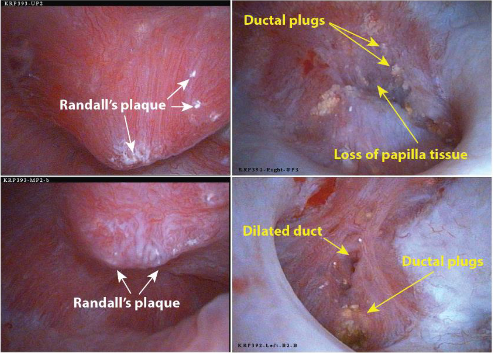
Endoscopic views of representative papillae from two patients. Both patients had majority CaOx stones and no systemic cause for their disease. The patient on the left was a 50 year-old female, and the patient on the right was a 40 year-old female. Left: Two papillae show normal shape, but both have some deposition of Randall’s (interstitial) plaque. Right: The two papillae show many plugs of mineral (yellowish in color). The upper right panel shows a papilla with a large amount of tissue loss at its tip. Lower right panel shows a compound papilla with a dilated duct. (A typical human papilla is about 1 cm wide at its base.) [add scores to the images]
These two phenotypes are both commonly seen
In our previous research, we identified groups of calcium oxalate (CaOx) stone formers characterized by either high levels of Randall’s plaque or extensive ductal plugging[4]. We have now extended these observations to include 101 idiopathic CaOx patients. Papillary scoring revealed an inverse correlation between the amount of Randall’s plaque and the score for plugging/dilated ducts (Figure 2, r2=0.34, P<0.0001). Additionally, scores for plugging were positively correlated with loss of papilla mass (loss of contour, r2=0.27, P<0.0001). In contrast, higher scores for Randall’s plaque were associated with less loss of papillary mass (papillary contour, r2=0.13, P=0.0002).
Figure 2.
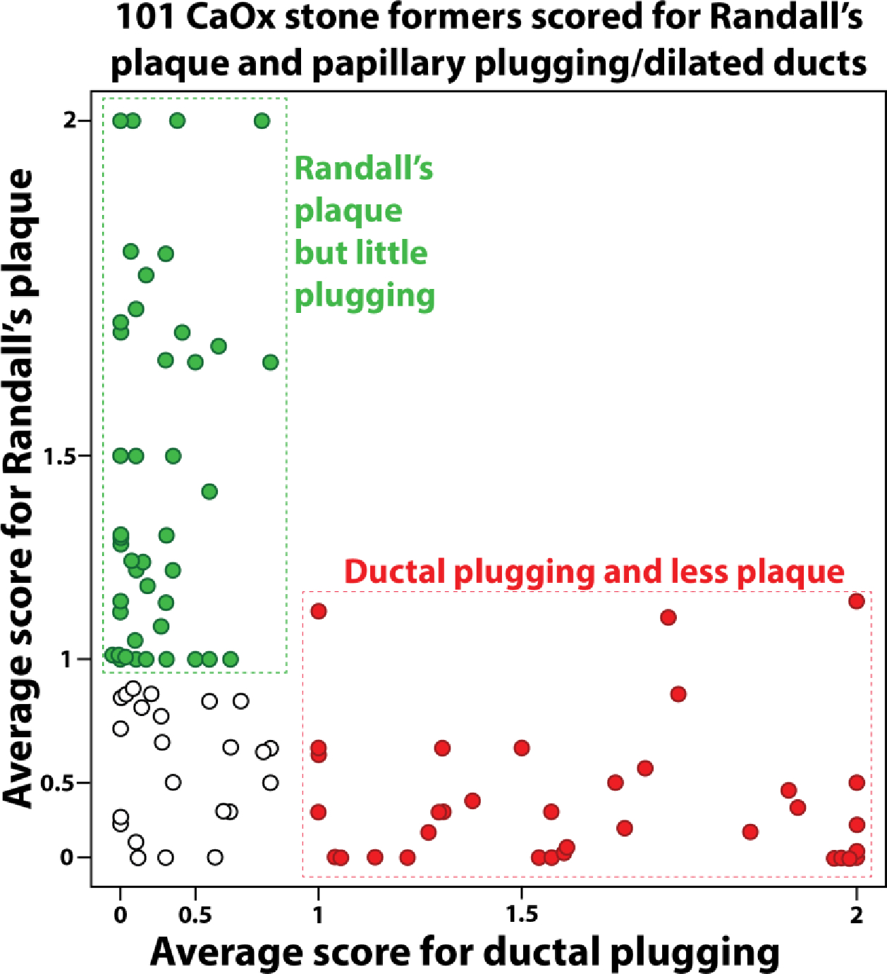
Idiopathic calcium oxalate (CaOx) stone formers tend to group into two papillary mineral patterns. Graph shows semi-quantitative grading of papillary mineral using endoscopic video in 101 CaOx stone formers. Axis scales are exponential. Note that a patient plugging score of 1.0 indicates that on average every papilla showed 1–5 plugs or dilated ducts; thus, a score <1.0 means that at least one papilla in that patient had no plugs or dilated ducts. In contrast, a Randall’s plaque score of 1.0 means that the average papilla was moderately covered with plaque, and a score of 0 means ‘mild’ coverage by plaque. Empty circles show patients with average scores <1 for both Randall’s plaque and plugging/dilated ducts (the Neither group in Table 1).
When we classified the 101 patients into high-plaque, high-plugging, or neither category, we found no significant differences in age, serum chemistries, or 24-hour urine values among the groups (Table 1). However, there were more females in the high-plugging group (P=0.04), and papillary pitting was more common in both the high-plaque and high-plugging groups compared to those with low mineral scores. Despite these differences, the patients were generally similar across most clinical measures.
Table 1.
Clinical characteristics of patient groups shown in Figure 2.
| High plugging scores | High Randall’s plaque scores | Neither | |
|---|---|---|---|
| N | 35 | 43 | 23 |
| Age | 52.6±13.0 | 50.4±16.6 | 50.7±16.7 |
| Sex (M/F) | 11/24* | 25/18 | 13/10 |
| Papillary plugging | 1.57±0.36 | 0.24±0.24 | 0.37±0.30 |
| Papillary pitting | 0.72±0.59 | 0.94±0.56 | 0.31±0.31 † |
| Loss of papillary contour | 0.46±0.40 ‡ | 0.13±0.22 | 0.23±0.25 |
| Papillary Randall’s plaque | 0.35±0.35 | 1.40±0.34 | 0.55±0.32 |
| Serum Ca (mg/dL) | 9.51±0.58 | 9.42±0.33 | 9.36±0.35 |
| Serum creatinine (mg/dL) | 0.97±0.29 | 0.93±0.20 | 0.91±0.16 |
| Urine vol (24 h, L) | 2.1±0.6 | 2.0±0.9 | 1.9±0.9 |
| Urine pH | 6.1±0.4 | 6.1±0.4 | 6.0±0.5 |
| Urine citrate (mg/day) | 568±341 | 627±355 | 724±376 |
| Urine Ca (mg/day) | 271±159 | 226±113 | 237±113 |
| Urine oxalate (mg/day) | 39±18 | 39±14 | 38±13 |
| Urine sodium (mmol/day) | 168±74 | 177±77 | 166±74 |
| Supersaturation for CaOx | 7.5±3.6 | 6.9±3.3 | 7.9±4.2 |
| Supersaturation for CaP | 1.3±1.1 | 1.3±0.9 | 1.4±1.1 |
Italics indicates scores used to distinguish groups.
Chi square test yields P=0.04 for sex distribution among these groups.
Different from both high plugging and high Randall’s plaque group, P<0.02 and P<0.001, respectively.
Different from both high Randall’s plaque and the ‘Neither’ group, P<0.001 and P<0.02, respectively.
The severe nature of ductal mineral plugs
It’s important to clarify that the plugging scored in this study refers to significantly dilated collecting ducts filled with mineral deposits. These are seen as yellow regions on the papilla tip, sometimes with bits of mineral extending into the urine space [17, 20]. The plugging score also includes dilated collecting duct openings, indicating a ductal mineral plug that had since fallen out [17]. These dilated ducts and plugs at the papilla tip are quite large—up to 20 times wider than normal collecting ducts.[20] This is in stark contrast to the occasional plugged tubule found in normal kidneys higher up in the medulla, which are about 40 micrometers in diameter [21, 22]. These smaller plugs, thinner than a human hair, would likely not be visible with an endoscope, even if they were on the papilla’s surface. Therefore, the ductal plugs observed in these CaOx stone formers represent a severe pathology.
Ductal plugging and the progressive loss of papilla tissue
The loss of papillary tissue associated with ductal plugging, as measured by changes in papillary contour scores, is explained by a hypothesis from 2005. This hypothesis suggests that the presence of a ductal mineral plug triggers destructive activity in nearby cells, leading to progressive tissue damage throughout the papilla [20]. According to this hypothesis, the process begins with the injury of collecting duct cells by luminal crystals, followed by the death of these cells. As the mineral plug expands, it presses into the surrounding tissue, provoking inflammation in the areas around the plugged duct[20]. Evidence for the last stages of this hypothesis is showing up in some newer work, as shown in Figure 3, which shows a collecting duct adjacent to a ductal mineral plug. The collecting duct appears normal in structure except for the presence of T-cells migrating up between the principal cells (arrows in Figure 3d). The invasion of lymphocytes into the renal tubular epithelium, known as tubulitis, is considered a sign of impending necrosis of the tubule [23].
Figure 3.
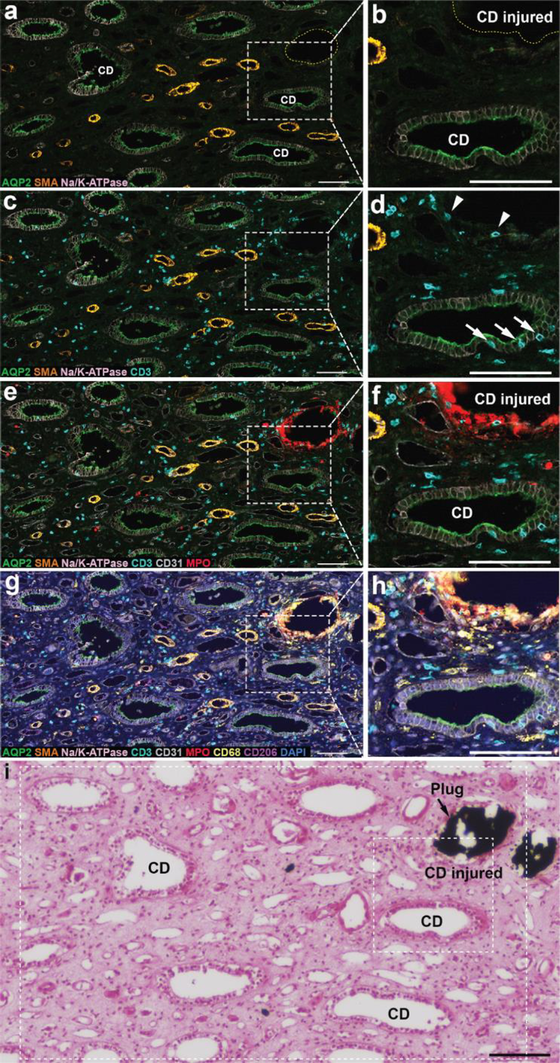
Multiplexed imaging on a biopsy from a CaOx stone former with ductal plugging. a. Antibody localization is shown for aquaporin 2 (green), alpha-SMA (orange), and NaK-ATPase (light pink). b. Blow-up showing injured collecting duct (which contained mineral, dashed yellow line), along with a normal appearing collecting duct. c, d. The same regions but with staining for T-cells revealed (cyan). Note T-cells invading the epithelium of the healthy collecting duct (arrows). e, f. Same regions but with visualization of CD31 (endothelium, light gray) and myeloperoxidase (neutrophils, red). Note coating of neutrophils around mineral plug. g, h. Visualization of macrophages (CD68, light yellow) and CD206 (marker of M2 macrophages, pink). Note that many macrophages also surround the mineral plug. i. Yasue stain of consecutive section, verifying the mineral plug in the collecting duct marked ‘injured’ in b and f. Dashed rectangles indicate approximate regions of panels a-h in this consecutive section. Scale bars are 100 μm.
Panels a and b in Figure 3 show a healthy collecting duct (CD) situated near a collecting duct filled with mineral deposits and which shows very little staining for collecting duct cells (CD injured). In panels c and d, the healthy collecting duct is seen to have T-cells within its epithelium. (Note that this phenomenon of tubulitis in collecting ducts is difficult to detect without special stains.) There are also a few T-cells next to the mineral plug. Panels e and f add visualization of neutrophils, which are seen to completely surround the mineral plug. Panels g and h add visualization of macrophages, which are also surrounding the mineral plug, so that the mineral plug is thoroughly enveloped by immune cells.
This focal investment of immune cells against the mineral plug is quite dramatic and points to a general increase in immune cell activity in the tissue. Note that the ductal plug in this case has not yet grown to a size that would press against neighboring tissues, but still there is T-cell activity attacking an adjacent collecting duct. The idea that a ductal plug could lead to destruction of nearby tissues, was first proposed for the profound papillary injury seen in papillary biopsies from brushite stone formers [20], but has also been proposed for other kinds of stone former in which ductal mineral plugs are also found [24]. These investigators also proposed that injury of a collecting duct epithelium might reduce its ability to acidify the tubular fluid and thus lead to the formation of a new calcium phosphate plug [20]. Thus, an initial ductal mineral plug would lead to injury to surrounding tubules which would then also become plugged with mineral, and the progression of tissue injury could spread through the papilla. There may also be a role for reactive oxygen species in all of this, as crystals interacting with cells can induce the production of reactive oxygen species and so contribute to the inflammatory state [16, 25].
Randall’s plaque and mild inflammation of the papilla tissue
Contrast the picture of a ductal plug associated with extensive investment of inflammatory cells with that shown in Figure 4, in which a biopsy from a CaOx stone former shows Randall’s plaque and no mineral plugs. The plaque regions—which are interstitial, beginning in the basement membranes of thin limbs [26]—have macrophages associated with them, but no tubulitis seen in any surrounding tubules. The association of Randall’s plaque with macrophages has been reported before [18]. An absence of tubular injury has been reported repeatedly for CaOx biopsies containing only Randall’s plaque [24, 26, 27].
Figure 4.

Papillary biopsy from a patient who had extensive deposition of Randall’s plaque, as seen by endoscopy. a. H&E staining of the multiplexed-imaged section after all the cycling of reagents and fluorescence imaging was complete; note remarkable preservation of the tissue. b. Multiplex image from that section: DAPI, stain for nuclei; AQP2, aquaporin-2 for collecting duct; aSMA, alpha-smooth muscle actin; CD11c, for antigen-presenting cells; CD206, for resident macrophages; CD31, labelling endothelium; CD3, labelling T-cells; and CD68, a general macrophage marker that is highly expressed in inflammatory states. c, d. Consecutive sections stained with Yasue and H&E, respectively. e. Higher power view of inset shown in b; arrows indicate CD68+ macrophages. f. Same region as e from Yasue stained section (c), showing that macrophages were associated with mineralization (Randall’s plaque, brown-to-black staining; note nearby thin limbs that have only dots of mineral). g. Higher power view of the other inset in b; V is a vessel surrounded by alpha-SMA-positive pericytes (blue), and arrows indicate CD68+ macrophages. h. Same region as g from Yasue stained section (c); again, locations of macrophages (arrows) are at regions of developing Randall’s plaque (brown; again, note variation in mineral deposition among thin limb segments). CD, collecting duct. TL, thin limb. Bars in a-d, 300 um; bars in e and g, 50 um.
Furthermore, the inverse relationship of Randall’s plaque scores with scores for loss of papilla mass suggests that the presence of plaque is consistent with the overall health of the papillary tissue. Whereas the presence of ductal plugs of mineral is associated with loss of papilla tissue, an abundance of Randall’s plaque correlates with a healthier papilla appearance. When we see a patient with Randall’s plaque, the kidney often looks very healthy and the papillae show only Randall’s plaque and the very shallow erosion pits left from previous stone events [28].
Hypothesis
Putting these data together, we see that there are two phenotypes of CaOx stone former and it seems likely that long-term prognosis of these phenotypes may not be the same (Figure 5). We hypothesize that the typical CaOx stone former making stones on Randall’s plaque has what is in the long term a disease that does not lead to destruction of papillary tissue, and which is unlikely to lead to loss of many nephrons. While this condition does result in a large number of stones [4] which are especially recurrent [2], the size of stones in this phenotype is relatively small [4]. The Randall’s plaque lesion is interstitial and the collecting ducts are free from luminal obstruction. The loss of calcified papillary tissue (Randall’s plaque) with a stone [4] must cause the breakage of loops of Henle, but with each stone that is pealed away only a few nephrons will be damaged. The immune cell infiltrate appears to consist mainly of macrophages collected near the region of interstitial mineral, with minimal T-cell activity [18]. All of this points to a form of stone disease that will not result in long-term degradation of renal function.
Figure 5.

Extended hypotheses based on the observations that CaOx stone formers tend to have either Randall’s plaque or ductal plugging. The obstructive nature of ductal plugging suggests that it is more likely to lead to significant nephron loss. In addition, preliminary data on cellular inflammation suggests that ductal plugging is associated with an aggressive T-cell infiltrate that could also lead to nephron loss.
In contrast, CaOx stone formers who produce luminal mineral plugs in the papillae exhibit several features that could lead to renal function loss over time. The plugging of collecting ducts indicates profound obstruction of urine flow. Since each terminal collecting duct serves thousands of glomeruli, blockage of these could lead to loss of many nephrons and the resultant decline in renal function. Additionally, if active inflammation accompanies these massive tubular mineral plugs, further damage may occur to papillary tissue. This additional damage could include injury to surrounding collecting ducts, injury which can make them also susceptible to the precipitation of apatite in the lumen to plug up additional trees of nephrons. The visual observation of loss of papillary mass in kidneys with plugged collecting ducts aligns with this depiction of plugging being detrimental to papillary tissue. If papillary tissue is damaged in a way that destroys collecting ducts, renal cortical damage may also follow.
Currently, the only way to distinguish CaOx stone formers as being Randall’s plaque or plugging types is via endoscopic observation. Thus, testing these hypotheses of progression to chronic kidney disease will initially be limited to patient populations undergoing endoscopic stone removal. We predict that CaOx patients who show extensive ductal plugging in their papillae will also have, over time, an increased rate of decline in renal filtration function [29]. In contrast, CaOx patients without ductal plugging should show a decline in renal filtration function with age that is similar to non-stone formers.
Only two phenotypes?
In this paper we stress two phenotypes of CaOx stone formers: one with extensive Randall’s plaque and the other with ductal mineral plugs. However, our data in Figure 2 also show a smaller group of CaOx stone formers with both low plaque and low plug. Do these non-plaque, non-plug stone formers represent yet another phenotype? Or will these stone formers transition into high-plaque or high-plug phenotypes over time? We have no data on these possibilities. It is notable that out of the 101 stone formers shown in Figure 2, none had both high-plaque and high-plug scores, suggesting that these two phenotypes are mutually exclusive. Nevertheless, although we highlight two phenotypes to drive home the possibilities of different long-term consequences for ductal plugging and Randall’s plaque, we recognize that there may well be more than two pathological phenotypes for CaOx stone formation.
Conclusion
Our findings highlight the existence of distinct phenotypes among CaOx stone formers, characterized by either Randall’s plaque or ductal plugging in the papillae. These phenotypes exhibit dramatic differences in papillary appearance, nature of the inflammatory infiltrate, and in potential long-term prognosis. Ductal mineral plugs present as the more severe pathology, potentially leading to renal function decline over time. Further research is warranted to validate these observations and explore their implications for patient management and prognosis prediction in CaOx stone disease.
Funding:
NIH P01 DK056788; NIH R01 DK124776
Footnotes
Ethics approval: Local Internal Review Board approved patient study.
Availability of data and material:
on request
Reference
- 1.Roberson D, Sperling C, Shah A, Ziemba J (2020) Economic Considerations in the Management of Nephrolithiasis. Curr Urol Rep 21: 18. [DOI] [PubMed] [Google Scholar]
- 2.Daudon M, Jungers P, Bazin D, Williams JC Jr. (2018) Recurrence rates of urinary calculi according to stone composition and morphology. Urolithiasis 46: 459–470 [DOI] [PMC free article] [PubMed] [Google Scholar]
- 3.Coe FL, Evan AP, Worcester EM (2005) Kidney stone disease. J Clin Invest 115: 2598–2608 [DOI] [PMC free article] [PubMed] [Google Scholar]
- 4.Williams JC, Al-Awadi H, Muthenini M, Bledsoe SB, El-Achkar T, Evan AP, Coe F, Lingeman JE, Worcester EM (2022) Stone morphology distinguishes two pathways of idiopathic calcium oxalate stone pathogenesis. J Endourol 36: 694–702 [DOI] [PMC free article] [PubMed] [Google Scholar]
- 5.Coe FL, Evan AP, Lingeman JE, Worcester EM (2010) Plaque and deposits in nine human stone diseases. Urol Res 38: 239–247 [DOI] [PMC free article] [PubMed] [Google Scholar]
- 6.Kriz W, Kaissling B (2008) Structural Organization of the Mammalian Kidney. In: Alpern RJ, Hebert SC (eds) Seldin and Giebisch’s The Kidney. Elsevier, New York, pp. 479–563 [Google Scholar]
- 7.Charlton JR, Baldelomar EJ, Hyatt DM, Bennett KM (2021) Nephron number and its determinants: a 2020 update. Pediatr Nephrol 36: 797–807 [DOI] [PMC free article] [PubMed] [Google Scholar]
- 8.Grande MT, Pérez-Barriocanal F, López-Novoa JM (2010) Role of inflammation in túbulo-interstitial damage associated to obstructive nephropathy. J Inflamm (Lond) 7: 19. [DOI] [PMC free article] [PubMed] [Google Scholar]
- 9.Capolongo G, Ferraro M, Unwin R (2023) Inflammation and kidney stones: cause and effect? Current Opinion in Urology 33: 129–135 [DOI] [PubMed] [Google Scholar]
- 10.Shoag J, Eisner BH (2014) Relationship between C-reactive protein and kidney stone prevalence. J Urol 191: 372–375 [DOI] [PubMed] [Google Scholar]
- 11.Liang D, Liu C, Yang M (2024) The association between C-reactive protein levels and the risk of kidney stones: a population-based study. BMC Nephrology 25: 39. [DOI] [PMC free article] [PubMed] [Google Scholar]
- 12.Dejban P, Wilson E, Jayachandran M, Herrera Hernandez L, Haskic Z, Wellik L, Sinha S, Rule A, Denic A, Koo K, Potretzke A, Lieske J (2022) Inflammatory Cells in Nephrectomy Tissue from Patients without and with a History of Urinary Stone Disease. Clin J Am Soc Nephrol 17: 414–422 [DOI] [PMC free article] [PubMed] [Google Scholar]
- 13.Taguchi K, Hamamoto S, Okada A, Unno R, Kamisawa H, Naiki T, Ando R, Mizuno K, Kawai N, Tozawa K, Kohri K, Yasui T (2017) Genome-Wide Gene Expression Profiling of Randall’s Plaques in Calcium Oxalate Stone Formers. J Am Soc Nephrol 28: 333–347 [DOI] [PMC free article] [PubMed] [Google Scholar]
- 14.Khan SR (2004) Crystal-induced inflammation of the kidneys: results from human studies, animal models, and tissue-culture studies. Clinical & Experimental Nephrology 8: 75–88 [DOI] [PubMed] [Google Scholar]
- 15.Khan SR, Canales BK (2015) Unified theory on the pathogenesis of Randall’s plaques and plugs. Urolithiasis 43: 109–123 [DOI] [PMC free article] [PubMed] [Google Scholar]
- 16.Khan SR, Canales BK (2023) Proposal for pathogenesis-based treatment options to reduce calcium oxalate stone recurrence. Asian J Urol 10: 246–257 [DOI] [PMC free article] [PubMed] [Google Scholar]
- 17.Borofsky MS, Paonessa JE, Evan AP, Williams JC Jr., Coe FL, Worcester EM, Lingeman JE (2016) A Proposed Grading System to Standardize the Description of Renal Papillary Appearance at the Time of Endoscopy in Patients with Nephrolithiasis. J Endourol 30: 122–127 [DOI] [PMC free article] [PubMed] [Google Scholar]
- 18.Canela VH, Bowen WS, Ferreira RM, Lingeman JE, Sabo AR, Barwinska D, Winfree S, Lake B, Cheng Y-H, LaFavers KA, Zhang K, Coe FL, Worcester E, Jain S, Eadon MT, James C. Williams J, El-Achkar TM (2023) A spatially anchored transcriptomic atlas of the human kidney papilla identifies significant immune injury and matrix remodeling in patients with stone disease. Nature Communications 14: 4140. [DOI] [PMC free article] [PubMed] [Google Scholar]
- 19.Winfree S, McNutt AT, Khochare S, Borgard TJ, Barwinska D, Sabo AR, Ferkowicz MJ, Williams JC Jr., Lingeman JE, Gulbronson CJ, Kelly KJ, Sutton TA, Dagher PC, Eadon MT, Dunn KW, El-Achkar TM (2023) Integrated Cytometry With Machine Learning Applied to High-Content Imaging of Human Kidney Tissue for In Situ Cell Classification and Neighborhood Analysis. Lab Invest 103: 100104. [DOI] [PMC free article] [PubMed] [Google Scholar]
- 20.Evan AP, Lingeman JE, Coe FL, Shao Y, Parks JH, Bledsoe SB, Phillips CL, Bonsib S, Worcester EM, Sommer AJ, Kim SC, Tinmouth WW, Grynpas M (2005) Crystal-associated nephropathy in patients with brushite nephrolithiasis. Kidney Int 67: 576–591 [DOI] [PubMed] [Google Scholar]
- 21.Chen L, Hsi RS, Yang F, Sherer BA, Stoller ML, Ho SP (2017) Anatomically-specific intratubular and interstitial biominerals in the human renal medullo-papillary complex. PLoS One 12: e0187103. [DOI] [PMC free article] [PubMed] [Google Scholar]
- 22.Huguet L, Dudal ML, Livrozet M, Bazin D, Frochot V, Perez J, Haymann JP, Brocheriou I, Daudon M, Letavernier E (2018) High frequency and wide range of human kidney papillary crystalline plugs. Urolithiasis 46: 333–341 [DOI] [PubMed] [Google Scholar]
- 23.Iványi B, Olsen S (1995) Tubulitis in Renal Disease. In: Dodd SM (ed) Tubulointerstitial and Cystic Disease of the Kidney. Springer Berlin Heidelberg, Berlin, Heidelberg, pp. 117–143 [Google Scholar]
- 24.Evan AP, Lingeman JE, Worcester EM, Sommer AJ, Phillips CL, Williams JC Jr., Coe FL (2014) Contrasting histopathology and crystal deposits in kidneys of idiopathic stone formers who produce hydroxyapatite, brushite, or calcium oxalate stones. Anat Rec 297: 731–748 [DOI] [PMC free article] [PubMed] [Google Scholar]
- 25.Khan SR (2013) Reactive Oxygen Species as the Molecular Modulators of Calcium Oxalate Kidney Stone Formation: Evidence from Clinical and Experimental Investigations. J Urol 189: 803–811 [DOI] [PMC free article] [PubMed] [Google Scholar]
- 26.Evan AP, Lingeman J, Coe FL, Worcester E (2006) Randall’s plaque: Pathogenesis and role in calcium oxalate nephrolithiasis. Kidney Int 69: 1313–1318 [DOI] [PubMed] [Google Scholar]
- 27.Evan AP, Lingeman JE, Coe FL, Parks JH, Bledsoe SB, Shao Y, Sommer AJ, Paterson RF, Kuo RL, Grynpas M (2003) Randall’s plaque of patients with nephrolithiasis begins in basement membranes of thin loops of Henle. J Clin Invest 111: 607–616 [DOI] [PMC free article] [PubMed] [Google Scholar]
- 28.Borofsky MS, Williams JC Jr., Dauw CA, Cohen A, Evan AP, Coe FL, Worcester E, Lingeman JE (2019) Association Between Randall’s Plaque Stone Anchors and Renal Papillary Pits. J Endourol 33: 337–342 [DOI] [PMC free article] [PubMed] [Google Scholar]
- 29.Rule AD, Krambeck AE, Lieske JC (2011) Chronic Kidney Disease in Kidney Stone Formers. Clinical Journal of the American Society of Nephrology 6: 2069–2075 [DOI] [PMC free article] [PubMed] [Google Scholar]
Associated Data
This section collects any data citations, data availability statements, or supplementary materials included in this article.
Data Availability Statement
on request