Abstract
When expressed in vitro, the neuraminidase (NA) of A/WSN/33 (WSN) virus binds and sequesters plasminogen on the cell surface, leading to enhanced cleavage of the viral hemagglutinin. To obtain direct evidence that the plasminogen-binding activity of the NA enhances the pathogenicity of WSN virus, we generated mutant viruses whose NAs lacked plasminogen-binding activity because of a mutation at the C terminus, from Lys to Arg or Leu. In the presence of trypsin, these mutant viruses replicated similarly to wild-type virus in cell culture. By contrast, in the presence of plasminogen, the mutant viruses failed to undergo multiple cycles of replication while the wild-type virus grew normally. The mutant viruses showed attenuated growth in mice and failed to grow at all in the brain. Furthermore, another mutant WSN virus, possessing an NA with a glycosylation site at position 130 (146 in N2 numbering), leading to the loss of neurovirulence, failed to grow in cell culture in the presence of plasminogen. We conclude that the plasminogen-binding activity of the WSN NA determines its pathogenicity in mice.
Influenza A viruses possess two virion surface glycoproteins, a hemagglutinin (HA) and a neuraminidase (NA). The HA binds to cell surface receptors and mediates fusion between the endosomal membrane and the viral envelope. The latter event requires cleavage of the HA into HA1 and HA2 subunits, thereby exposing the N-terminal hydrophobic region, which is thought to interact with the host membrane and trigger membrane fusion (32). Thus, influenza A viruses cannot infect host cells unless the HA is proteolytically cleaved (14, 15).
Although the virulence of influenza A viruses is controlled polygenically, the HA plays a pivotal role in determining the severity of infection in avian strains (8, 11, 26). The HA cleavage site sequences in virulent and avirulent avian influenza viruses differ; the former possess a series of basic amino acids at this site, while the latter do not (10, 13). The ubiquitous host proteases furin and PC6, which specifically recognize these multiple basic residues, cleave the HAs of virulent viruses, leading to systemic infection (12, 27). By contrast, the HAs of avirulent viruses are not cleaved by these proteases because they lack the requisite series of basic residues at their cleavage sites. Instead, they are susceptible to proteases that are presumably localized in the respiratory and/or intestinal tract, thus leading to localized viral infection.
All mammalian influenza viruses, excluding equine H7N7 viruses, have a single Arg residue at the HA cleavage site. Thus, the HAs cannot be cleaved by ubiquitous furin or PC6 protease, resulting in a localized infection. However, a mouse-adapted human isolate, A/WSN/33 (WSN; H1N1), which is recognized as a neurovirulent strain, causes systemic infection when inoculated intranasally into mice (3). Studies with WSN-A/Hong Kong/68 (H3N2) reassortant viruses indicated that the NA gene determines WSN neurovirulence in mice by facilitating HA cleavage (24). Only a single Arg is present at the HA cleavage site of WSN virus, suggesting that the mechanism of HA cleavage mediated by the WSN NA differs from that in pathogenic avian viruses.
The NA is a type II membrane protein with its C terminus in the ectodomain. We previously demonstrated that the WSN NA serves as a plasminogen receptor on the cell surface and participates in the activation of plasminogen to plasmin, leading to HA cleavage by plasmin (9). Two structural features of the NA, the presence of a C-terminal Lys and the lack of glycosylation at position 130 (146 in N2 numbering), are required for binding of the NA to plasminogen. Since plasminogen circulates in the bloodstream and is therefore widely distributed throughout the body, we proposed a novel mechanism by which the WSN virus might acquire virulence in mice—i.e., that the acquisition of plasminogen-binding activity by the NA leads to HA cleavage in multiple organs (including the brain), thereby enhancing virulence. However, previous research was based on in vitro expression of the HA and NA from plasmids followed by testing of HA cleavage in cell culture. Thus, the role of plasminogen binding by the NA during viral infection has not been investigated. To directly demonstrate that the plasminogen-binding activity of the NA correlates with WSN virus pathogenicity in animals, we generated mutant WSN viruses with NAs lacking plasminogen-binding activity by plasmid-based reverse genetics (4, 21) and characterized their biological properties.
MATERIALS AND METHODS
Cells.
Madin-Darby bovine kidney (MDBK) cells and Madin-Darby canine kidney (MDCK) cells were maintained in Eagle's minimal essential medium containing 10% fetal calf serum (FCS) and 5% newborn calf serum, respectively. 293T human embryonic kidney cells were grown in Dulbecco's modified Eagle's medium supplemented with 10% FCS. All cells were maintained at 37°C in a 5% CO2 atmosphere.
Plasmid construction.
To generate influenza virus RNA in cells, we used a previously described RNA polymerase I system (21) in which viral cDNA is placed between the human RNA polymerase I promoter and murine terminator so that viral RNA is produced by RNA polymerase I in the nucleus. The C-terminal residue in the WSN NA gene (codon 453) in pPolI-WSN-NA was changed from AAG (for Lys) to CGC (for Arg) or CTA (for Leu) by PCR mutagenesis, resulting in pPolI-NA453R and pPolI-NA453L, respectively. Similarly, codon 130 (146 in N2 numbering) in the WSN NA gene in pPolI-WSN-NA was changed from AGG to AAC to create a glycosylation site (130-Asn-Gly-Thr), resulting in pPolI-NA146N.
Generation of mutant viruses.
The production of influenza virus by plasmid-based reverse genetics was described previously (21). Briefly, 293T cells were transfected with eight pPolI plasmids (for synthesis of WSN virus RNA) and with nine protein expression plasmids and incubated at 37°C for 48 h. N-tosyl-l-phenylalanine chloromethyl ketone (TPCK)-trypsin was then added at a concentration of 1 μg/ml, and the cells were incubated for 30 min prior to harvesting of the culture medium. Virus was then plaque purified in MDCK cells. To make virus stocks, a single plaque was inoculated onto MDCK cells and the cells were incubated in Eagle's minimal essential medium containing 0.3% bovine serum albumin (MEM/BSA) and TPCK-trypsin (0.5 μg/ml). The aliquots of stock virus were frozen and stored at −80°C.
Virus growth in cell culture.
MDBK cells were cultured in 1.6-cm-diameter dishes. Confluent cells were washed with MEM/BSA five times, incubated with virus (100 PFU in 100 μl) for 60 min at 37°C, and then washed with MEM/BSA five times. The cells were then incubated in 0.5 ml of MEM/BSA containing 0.5 μg of TPCK-trypsin/ml, 0.1% FCS, or 5 μg of human plasminogen (Sigma)/ml. The culture medium was harvested at 0, 12, 24, 48, and 72 h.
Metabolic labeling and radioimmunoprecipitation of HA protein.
MDBK cells, grown in 2.2-cm-diameter dishes, were infected with virus (wild-type WSN, 4.2 × 10⁶ PFU/ml; WSN-NA453R, 1 × 10⁷ PFU/ml; or WSN-NA453L, 7.5 × 10⁶ PFU/ml). The cells were washed with MEM/BSA five times before and after infection. The infected cells were incubated at 37°C in MEM/BSA containing 50 μCi of Tran35S-Label (ICN)/ml and 0.5 μg of TPCK-trypsin/ml, 0.1% FCS, or 5 μg of plasminogen/ml. Twenty-one hours after infection, virions in the culture supernatant were purified through 25% sucrose, lysed with 100 μl of lysis buffer (50 mM Tris-HCl [pH 7.2], 0.6 M KCl, and 0.5% Triton X-100), and then incubated with a cocktail of five monoclonal antibodies to the WSN HA (162/3, 189/2, 410/2, 523/6, and 524/2) and protein A-Sepharose 4B (Sigma) coated with rabbit anti-mouse immunoglobulin (Zymed). The immunoprecipitated HA was separated by sodium dodecyl sulfate-polyacrylamide gel electrophoresis, and the gels were processed for fluorography.
Experimental infection.
The minimum dose lethal to 50% of mice (MLD50) was determined by intranasal infection of 3-week-old BALB/c mice with serial log dilutions of 100 μl of virus (four mice per dilution). To determine the effect of viral replication in the brain, we anesthetized mice by isofluorane inhalation, infected them intracerebrally with 100 μl of virus, and determined virus titers in that site 3 days later, using MDCK cells. Five 3-week-old BALB/c mice were intranasally infected with 50 μl (2.1 × 10⁶ PFU) of virus after isofluorane anesthetization to determine viral replication in respiratory organs. Tissues were harvested on day 4 postinfection, and virus titers were determined on MDCK cells in the presence of 0.5 μg of trypsin/ml.
RESULTS
Generation of viruses with a mutation at the C terminus of NA.
We previously showed that a mutation at the C terminus of the WSN NA abolished its plasminogen-binding activity (9). However, the role of the C-terminal Lys in virulence remained unknown, since studies were performed in an in vitro expression system without the use of virus. We therefore generated viruses with a mutation at the C terminus of NA by using a plasmid-based reverse-genetics system (21). The codon for the C-terminal Lys at position 453 in the wild-type NA gene in pPolI-WSN-NA was replaced with that for Arg or Leu, resulting in pPolI-NA453R and pPolI-NA453L, respectively (Table 1). pPolI-NA453R or pPolI-NA453L was transfected into 293T cells together with plasmids for the generation of the remaining seven viral RNA segments and with those for the expression of nine structural proteins of the virus. The culture supernatant of the transfected cells was harvested 2 days after transfection and inoculated onto MDCK cells to determine if the mutant viruses formed plaques. Plaques were produced in both samples, indicating that infectious virus containing a mutant NA possessing either the Lys-to-Arg or the Lys-to-Leu substitution (designated WSN-NA453R and WSN-NA453L, respectively) had been generated. To make virus stocks for the subsequent studies, we propagated each virus isolated from a single plaque in MDCK cells and determined its titer. The mutant viruses replicated to levels similar to that of the wild-type WSN virus (Table 1).
TABLE 1.
Mutant WSN virus replication in cell culture and their pathogenicity in mice
| Virus | Amino acid residue at position 453 (codon) | Titer of stock virus grown in MDCK cells (log10 PFU/ml) | MLD50a (log10 PFU) | Viral replication in respiratory organs (mean log10 PFU/g ± SD)b
|
|
|---|---|---|---|---|---|
| Nasal turbinate | Lung | ||||
| Wild-type WSN | Lys (AAG) | 7.6 | 3.4 | 3.0 ± 0.8 | 6.1 ± 0.2 |
| WSN-NA453R | Arg (CGC) | 8.0 | 6.3 | 3.8 ± 0.9 | 5.3 ± 0.2 |
| WSN-NA453L | Leu (CTA) | 7.7 | 7.7 | 2.8c | 4.4 ± 0.2 |
BALB/c mice were infected intranasally with 100 μl of virus and observed for 2 weeks.
Five BALB/c mice were infected intranasally with 50 μl (2.1 × 10⁶ PFU) of virus. Organs were harvested on day 4 postinfection, and virus was titrated on MDCK cells in the presence of 0.5 μg of trypsin/ml.
Virus was isolated from one of five mice.
Viruses with a C-terminal mutation in NA lose the ability to replicate in MDBK cells.
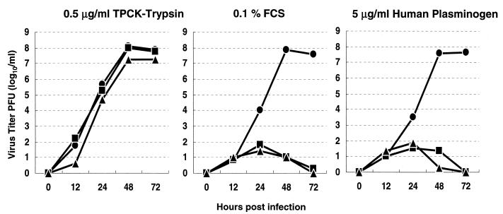
To test whether the C-terminal Lys is essential for plasminogen-mediated WSN virus growth, we infected MDBK cells with 100 PFU of mutant or wild-type WSN virus (multiplicity of infection, 2 × 10⁻⁴) and incubated them in the presence of FCS (0.1%, a proportion determined in a previous in vitro study to be appropriate [9]), human plasminogen (5 μg/ml), or TPCK-trypsin (0.5 μg/ml). Although WSN-NA453L grew slightly more slowly than the other mutant and the wild-type virus, all viruses replicated in the presence of trypsin, reaching their highest titers at 48 h postinfection (Fig. 1). In the presence of plasminogen, however, only the wild-type virus replicated. The titers of the two mutant viruses increased for 24 h after infection but then declined. The fact that all viruses showed essentially the same titer at 12 h postinfection indicated that the mutant viruses replicated for only a single cycle while the wild-type virus underwent multiple growth cycles.
FIG. 1.
Loss of plasminogen-mediated viral growth due to C-terminal mutations in NA. MDBK cells were infected with 100 PFU of the wild-type WSN (●) or mutant WSN-453R (▪) or WSN-453L (▴) virus and incubated in the presence of 0.5 μg of TPCK-trypsin/ml, 0.1% FCS, or 5 μg of human plasminogen/ml. At 0, 12, 24, 48, and 72 h postinfection, virus titers in the culture supernatants were determined.
HA cleavage in the presence of plasminogen.
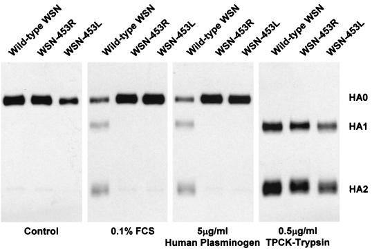
To determine the mechanism of differential growth distinguishing the wild-type virus from the mutant viruses, we compared the HA proteins in virions produced in the presence of plasminogen with those produced in the presence of trypsin (Fig. 2). With trypsin, the HAs of all viruses tested were cleaved into HA1 and HA2 subunits, whereas with plasminogen, the HA of the wild-type virus was cleaved into HA1 and HA2 but those of the mutant viruses were not. Thus, the ability of the WSN virus to grow in the presence of plasminogen correlates with HA cleavage. These results, together with our previous finding that alteration of the C-terminal Lys of NA abolishes plasminogen binding, indicate that the plasminogen-binding activity of NA is essential for viral growth mediated by plasminogen.
FIG. 2.
HA cleavage in the presence of plasminogen. MDBK cells infected with wild-type WSN or mutant WSN-453R or WSN-453L virus were incubated in medium containing 0.1% FCS, 5 μg of human plasminogen/ml, or 0.5 μg of TPCK-trypsin/ml. Viral proteins were labeled with [35S]Met-[35S]Cys (50 μCi/ml). After lysis of purified virions, HA proteins were immunoprecipitated with anti-HA monoclonal antibodies and separated by sodium dodecyl sulfate-polyacrylamide gel electrophoresis.
C-terminal Lys of NA is critical for virulence.
The WSN-NA453R and WSN-NA453L viruses failed to undergo multiple cycles of replication in cell culture in the presence of plasminogen, suggesting that they might be attenuated because of their inability to use systemically available plasminogen for HA cleavage. We therefore compared the virulence of the mutant viruses with that of the wild-type virus by determining the MLD50s after intranasal inoculation (Table 1). The MLD50s for WSN-NA453R (106.3 PFU) and WSN-NA453L (107.7 PFU) were more than 600 times higher than that of the wild-type virus (103.4 PFU), indicating that the C-terminal Lys is critical for virulence.
To determine the effect of the alteration of C-terminal Lys on viral replication in respiratory organs, we infected mice intranasally with wild-type or mutant virus and determined virus titers in nasal turbinates and lungs (Table 1). In accord with the mouse lethality data, both mutants were attenuated in their replication, especially in lungs, compared with the wild-type virus. To assess the importance of the C-terminal Lys in viral neurotropism, we inoculated mice intracerebrally with different dilutions of virus (Table 2). Wild-type virus was recovered from the brains of mice at all doses of inocula, while WSN-NA453R and WSN-NA453L were not recovered at any dose. These results indicate that the C-terminal Lys, which supports plasminogen-binding activity, is critical for WSN virus neurotropism.
TABLE 2.
Recovery of virus from mouse brains infected with wild-type WSN or mutant WSN virusa
| Virus | Amt of virus inoculated into brain (log10 PFU) | Virus titer (mean log10 PFU/g ± SD) |
|---|---|---|
| Wild-type WSN | 4.6 | 5.30 ± 0.32 |
| 5.6 | 4.76 ± 0.43b | |
| 6.6 | 4.83 ± 0.26 | |
| WSN-453R | 4.6 | NRc |
| 5.6 | NR | |
| 6.6 | NR | |
| WSN-453L | 4.6 | NR |
| 5.6 | NR | |
| 6.6 | NR |
Five BALB/c mice were inoculated intracerebrally for each dilution of virus. Three days after infection, viruses in brains were titrated in MDCK cells.
Virus was recovered from three of the five mice infected.
NR, virus was not recovered (<5.0 × 101 PFU/g).
Inability to use plasminogen for multiple growth cycles restricts neurovirulence of an NA glycosylation mutant.
We previously showed that the presence of an oligosaccharide at position 130 (146 in N2 numbering) of the WSN NA decreased binding of plasminogen to this molecule (9). Li et al. (16) showed that the presence of an oligosaccharide chain at position 130 in the WSN NA decreased the neurovirulence of WSN virus. Thus, we generated a WSN-NA146N virus with an oligosaccharide chain at position 130 of the NA and studied its growth in the presence of plasminogen. There was no recovery of virus after intracerebral inoculation of 4.2 × 10⁶ PFU into mice (data not shown), confirming the observation of Li and colleagues (16). To determine the growth properties of the virus in the presence of plasminogen, we infected MDBK cells with WSN-NA146N and assessed HA activity in the culture supernatant at 48 h postinfection. As shown in Table 3, plasminogen did not support the growth of WSN-NA146N, even at a concentration of 50 μg/ml, although the virus grew well in the presence of trypsin. These results indicate that the loss of neurovirulence by WSN-NA146N virus stemmed from its inability to use plasminogen for growth.
TABLE 3.
Lack of plasminogen-mediated growth of a mutant WSN virus possessing the glycosylation site at position 130 in the NAa
| Virus | Reciprocal of HA titer in culture supernatant
|
|||
|---|---|---|---|---|
| Plasminogen (μg/ml)
|
Trypsin (0.5 μg/ml) | |||
| 50 | 5 | 0.5 | ||
| Wild-type WSN | >2,048 | >2,048 | >2,048 | >2,048 |
| WSN-146N | <b | < | < | 1,024 |
| WSN-453R | < | < | < | 1,024 |
| WSN-453L | < | < | < | 1,024 |
Viral growth was measured by determining the HA titer in the culture supernatant. A negative control, for which protease was not added to culture medium, was run in each case; virus was never detected in undiluted culture supernatant.
<, virus was not detected in undiluted culture supernatant.
DISCUSSION
We have directly demonstrated that the C-terminal Lys of the WSN NA is essential for the virulence of this virus and for its ability to replicate in the mouse brain, and we confirmed the importance of glycosylation at position 130 of the NA (16) in the attenuated growth of this virus. Taken together, the results support our original hypothesis that the pathogenicity of WSN virus in mice depends on the ability of the viral NA to utilize plasminogen for proteolytic activation of the HA.
Of the two structural features that permit binding of plasminogen to NA—the presence of the C-terminal Lys and the lack of an oligosaccharide chain at position 130—the former is conserved among N1 NAs. Thus, viruses with the N1 NA have a greater potential for virulence than do those with other NA subtypes, which lack the C-terminal Lys. However, only the NAs of the WSN virus and its close relative NWS (28) lack a glycosylation site at position 130 (31), raising the intriguing question of why there are not any natural isolates with this structural feature. In this regard, it is interesting that the WSN and NWS viruses were both derived from repeated passage of the WS virus in mouse brains (28); similar passages with other viruses, even those with N1 NAs, failed to yield neurovirulent viruses (28). These observations suggest that there may be structural features unique to the WS NA that led to its loss of the oligosaccharide at position 130. In support of this hypothesis, we were unable to alter the plasminogen-binding property of the PR8 NA (which is also an N1 subtype), even when we abolished the glycosylation site (H. Goto and Y. Kawaoka, unpublished data). Nonetheless, although only two laboratory strains of influenza A virus with this activity have been reported thus far, one cannot rule out the possibility of pathogenic strains emerging by this mechanism in the future.
The influenza virus responsible for the 1918 pandemic was noted for its exceptional virulence (30), although the causative mechanism remains unsolved. Because WSN virus descended from this pandemic strain (25), it may be worth discussing the relevance of a virulence mechanism involving binding of plasminogen to NA to resolve this issue. Taubenberger and colleagues (22, 29) showed that the 1918 pandemic strain did not contain a series of basic amino acids at the HA cleavage site, thus excluding the virulence mechanism employed by pathogenic avian influenza viruses. Subsequent studies indicated that the NA of the pandemic strain possessed the C-terminal Lys and the glycosylation signal at position 130, suggesting that the NA of the 1918 virus most likely did not serve as a plasminogen-binding protein (23). A recent study also excluded the NS1 protein, an interferon antagonist (6, 7), from being responsible for the extreme virulence of this strain (1). Hence, we will need to consider other mechanisms of conversion to extreme virulence, which might be deduced from further sequencing of viruses associated with the 1918 outbreak.
Plasminogen is essential for homeostasis, as demonstrated by its activity during fibrinolysis, for example (17). However, the proteolytic activity of plasmin has been implicated in a number of pathological processes (2), including the invasion of tissues by bacteria (5, 18, 19). Although WSN virus provides the first example of a viral protein whose plasminogen-binding activity is required for the acquisition of virulence, we suggest that other viruses also take advantage of plasmin-induced proteolysis for enhanced virulence. In fact, Monroy and Ruiz (20) recently found that the dengue virus-associated glycoprotein, E protein, bound and activated plasminogen, suggesting that an increased level of plasmin at viral replication sites may be related to the hemorrhagic manifestation of dengue virus infection. Thus, the host plasmin/plasminogen system may become a fertile new topic to investigate in viral pathogenesis.
ACKNOWLEDGMENTS
We thank Robert G. Webster for providing the antibodies used in this study. We also thank John Gilbert for editing the manuscript.
This work was supported by NIAID Public Health Service research grants and by grants-in-aid from the Ministry of Education, Culture, Sports, Science and Technology and the Ministry of Health, Labor and Welfare, Japan.
REFERENCES
- 1.Basler C F, Reid A H, Dybing J K, Janczewski T A, Fanning T G, Zheng H Y, Salvatore M, Perdue M L, Swayne D E, Garcia-Sastre A, Palese P, Taubenberger J K. Sequence of the 1918 pandemic influenza virus nonstructural gene (NS) segment and characterization of recombinant viruses bearing the 1918 NS genes. Proc Natl Acad Sci USA. 2001;98:2746–2751. doi: 10.1073/pnas.031575198. [DOI] [PMC free article] [PubMed] [Google Scholar]
- 2.Boyle M D, Lottenberg R. Plasminogen activation by invasive human pathogens. Thromb Haemost. 1997;77:1–10. [PubMed] [Google Scholar]
- 3.Castrucci M R, Kawaoka Y. Biological importance of neuraminidase stalk length in influenza A virus. J Virol. 1993;67:759–764. doi: 10.1128/jvi.67.2.759-764.1993. [DOI] [PMC free article] [PubMed] [Google Scholar]
- 4.Fodor E, Devenish L, Engelhardt O G, Palese P, Brownlee G G, Garcia-Sastre A. Rescue of influenza A virus from recombinant DNA. J Virol. 1999;73:9679–9682. doi: 10.1128/jvi.73.11.9679-9682.1999. [DOI] [PMC free article] [PubMed] [Google Scholar]
- 5.Fuchs H, Wallich R, Simon M M, Kramer M D. The outer surface protein A of the spirochete Borrelia burgdorferi is a plasmin(ogen) receptor. Proc Natl Acad Sci USA. 1994;91:12594–12598. doi: 10.1073/pnas.91.26.12594. [DOI] [PMC free article] [PubMed] [Google Scholar]
- 6.Garcia-Sastre A. Inhibition of interferon-mediated antiviral responses by influenza A viruses and other negative-strand RNA viruses. Virology. 2001;279:375–384. doi: 10.1006/viro.2000.0756. [DOI] [PubMed] [Google Scholar]
- 7.Garcia-Sastre A, Egorov A, Matassov D, Brandt S, Levy D E, Durbin J E, Palese P, Muster T. Influenza A virus lacking the NS1 gene replicates in interferon-deficient systems. Virology. 1998;252:324–330. doi: 10.1006/viro.1998.9508. [DOI] [PubMed] [Google Scholar]
- 8.Garten W, Klenk H D. Understanding influenza virus pathogenicity. Trends Microbiol. 1999;7:99–100. doi: 10.1016/s0966-842x(99)01460-2. [DOI] [PubMed] [Google Scholar]
- 9.Goto H, Kawaoka Y. A novel mechanism for the acquisition of virulence by a human influenza A virus. Proc Natl Acad Sci USA. 1998;95:10224–10228. doi: 10.1073/pnas.95.17.10224. [DOI] [PMC free article] [PubMed] [Google Scholar]
- 10.Horimoto T, Kawaoka Y. Reverse genetics provides direct evidence for a correlation of hemagglutinin cleavability and virulence of an avian influenza A virus. J Virol. 1994;68:3120–3128. doi: 10.1128/jvi.68.5.3120-3128.1994. [DOI] [PMC free article] [PubMed] [Google Scholar]
- 11.Horimoto T, Kawaoka Y. Pandemic threat posed by avian influenza A viruses. Clin Microbiol Rev. 2001;14:129–149. doi: 10.1128/CMR.14.1.129-149.2001. [DOI] [PMC free article] [PubMed] [Google Scholar]
- 12.Horimoto T, Nakayama K, Smeekens S P, Kawaoka Y. Proprotein-processing endoproteases PC6 and furin both activate hemagglutinin of virulent avian influenza viruses. J Virol. 1994;68:6074–6078. doi: 10.1128/jvi.68.9.6074-6078.1994. [DOI] [PMC free article] [PubMed] [Google Scholar]
- 13.Kawaoka Y, Webster R G. Sequence requirements for cleavage activation of influenza virus hemagglutinin expressed in mammalian cells. Proc Natl Acad Sci USA. 1988;85:324–328. doi: 10.1073/pnas.85.2.324. [DOI] [PMC free article] [PubMed] [Google Scholar]
- 14.Klenk H D, Rott R, Orlich M, Blodorn J. Activation of influenza A viruses by trypsin treatment. Virology. 1975;68:426–439. doi: 10.1016/0042-6822(75)90284-6. [DOI] [PubMed] [Google Scholar]
- 15.Lazarowitz S G, Choppin P W. Enhancement of the infectivity of influenza A and B viruses by proteolytic cleavage of the hemagglutinin polypeptide. Virology. 1975;68:440–454. doi: 10.1016/0042-6822(75)90285-8. [DOI] [PubMed] [Google Scholar]
- 16.Li S, Schulman J, Itamura S, Palese P. Glycosylation of neuraminidase determines the neurovirulence of influenza A/WSN/33 virus. J Virol. 1993;67:6667–6673. doi: 10.1128/jvi.67.11.6667-6673.1993. [DOI] [PMC free article] [PubMed] [Google Scholar]
- 17.Lijnen H R, Collen D. Interaction of plasminogen activators and inhibitors with plasminogen and fibrin. Semin Thromb Hemost. 1982;8:2–10. doi: 10.1055/s-2007-1005038. [DOI] [PubMed] [Google Scholar]
- 18.Lottenberg R, Minning-Wenz D, Boyle M D. Capturing host plasmin(ogen): a common mechanism for invasive pathogens? Trends Microbiol. 1994;2:20–24. doi: 10.1016/0966-842x(94)90340-9. [DOI] [PubMed] [Google Scholar]
- 19.Monroy V, Amador A, Ruiz B, Espinoza-Cueto P, Xolalpa W, Mancilla R, Espitia C. Binding and activation of human plasminogen by Mycobacterium tuberculosis. Infect Immun. 2000;68:4327–4330. doi: 10.1128/iai.68.7.4327-4330.2000. [DOI] [PMC free article] [PubMed] [Google Scholar]
- 20.Monroy V, Ruiz B H. Participation of the Dengue virus in the fibrinolytic process. Virus Genes. 2000;21:197–208. doi: 10.1023/a:1008191530962. [DOI] [PubMed] [Google Scholar]
- 21.Neumann G, Watanabe T, Ito H, Watanabe S, Goto H, Gao P, Hughes M, Perez D R, Donis R, Hoffmann E, Hobom G, Kawaoka Y. Generation of influenza A viruses entirely from cloned cDNAs. Proc Natl Acad Sci USA. 1999;96:9345–9350. doi: 10.1073/pnas.96.16.9345. [DOI] [PMC free article] [PubMed] [Google Scholar]
- 22.Reid A H, Fanning T G, Hultin J V, Taubenberger J K. Origin and evolution of the 1918 “Spanish” influenza virus hemagglutinin gene. Proc Natl Acad Sci USA. 1999;96:1651–1656. doi: 10.1073/pnas.96.4.1651. [DOI] [PMC free article] [PubMed] [Google Scholar]
- 23.Reid A H, Fanning T G, Janczewski T A, Taubenberger J K. Characterization of the 1918 “Spanish” influenza virus neuraminidase gene. Proc Natl Acad Sci USA. 2000;97:6785–6790. doi: 10.1073/pnas.100140097. [DOI] [PMC free article] [PubMed] [Google Scholar]
- 24.Schulman J L, Palese P. Virulence factors of influenza A viruses: WSN virus neuraminidase required for plaque production in MDBK cells. J Virol. 1977;24:170–176. doi: 10.1128/jvi.24.1.170-176.1977. [DOI] [PMC free article] [PubMed] [Google Scholar]
- 25.Smith W, Andrewes C H, Laidlaw P P. A virus obtained from influenza patients. Lancet. 1933;i:66–68. [Google Scholar]
- 26.Steinhauer D A. Role of hemagglutinin cleavage for the pathogenicity of influenza virus. Virology. 1999;258:1–20. doi: 10.1006/viro.1999.9716. [DOI] [PubMed] [Google Scholar]
- 27.Stieneke-Grober A, Vey M, Angliker H, Shaw E, Thomas G, Roberts C, Klenk H D, Garten W. Influenza virus hemagglutinin with multibasic cleavage site is activated by furin, a subtilisin-like endoprotease. EMBO J. 1992;11:2407–2414. doi: 10.1002/j.1460-2075.1992.tb05305.x. [DOI] [PMC free article] [PubMed] [Google Scholar]
- 28.Stuart-Harris C H. Neurotropic strain of human influenza virus. Lancet. 1939;i:497–499. [Google Scholar]
- 29.Taubenberger J K, Reid A H, Krafft A E, Bijwaard K E, Fanning T G. Initial genetic characterization of the 1918 “Spanish” influenza virus. Science. 1997;275:1793–1796. doi: 10.1126/science.275.5307.1793. [DOI] [PubMed] [Google Scholar]
- 30.Taubenberger J K, Reid A H, Fanning T G. The 1918 influenza virus: a killer comes into view. Virology. 2000;274:241–245. doi: 10.1006/viro.2000.0495. [DOI] [PubMed] [Google Scholar]
- 31.Ward A C. Changes in the neuraminidase of neurovirulent influenza virus strains. Virus Genes. 1995;10:253–260. doi: 10.1007/BF01701815. [DOI] [PubMed] [Google Scholar]
- 32.Wiley D C, Skehel J J. The structure and function of the hemagglutinin membrane glycoprotein of influenza virus. Annu Rev Biochem. 1987;56:365–394. doi: 10.1146/annurev.bi.56.070187.002053. [DOI] [PubMed] [Google Scholar]


