Abstract
Background:
Microvascular decompression (MVD) through a retrosigmoid approach is considered the treatment of choice in cases of hemifacial spasm (HFS) due to neurovascular conflict (NVC). Despite the widespread of neuronavigation and intraoperative neuromonitoring (IONM) techniques in neurosurgery, their contemporary application in MVD for HFS has been only anecdotally reported.
Methods:
Here, we report the results of MVD performed with a combination of neuronavigation and IONM, including lateral spread response (LSR) in 20 HFS patients. HFS clinical outcome and different surgical-related factors, such as craniotomy size, surgical duration, mastoid air cell (MAC) opening, postoperative cerebral spinal fluid (CSF) leakage, sinus injury, and other complications occurrence, and the length of hospitalization (LOS) were studied.
Results:
Postoperatively, residual spasm persisted only in two patients, but at the latest follow-up (FU) (mean: 12.5 ± 8.98 months), all patients had resolution of symptoms. The mean surgical duration was 103.35 ± 19.36 min, and the mean LOS was 2.21 ± 1.12 days. Craniotomy resulted in 4.21 ± 1.21 cm2 in size. Opening of MAC happened in two cases, whereas no cases of CSF leak were reported as well as no other complications postoperatively and during FU.
Conclusion:
MVD for HFS is an elective procedure, and for this reason, surgery should integrate all technologies to ensure safety and efficacy. The disappearance of LSR is a crucial factor for identifying the vessel responsible for NVC and for achieving long-term resolution of HFS symptoms. Simultaneously, the benefits of using neuronavigation, including the ability to customize the craniotomy, contribute to reduce the possibility of complications.
Keywords: Hemifacial spasm, Intraoperative neuromonitoring, Lateral spread response, Microvascular decompression, Neuronavigation

INTRODUCTION
Hemifacial spasm (HFS) is a neurological disorder characterized by recurrent, involuntary twitching of facial muscles located on one side of the face, receiving innervation from the ipsilateral facial nerve. Classified within the peripheral neuromuscular movement disorder category, the spasmodic contractions typically originate in the orbicularis oculi muscle and gradually extend to encompass other muscles innervated by the facial nerve on that side. Etiologically, HFS is classified into (1) a primary form, linked to neurovascular conflict (NVC); (2) hereditary, marked by a robust family history of HFS in first-degree relatives; (3) secondary to an identifiable cause (such as Bell’s palsy, facial nerve injury, demyelinating pathologies, or vascular insults); and (4) HFS mimickers, encompassing psychogenic, tics, dystonia, myoclonus, myokymia, myorhythmia, and hemimasticatory spasm.[28] The estimated global prevalence of HFS is 14.5/100,000 women and 7.4/100,000 men, indicating that females are twice as susceptible to HFS as males.[28] A systematic review by Sharma et al. identified the anterior inferior cerebellar artery (AICA) as the most common offending vessel, followed by the posterior inferior cerebellar artery, the vertebrobasilar system, and veins. Multiple vessel involvement has been reported in about 27% of patients.[25] Furthermore, a significant association between the lateral deviation of the vertebral artery (VA) and the symptomatic side of primary HFS has been reported.[8] The established medical approach for HFS involves the use of antiepileptic drugs and botulinum neurotoxin (BoNT) injections, offering low risk but somewhat restricted symptomatic relief. The only etiologic procedure for HFS is microvascular decompression (MVD), a surgical procedure aiming at alleviating compression on the facial nerve root entry zone (REZ) by resolving the NVC.[16] While numerous studies in the literature highlight the efficacy of this approach in achieving clinical resolution of HFS, the occurrence of both minor and major complications after MVD remains a significant issue. These complications, ranging from transient or permanent sensorineural hearing loss and tinnitus to seventh nerve motor deficits, facial numbness, postoperative meningitis, stroke, hematoma, pseudomeningocele, and cerebral spinal fluid (CSF) leakage, can affect patients’ overall well-being and satisfaction, potentially influencing the overall clinical outcome.[4,19,33] MVD is usually performed through a retrosigmoid approach based on anatomical landmarks that are used for identifying the position of the transverse sinus, sigmoid sinuses, and the transversesigmoid sinus junction (TSSJ). Nonetheless, several studies have highlighted the lack of a precise correspondence between anatomical landmarks and sinus anatomy, thereby elevating the risk of inadvertent venous sinus injury.[1,12] The integration of neuronavigation into the surgical protocol for MVD in trigeminal neuralgia has been shown to be useful in minimizing craniotomy size and, consequently, reducing the complication rate and the overall surgical duration.[15] In the last few years, intraoperative neuromonitoring (IONM) techniques have been implemented in different neurosurgical procedures. Lateral spread response (LSR) represents an electrophysiological aberration characteristic of primary HFS that can be effectively monitored during surgery[20,30], although the prognostic value of LSR remains a matter of debate.[5,9] Due to the elective nature of this kind of surgery (MVD for HSF is not a life-saving procedure but a surgery performed to improve quality of life), at least in theory, all the available surgical tools should be implemented. Thus, this paper aimed to report the results of a surgical series of HFS patients in which MVD was performed with the contemporary use of neuronavigation and IONM with the primary focus of evaluating the clinical outcome and the complication occurrence.
MATERIALS AND METHODS
Patients
The present series includes 20 consecutive patients suffering from medically intractable HFS who underwent MVD at our institution between January 2022 and July 2023. The inclusion criteria were the following: (1) confirmed clinical and neurophysiological diagnosis of HFS and (2) a preoperative brain magnetic resonance imaging (MRI) comprising time-of-flight and fast imaging employing steady-state acquisition sequences showing NVC at the facial REZ. The exclusion criteria were the following: (1) MRI showing structural lesions such as tumors, arteriovenous malformation, and Chiari I malformation and (2) patients with blepharospasm, facial myokymia, or Meige syndrome.
All patients were operated by the senior author (NM). All patients gave informed consent both to the surgical procedure and to the present study. Ethics committee review/approval was not needed due to the retrospective nature of this study.
Surgical procedure
All procedures were performed under general anesthesia in a park bench position that, in our experience, provides easy and comfortable access to the cerebellopontine angle.[11,15] MRI sequences available for navigation were loaded into the Medtronic Stealth Station S8 navigation system (Minneapolis, Minnesota, USA). After careful registration of the patient, the skin incision was placed, and the “keyhole” for the craniotomy was identified just posteriorly and below the TSSJ [Figure 1].[15] The dura was opened under the operative microscope with a Y-shaped incision, creating dural leaves based on the transverse sinus and the sigmoid sinus. After a gentle CSF drainage, the suprameatal tuberculum was identified. After a fine dissection of the arachnoid plane, the facial nerve was identified both anatomically and by direct electrical stimulation (DES). The NVC between the facial nerve and the arterial loop was identified and finally resolved with the interposition of a Teflon patch. In all cases, the closure was performed with a duroplasty using a suturable dural substitute reinforced with Hemopatch® and fibrin glue according to a standardized technique previously reported.[2,21,23]
Figure 1:
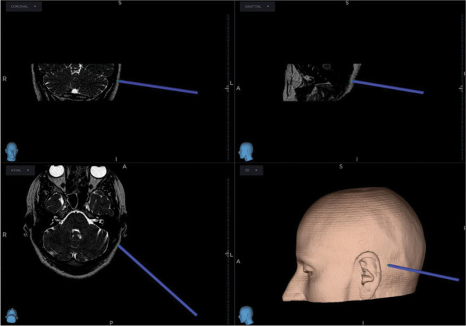
Intraoperative neuronavigation showing that the “keyhole” for the craniotomy is identified just posteriorly and below the transverse-sigmoid sinus junction.
IONM
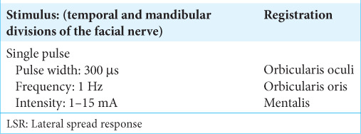
NIM eclipse system (Medtronic) was used for IONM. DES of facial cranial nerves was used to identify the neural structures correctly. A monopolar constant-current low-frequency stimulation was used for DES in all cases (monopolar probe [cathodal] 300 μs, 1 Hz, 0.1–0.5 mA). Usually, an intensity of stimulus at 0.1 mA, delivered on the nerve surface, is enough to record a compound muscle action potential. The need for higher stimulus intensity might indicate a relative distance to the neural structure or the presence of a relevant tissue barrier between the probe and the nerve. The more motor response is evoked with low intensity, the more the surgeon is working close to the nerve. At the end of the surgery, we always perform a proximal stimulation at a low threshold (0.1–0.3 mA) to confirm the functional facial nerve integrity. Entire cranial nerves motor pathways integrity was evaluated using corticobulbar motor evoked potentials.[3] The neurophysiological parameters for LSR recording are reported in Table 1. LSR disappearance indicates a successful MVD.[6]
Table 1:
Neurophysiological parameters for LSR recording.
Outcome indicators
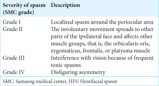
The following parameters were recorded: craniotomy size, surgical duration, mastoid air cell (MAC) opening, postoperative CSF leakage, sinus injury, and other complications occurrence, length of hospitalization (LOS), and HFS outcome at hospital admission at discharge and latest follow-up (FU) according to Samsung Medical Center (SMC) grading system for severity of HFS spasms [Lee’s scale; Table 2].[14] The craniotomy size was assessed on postoperative computed tomography (CT) examination.[31]
Table 2:
SMC grading system for severity of HFS spasms (Lee’s scale).
RESULTS
Demographic data
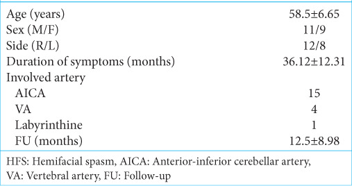
Demographic data are listed in Table 3. Twenty patients were finally enrolled, of whom 55% were males and 45% females. The mean age of the patients at the time of surgery was 58.50 ± 6.65 years, and the mean duration of FU was 12.50 ± 8.98 months. The right facial nerve was most commonly affected (60% of patients). Symptom duration before surgery was 36.12 ± 12.31 months. AICA was the most common arterial vessel responsible for the NVC (75%), whereas the VA was involved in 20% of patients. A rare case of labyrinthine artery NVC was evident [Table 2].
Table 3:
Preoperative findings of 20 HFS patients.
Outcome data
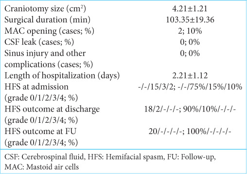
The outcome results are reported in Table 4. The mean surgical duration was 103.35 ± 19.36 min, and the mean LOS was 2.21 ± 1.12 days. Craniotomy resulted in 4.21 ± 1.21 cm2 in size. Opening of MAC happened in two cases, whereas no cases of CSF leak were reported as well as no other complications. On admission (before surgery), 15 patients had a SMC grade II, three patients had a SMC grade III, and two patients had a SMC grade IV, respectively [Tables 2 and 4]. At discharge, 18 patients experienced a complete disappearance of HFS, whereas two had residual spasms limited to orbicularis oculi (SMC grade I) while ameliorated compared to preoperative severity [Table 4]. At the latest FU, no cases of residual spasm or recurrent spasm were recorded, and all patients experienced symptom resolution. No late complications were recorded.
Table 4:
Clinical and surgical outcomes of HFS patients.
Cases illustration
Case 1
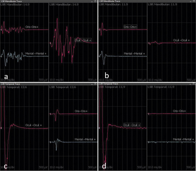
A 59-year-old male patient presented at our institution’s outpatient clinic with a 3-year history of left HFS. The symptoms initially manifested as spasms in the left orbicularis oculi muscle, gradually progressing to involve the remaining left facial muscles, excluding the platysma. In addition, the frequency of spasm episodes increased to 10 per day. Electromyography revealed spontaneous sporadic bursts in the left orbicularis oculi. Brain MRI identified a dolichoectatic left VA compressing the left seventh nerve at its REZ [Figure 2]. The patient had undergone BoNT infiltration without benefit; moreover, as a side effect, he had transient left hemifacial paralysis. The neurologic examination did not reveal any focal deficits. Subsequently, the patient underwent a left neuronavigated MVD. The NVC was identified with a dolichoectasic VA, meticulously dissected, and resolved by interposing a Teflon patch [Figure 3]. Following the anatomical resolution of the NVC, LSR disappeared, confirming the adequacy of the decompression [Figure 4]. The postoperative course was uneventful [Figure 5]. Upon discharge, the patient exhibited no residual spasm, and this benefit was maintained at FU (15 months).
Figure 2:
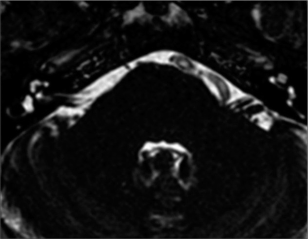
Preoperative axial fast imaging employing steady-state acquisition magnetic resonance imaging. The neurovascular conflict is evident between a dolichoectasic vertebral artery and the left facial nerve root entry zone.
Figure 3:
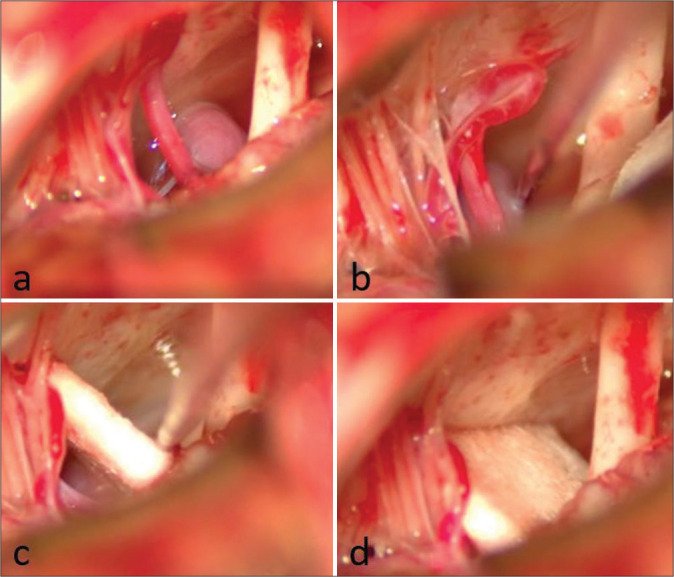
Intraoperative findings of Case 1. (a) Neurovascular conflict between the left vertebral artery (VA) and the left seventh nerve. (b) Nerve hook is utilized for dissecting the VA from the nerve. (c) Following Teflon positioning, the nerve hook is employed to push the VA away from the nerve. (d) Final result after Teflon release.
Figure 4:
Lateral spread response (LSR) intraoperative recording of Case 1. The upper figure (a and b) displays the LSR mandibular recording, while the lower one (c and d) shows the LSR temporal recording. LSR records on the left (a and c) are before decompression and Teflon placement, while those on the right (b and d) are after decompression and Teflon placement. Immediately after Teflon placement between the vessel culprit and the seventh nerve, the disappearance of pathological LSR is evident (b and d). (b) Disappearance of LSR at the orbicularis oculi. (d) Disappearance of LSR at the orbicularis oris and mentalis muscle.
Figure 5:
Postoperative fast imaging employing steady-state acquisition magnetic resonance imaging (a) and computed tomography-scan (b and c) of Case 1. (a and b) The Teflon (red arrow) is evident, separating the vertebral artery and facial nerve root entry zone. (c) The extent of the craniotomy and reconstruction is depicted in 3D reconstruction.
Case 2
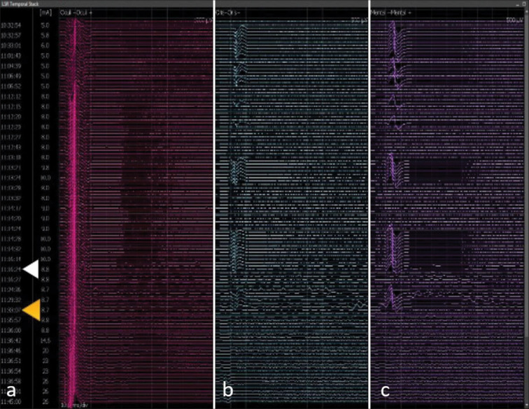
A 54-year-old male patient was admitted to our department with a 10-year history of right HFS, initially involving the right orbicularis oculi and subsequently extending to the remaining right face, excluding the platysma. A brain MRI revealed a NVC at the REZ of the right facial nerve with the ipsilateral AICA [Figure 6]. Subsequently, a neuronavigated right retrosigmoid approach was undertaken. During the procedure, the NVC was identified at the right facial nerve REZ with the ipsilateral AICA. Following the dissection and placement of a Teflon patch [Figure 7], the LSR remained unchanged [Figure 8]. Consequently, we went on with a further exploration, revealing a second NVC due to the right labyrinthine artery that was resolved with a second Teflon pad [Figure 9]. At this point, LSR disappeared, confirming the adequacy of the decompression [Figure 8]. At discharge, the spasm had completely disappeared, and that benefit was maintained at the last FU (13 months).
Figure 6:
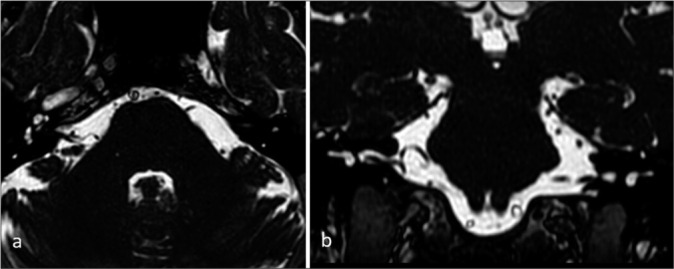
Preoperative brain magnetic resonance imaging of Case 2. (a) Axial and (b) coronal fast imaging employing steady-state acquisition sequences showing a neurovascular conflict between the anterior inferior cerebellar artery and the right facial nerve.
Figure 7:
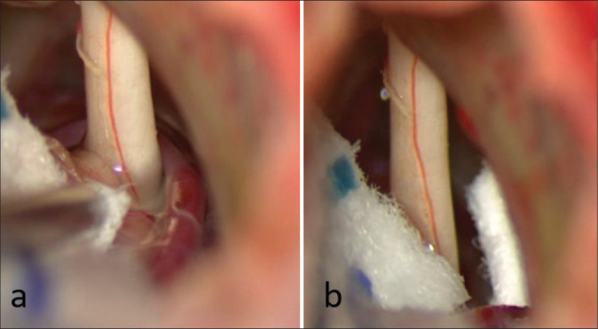
Intraoperative view of Case 2. (a) Neurovascular conflict at the root entry zone of the right facial nerve with the ipsilateral anterior inferior cerebellar artery before and (b) after decompression and Teflon placement. A persistence of lateral spread response was noted, and further exploration was needed.
Figure 8:
Lateral spread response (LSR) recording of Case 2. (a) The figure shows the LSR of the temporal branch. The normal response is one recorded in the orbicularis oculi muscle, (b) while the pathological response is one recorded in the orbicularis oris and (c) mentalis muscles. Immediately after Teflon placement between the anterior inferior cerebellar artery and the right facial nerve (11:16 on the timeline, white triangle), the persistence of pathological LSR in the orbicularis oris (b) and mentalis (c) muscles was noted. Thus, further exploration of the right seventh cranial nerve was continued identifying a second neurovascular conflict between the right facial nerve and the labyrinthine artery. Only after decompression and Teflon placement between the labyrinthine artery and right facial nerve (11:36 on the timeline, yellow triangle) the disappearance of pathological LSR on the orbicularis oris (b) and mentalis (c) muscles was evident. The normal response in the orbicularis oculi muscle (a) is always retained.
Figure 9:
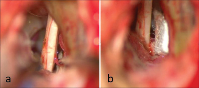
Intraoperative view of case 2. A second neurovascular conflict was appreciated with the labyrinthine artery. Findings (a) before and (b) after decompression and Teflon interposition.
DISCUSSION
HFS significantly affects patients’ quality of life, causing disruptions in sleep, social embarrassment, and potential vision impairment in the affected eye.[28] While effective, BoNT injections offer limited and temporary symptomatic relief. A study by Mazlout et al. reported an average total therapeutic effect duration of 9.35 ± 3.64 weeks.[17] Moreover, a meta-analysis by Zagui et al. on 1003 subjects identified side effects in 182 cases (18.4%), with eyelid ptosis being the most commonly reported (3.39%), along with other issues such as dry eyes, local edema, erythema, facial paresis, headache, and eyebrow ptosis.[32] MVD represents the only etiological treatment for primary HSF.[7,13] Nonetheless, there are several risks associated with MVD, such as CSF leak, neurological deficits, infections as well as inadvertent injury to venous sinuses during the retrosigmoid craniotomy, potentially leading to severe postoperative consequences such as venous sinus thrombosis, pseudomeningocele, dysphagia necessitating gastrostomy, or cerebellar stroke. Early postoperative initiation of systemic anticoagulation is also required to prevent thrombosis progression and the ensuing complications.[22] Consequently, several studies have focused on precisely describing anatomical landmarks to enhance the safety of retrosigmoid procedures. While asterion is generally considered the key landmark for identifying the TSSJ, studies have shown its inconsistency, with a very low correspondence (23.3%) between these structures.[1] Jian et al. proposed an alternative keyhole location, defined by a vertical line from the top point of the mastoid groove intersecting with a horizontal line connecting the infraorbital margin and the upper edge of the external auditory canal.[12] Despite being more reliable, the digastric point was found to overlap with the sigmoid sinus in 38% of cadaveric specimens.[24] Neuronavigation use in retrosigmoid approaches has been associated with a reduced incidence of complications, as evidenced by a comparative analysis by Legninda Sop et al. on patients undergoing MVD for trigeminal neuralgia.[15] In that study, the neuronavigation group exhibited a decreased incidence of CSF leak (0% vs. 27%), a trend toward reduced MAC opening (26.31% vs. 54.54%), as well as smaller craniotomy size (4.80 ± 1.13 cm2 vs. 6.04 ± 1.97 cm2) and shorter surgical duration (107.63 ± 18.13 min vs. 148.18 ± 41.48 min). In our series focused on MVD for HFS, the mean craniotomy size was even smaller compared to the study by Legninda Sop et al. focused on trigeminal neuralgia[15], probably due to the fact that we performed our burr hole farer from TSJJ to approach the facial nerve. Considering the MAC opening, we had only two cases in which this event occurred. A retrospective study by Yanagawa et al. on 210 consecutive MVDs [29] identified the risk of MAC opening based on the classification of MAC anatomy and their relationship with the ipsilateral sigmoid sinus, as assessed on preoperative head CT-scan. That study suggested that the more medially the extension of MAC in relation to the lateral edge of the ipsilateral sigmoid sinus (MAC type 3 and type 4), the higher the risk of inadvertent opening. In that study, the burr hole site was designed at the intersection between the external acoustic meatus line-inion and the posterior edge of the mastoid bone, and they had a MAC opening in 30% of type 3 MAC and 76% of type 4 MAC, respectively.[29] Thus, it is intuitive that exposing the sigmoid sinus during the craniotomy (as requested by MVD performed on the anatomical landmarks) can increase the risk of MAC opening. In our series, we had a significantly lower incidence of MAC opening (10%). Although the anatomy of MAC can play a significant role, the customization of the craniotomy and the inutility to expose the sigmoid sinus with neuronavigation might contribute to reducing MAC opening in MVD performed with the neuronavigated technique. Nonetheless, further comparative studies on the incidence of MAC opening between anatomical and neuronavigated retrosigmoid craniotomies are advocated. Despite higher reported risks of CSF leakage when MAC opening occurs, in our series, no cases of CSF leakage were recorded. These data are probably explained by the smaller craniotomy that can be performed using neuronavigation that prevents the opening of a larger MAC. We acknowledge that an experienced neurosurgeon can perform the craniotomy without neuronavigation. Nonetheless, the use of neuronavigation allows to customize of the craniotomy, making it useless to expose the medial edge of the sigmoid sinus to reach the REZ of the facial nerve (as shown by our intraoperative pictures). This carries the undeniable advantage of avoiding the possibility of provoking a sinus injury [no cases in our series; Table 4].
In the context of HFS management with MVD, IONM, which includes LSR, plays a significant role. LSR is defined as a pathological latent, abnormal response elicited by the stimulation of one branch of the facial nerve in patients with HFS, resulting in the contraction of the facial muscles innervated by the other branch of the facial nerve. This phenomenon likely arises from the cross-transmission of antidromic activity from the stimulated branch of the facial nerve. A retrospective analysis by Song et al. on 73 patients who underwent MVD for HFS demonstrated significantly higher short-term and long-term spasm resolution in the group where LSR disappeared compared to the group where LSR persisted.[26] When investigating long-term HFS recurrence, a recent meta-analysis[18] revealed that LSR persistence at the end of the MVD procedure is the only factor correlated with spasm recurrence. In addition, a shorter preoperative symptom duration is associated with a lower incidence of long-term HFS recurrence. The type of offending vessel does not affect HFS recurrence.[18] Considering these data, we always try to obtain LSR disappearance during the MVD procedure, as reported in illustrative case 2. In that case, following the resolution of NVC involving the AICA, as revealed in the preoperative MRI, the disappearance of LSR was not observed. In light of this, we went on with the exploration of the facial nerve and we were able to find another NVC arising from the labyrinthine artery. We advise investigating uncommon causes of NVC even when not apparent in preoperative MRI until the disappearance of LSR is achieved. In our case series, LSR disappeared in all cases at the end of the procedure. Although two patients had reduced but still present HFS at discharge, all patients achieved complete resolution of HFS at the FU. In our opinion, this data further highlights the importance of obtaining LSR resolution during the MVD procedure, even if the literature data are inconclusive on this aspect.[5,9,10]
To the best of our knowledge, this is the largest series focusing on neuronavigated MVD for HFS. The only previously published series on this topic included only 12 HFS patients.[27] Although our paper was the only one in which both techniques (neuronavigation and IONM) were systematically used in this clinical setting, we recognize that the main limitations of our study are the small sample size, the lack of a control group, and the relatively short FU. For these reasons, we acknowledge that our study alone is not enough to definitively conclude that both neuronavigation and IONM must be used during MVD for HFS. Nonetheless, we should always keep in mind that when approaching HFS surgery, we are performing an elective procedure to improve the quality of life (not a life-saving procedure), and all our efforts must be directed to improve the clinical outcome and reduce the possibility of having complications.
CONCLUSION
In conclusion, the contemporary use of neuronavigation and IONM with LSR in MVD for HFS seems to enhance the safety/efficacy balance of this procedure because the disappearance of LSR helps in identifying the vessel responsible for NVC to achieve long-term resolution of HFS symptoms. Simultaneously, the benefits of using neuronavigation, including the ability to customize the craniotomy, contribute to reducing the possibility of complications.
Footnotes
How to cite this article: Battistelli M, Izzo A, D’Ercole M, D’Alessandris Q, Di Domenico M, Ioannoni E, et al. Optimizing surgical technique in microvascular decompression for hemifacial spasm – Results from a surgical series with contemporary use of neuronavigation and intraoperative neuromonitoring. Surg Neurol Int. 2024;15:319. doi: 10.25259/SNI_268_2024
Contributor Information
Marco Battistelli, Email: marco.battistelli23494@gmail.com.
Alessandro Izzo, Email: alessandro.izzo@policlinicogemelli.it.
Manuela D’Ercole, Email: manuela.dercole@policlinicogemelli.it.
Quintino Giorgio D’Alessandris, Email: quintinogiorgio.dalessandris@policlinicogemelli.it.
Michele Di Domenico, Email: michele.didomenico@policlinicogemelli.it.
Eleonora Ioannoni, Email: eleonora.ioannoni@policlinicogemelli.it.
Camilla Gelormini, Email: camilla.gelormini@policlinicogemelli.it.
Renata Martinelli, Email: renata.martinelli01@icatt.it.
Federico Valeri, Email: federicovaleri97@gmail.com.
Fulvio Grilli, Email: fulvio.grilli96@gmail.com.
Nicola Montano, Email: nicolamontanomd@yahoo.it.
Ethical approval
Institutional Review Board approval is not required.
Declaration of patient consent
The authors certify that they have obtained all appropriate patient consent.
Financial support and sponsorship
Nil.
Conflicts of interest
There are no conflicts of interest.
Use of artificial intelligence (AI)-assisted technology for manuscript preparation
The authors confirm that there was no use of artificial intelligence (AI)-assisted technology for assisting in the writing or editing of the manuscript and no images were manipulated using AI.
Disclaimer
The views and opinions expressed in this article are those of the authors and do not necessarily reflect the official policy or position of the Journal or its management. The information contained in this article should not be considered to be medical advice; patients should consult their own physicians for advice as to their specific medical needs.
REFERENCES
- 1.Da Silva EB, Jr, Leal AG, Milano JB, da Silva LF, Jr, Clemente RS, Ramina R. Image-guided surgical planning using anatomical landmarks in the retrosigmoid approach. Acta Neurochir (Wien) 2010;152:905–10. doi: 10.1007/s00701-009-0553-5. [DOI] [PubMed] [Google Scholar]
- 2.Della Pepa GM, Montano N, Lucantoni C, Alexandre AM, Papacci F, Meglio M. Craniotomy repair with the retrosigmoid approach: The impact on quality of life of meticulous reconstruction of anatomical layers. Acta Neurochir (Wien) 2011;153:2255–8. doi: 10.1007/s00701-011-1113-3. [DOI] [PubMed] [Google Scholar]
- 3.Della Pepa GM, Stifano V, D’Alessandris QG, Menna G, Burattini B, Di Domenico M, et al. Intraoperative corticobulbar motor evoked potential in cerebellopontine angle surgery: A clinically meaningful tool to predict early and late facial nerve recovery. Neurosurgery. 2022;91:406–13. doi: 10.1227/neu.0000000000002039. [DOI] [PubMed] [Google Scholar]
- 4.Di Carlo DT, Benedetto N, Perrini P. Clinical outcome after microvascular decompression for trigeminal neuralgia: A systematic review and meta-analysis. Neurosurg Rev. 2022;46:8. doi: 10.1007/s10143-022-01922-0. [DOI] [PubMed] [Google Scholar]
- 5.El Damaty A, Rosenstengel C, Matthes M, Baldauf J, Schroeder HW. The value of lateral spread response monitoring in predicting the clinical outcome after microvascular decompression in hemifacial spasm: A prospective study on 100 patients. Neurosurg Rev. 2016;39:455–66. doi: 10.1007/s10143-016-0708-9. [DOI] [PubMed] [Google Scholar]
- 6.Fernández-Conejero I, Ulkatan S, Sen C, Deletis V. Intra-operative neurophysiology during microvascular decompression for hemifacial spasm. Clin Neurophysiol. 2012;123:78–83. doi: 10.1016/j.clinph.2011.10.007. [DOI] [PubMed] [Google Scholar]
- 7.Gardner WJ. Concerning the mechanism of trigeminal neuralgia and hemifacial spasm. J Neurosurg. 1962;19:947–58. doi: 10.3171/jns.1962.19.11.0947. [DOI] [PubMed] [Google Scholar]
- 8.Guan HX, Zhu J, Zhong J. Correlation between idiopathic hemifacial spasm and the MRI characteristics of the vertebral artery. J Clin Neurosci. 2011;18:528–30. doi: 10.1016/j.jocn.2010.08.015. [DOI] [PubMed] [Google Scholar]
- 9.Hatem J, Sindou M, Vial C. Intraoperative monitoring of facial EMG responses during microvascular decompression for hemifacial spasm. Prognostic value for long-term outcome: A study in a 33-patient series. Br J Neurosurg. 2001;15:496–9. doi: 10.1080/02688690120105101. [DOI] [PubMed] [Google Scholar]
- 10.Helal A, Graffeo CS, Meyer FB, Pollock BE, Link MJ. Predicting long-term outcomes after microvascular decompression for hemifacial spasm according to lateral spread response and immediate postoperative outcomes: A cohort study. J Neurosurg. 2024;140:1664–71. doi: 10.3171/2023.11.JNS231299. [DOI] [PubMed] [Google Scholar]
- 11.Izzo A, Stifano V, Della Pepa GM, Di Domenico M, D’Alessandris QG, Menna G, et al. Tailored approach and multimodal intraoperative neuromonitoring in cerebellopontine angle surgery. Brain Sci. 2022;12:1167. doi: 10.3390/brainsci12091167. [DOI] [PMC free article] [PubMed] [Google Scholar]
- 12.Jian ZH, Sheng MF, Li JY, An DZ, Weng ZJ, Chen G. Developing a method to precisely locate the keypoint during craniotomy using the Retrosigmoid Keyhole approach: Surgical anatomy and technical nuances. Front Surg. 2021;8:700777. doi: 10.3389/fsurg.2021.700777. [DOI] [PMC free article] [PubMed] [Google Scholar]
- 13.Kaufmann AM, Price AV. A history of the Jannetta procedure. J Neurosurg. 2019;132:639–46. doi: 10.3171/2018.10.JNS181983. [DOI] [PubMed] [Google Scholar]
- 14.Lee JA, Jo KW, Kong DS, Park K. Using the new clinical grading scale for quantification of the severity of hemifacial spasm: Correlations with a quality of life scale. Stereotact Funct Neurosurg. 2012;90:16–9. doi: 10.1159/000330396. [DOI] [PubMed] [Google Scholar]
- 15.Legninda Sop FY, D’Ercole M, Izzo A, Rapisarda A, Ioannoni E, Caricato A, et al. The impact of neuronavigation on the surgical outcome of microvascular decompression for trigeminal neuralgia. World Neurosurg. 2021;149:80–5. doi: 10.1016/j.wneu.2021.02.063. [DOI] [PubMed] [Google Scholar]
- 16.Lu AY, Yeung JT, Gerrard JL, Michaelides EM, Sekula RF, Jr, Bulsara KR. Hemifacial spasm and neurovascular compression. ScientificWorldJournal. 2014;2014:349319. doi: 10.1155/2014/349319. [DOI] [PMC free article] [PubMed] [Google Scholar]
- 17.Mazlout H, Kamoun Gargouri H, Triki W, Kéfi S, Brour J, El Afrit MA, et al. Safety and efficacy of botulinum toxin in hemifacial spasm. J Fr Ophtalmol. 2013;36:242–6. doi: 10.1016/j.jfo.2012.01.011. [DOI] [PubMed] [Google Scholar]
- 18.Menna G, Battistelli M, Rapisarda A, Izzo A, D’Ercole M, Olivi A, et al. Factors related to hemifacial spasm recurrence in patients undergoing microvascular decompression-a systematic review and meta-analysis. Brain Sci. 2022;12:583. doi: 10.3390/brainsci12050583. [DOI] [PMC free article] [PubMed] [Google Scholar]
- 19.Moffat DA, Durvasula VS, Stevens King A, De R, Hardy DG. Outcome following retrosigmoid microvascular decompression of the facial nerve for hemifacial spasm. J Laryngol Otol. 2005;119:779–83. doi: 10.1258/002221505774481255. [DOI] [PubMed] [Google Scholar]
- 20.Møller AR, Jannetta PJ. Microvascular decompression in hemifacial spasm: Intraoperative electrophysiological observations. Neurosurgery. 1985;16:612–8. doi: 10.1227/00006123-198505000-00005. [DOI] [PubMed] [Google Scholar]
- 21.Montano N, Giordano M, Caccavella VM, Ioannoni E, Polli FM, Papacci F, et al. Hemopatch® with fibrin glue as a dural sealant in cranial and spinal surgery. A technical note with a review of the literature. J Clin Neurosci. 2020;79:144–7. doi: 10.1016/j.jocn.2020.07.011. [DOI] [PubMed] [Google Scholar]
- 22.Orlev A, Jackson CM, Luksik A, Garzon-Muvdi T, Yang W, Chien W, et al. Natural history of untreated transverse/sigmoid sinus thrombosis following posterior fossa surgery: Case series and literature review. Oper Neurosurg (Hagerstown) 2020;19:109–16. doi: 10.1093/ons/opz396. [DOI] [PubMed] [Google Scholar]
- 23.Rapisarda A, Orlando V, Izzo A, D’Ercole M, Polli FM, Visocchi M, et al. New tools and techniques to prevent CSF leak in cranial and spinal surgery. Surg Technol Int. 2022;40:399–403. doi: 10.52198/22.STI.40.NS1577. [DOI] [PubMed] [Google Scholar]
- 24.Raso JL, Gusmão SN. A new landmark for finding the sigmoid sinus in suboccipital craniotomies. Neurosurgery. 2011;68:1–6. doi: 10.1227/NEU.0b013e3182082afc. [DOI] [PubMed] [Google Scholar]
- 25.Sharma R, Garg K, Agarwal S, Agarwal D, Chandra PS, Kale SS, et al. Microvascular decompression for hemifacial spasm: A systematic review of vascular pathology, long term treatment efficacy and safety. Neurol India. 2017;65:493–505. doi: 10.4103/neuroindia.NI_1166_16. [DOI] [PubMed] [Google Scholar]
- 26.Song H, Xu S, Fan X, Yu M, Feng J, Sun L. Prognostic value of lateral spread response during microvascular decompression for hemifacial spasm. J Int Med Res. 2019;47:6120–8. doi: 10.1177/0300060519839526. [DOI] [PMC free article] [PubMed] [Google Scholar]
- 27.Wang J, Zhang W, Wang X, Luo T, Wang X, Qu Y. Application of neuronavigation in microvascular decompression: Optimizing craniotomy and 3D reconstruction of neurovascular compression. J Craniofac Surg. 2023;34:e620–3. doi: 10.1097/SCS.0000000000009388. [DOI] [PubMed] [Google Scholar]
- 28.Yaltho TC, Jankovic J. The many faces of hemifacial spasm: Differential diagnosis of unilateral facial spasms. Mov Disord. 2011;26:1582–92. doi: 10.1002/mds.23692. [DOI] [PubMed] [Google Scholar]
- 29.Yanagawa T, Hatayama T, Harada Y, Sato E, Yamashita K, Tanaka M, et al. Preoperative risk assessment for predicting the opening of mastoid air cells in lateral suboccipital craniotomy for microvascular decompression. Clin Neurol Neurosurg. 2020;189:105624. doi: 10.1016/j.clineuro.2019.105624. [DOI] [PubMed] [Google Scholar]
- 30.Ying TT, Li ST, Zhong J, Li XY, Wang XH, Zhu J. The value of abnormal muscle response monitoring during microvascular decompression surgery for hemifacial spasm. Int J Surg. 2011;9:347–51. doi: 10.1016/j.ijsu.2011.02.010. [DOI] [PubMed] [Google Scholar]
- 31.Yushkevich PA, Piven J, Hazlett HC, Smith RG, Ho S, Gee JC, et al. User-guided 3D active contour segmentation of anatomical structures: significantly improved efficiency and reliability. Neuroimage. 2006;31:1116–28. doi: 10.1016/j.neuroimage.2006.01.015. [DOI] [PubMed] [Google Scholar]
- 32.Zagui RM, Matayoshi S, Moura FC. Adverse effects associated with facial application of botulinum toxin: A systematic review with meta-analysis. Arq Bras Oftalmol. 2008;71:894–901. doi: 10.1590/s0004-27492008000600027. [DOI] [PubMed] [Google Scholar]
- 33.Zhang KW, Shun ZT. Microvascular decompression by the retrosigmoid approach for idiopathic hemifacial spasm: Experience with 300 cases. Ann Otol Rhinol Laryngol. 1995;104:610–2. doi: 10.1177/000348949510400804. [DOI] [PubMed] [Google Scholar]


