Abstract
Chimeric bovine respiratory syncytial viruses (BRSV) expressing glycoproteins of bovine parainfluenza virus type 3 (BPIV-3) instead of BRSV glycoproteins were generated from cDNA. In the BRSV antigenome cDNA, the open reading frames of the major BRSV glycoproteins, attachment protein G and fusion protein F, were replaced individually or together by those of the BPIV-3 hemagglutinin-neuraminidase (HN) and/or fusion (F) glycoproteins. Recombinant virus could not be recovered from cDNA when the BRSV F open reading frame was replaced by the BPIV-3 F open reading frame. However, cDNA recovery of the chimeric virus rBRSV-HNF, with both glycoproteins replaced simultaneously, and of the chimeric virus rBRSV-HN, with the BRSV G protein replaced by BPIV-3 HN, was successful. The replication rates of both chimeras were similar to that of standard rBRSV. Moreover, rBRSV-HNF was neutralized by antibodies specific for BPIV-3, but not by antibodies specific to BRSV, demonstrating that the BRSV glycoproteins can be functionally replaced by BPIV-3 glycoproteins. In contrast, rBRSV-HN was neutralized by BRSV-specific antisera, but not by BPIV-3 specific sera, showing that infection of rBRSV-HN is mediated by BRSV F. Hemadsorption of cells infected with rBRSV-HNF and rBRSV-HN proved that BPIV-3 HN protein expressed by rBRSV is functional. Colocalization of the BPIV-3 glycoproteins with BRSV M protein was demonstrated by confocal laser scan microscopy. Moreover, protein analysis revealed that the BPIV-3 glycoproteins were present in chimeric virions. Taken together, these data indicate that the heterologous glycoproteins were not only expressed but were incorporated into the envelope of recombinant BRSV. Thus, the envelope glycoproteins derived from a member of the Respirovirus genus can together functionally replace their homologs in a Pneumovirus background.
Bovine respiratory syncytial virus (BRSV) is a member of the Pneumovirus genus, family Paramyxoviridae, order Mononegavirales. Together with Bovine parainfluenza virus type 3 (BPIV-3), BRSV represents the most important viral etiological agent of respiratory tract infections of calves and, therefore, is of high economic impact (44).
The BRSV genome is a single-stranded RNA of negative polarity which comprises 10 genes from which 10 mRNAs are transcribed, coding for 11 proteins (6). The genomic RNA is contained in a ribonucleoprotein (RNP) complex, tightly encapsidated by the major nucleocapsid protein N and associated with the phosphoprotein P and the polymerase L (16, 49). The transcription elongation factor M2-1 (7, 8, 18) which is translated from the first of two open reading frames (ORFs) of the M2 gene is also part of the RNP complex. The matrix protein M is thought to connect the RNP complex and the viral envelope (41). BRSV contains three envelope-associated proteins, namely, fusion glycoprotein F (45), attachment glycoprotein G (24, 28), and the small hydrophobic protein SH of unknown function, which all are incorporated into the host cell-derived viral envelope. Finally, the BRSV genome encodes two nonstructural proteins, NS1 and NS2, which cooperatively mediate escape from the host cell interferon response (34).
Transcription initiates at the 3′ leader region of the genome. The BRSV polymerase is directed by conserved gene start and semiconserved gene end and polyadenylation signals which frame each gene (24, 50), resulting in a sequential start-stop mechanism of transcription (23). Later in the replicative cycle, the polymerase switches into a readthrough mode by a so far unknown mechanism and transcribes full-length antigenomic RNA, which serves as template for synthesis of genomic RNA.
Reverse genetic systems which allow the generation of negative-strand RNA viruses entirely from cDNA can be used to engineer recombinant viruses expressing heterologous sequences (10, 30). Thus, it has become possible to design chimeric or multivalent live vaccines on the basis of recombinant negative-strand RNA viruses expressing desired antigens. We have previously described recombinant chimeric BRSVs which express the human respiratory syncytial virus (HRSV) homologs of the BRSV G and F glycoproteins instead of the BRSV glycoproteins (4). By this approach, we were aiming at the generation of an attenuated live vaccine against HRSV infection which combines the antigenic determinants of HRSV and the replication features of BRSV to confer attenuation in the heterologous human host. BRSV and HRSV are closely related members of the Pneumovirus genus, and we found that the glycoproteins of HRSV were functional in a BRSV background. For several closely related members of the Paramyxovirinae subfamily, namely, the respiroviruses human parainfluenza virus type 1 (HPIV-1) and human parainfluenza virus type 3 (HPIV-3) (40), HPIV-1 and BPIV-3 (35), and the morbilliviruses rinderpest virus and peste des petits ruminants virus (12), it was recently shown that simultaneous cosubstitution of HN and F results in replication-competent chimeric viruses. However, all of the chimeras mentioned above are members of the identical genus of the Paramyxovirinae subfamily. In the work presented here, we describe the generation of a chimeric Pneumovirus, BRSV, expressing the glycoproteins of a Respirovirus. The BRSV attachment protein G and fusion protein F have been replaced individually or together, respectively, with the hemagglutinin-neuraminidase protein HN and fusion protein F of BPIV-3.
MATERIALS AND METHODS
Viruses and cells.
Recombinant BRSV (rBRSV) strain ATue51908 was described before (1). BPIV-3 strain INT2 (Intervet International, B.V.) is licensed as a live vaccine. Both viruses were propagated in monolayer cultures of MDBK cells. After infection at a multiplicity of infection (MOI) of 0.1, cells were incubated at 37°C in minimal essential medium supplemented with 3% fetal calf serum in a 5% CO2 atmosphere.
Monoclonal antibodies and antisera.
A rabbit serum against BRSV M was obtained after immunization with purified BRSV M protein expressed in Escherichia coli. Antibody specificity was tested in an indirect immunofluorescence assay performed on BSR T7/5 cells stably expressing T7 RNA polymerase after transient transfection with expression plasmid pMTit, which contains the BRSV M ORF downstream of the T7 promotor and an internal ribosome entry site sequence. Immunoblots were done using extracts from recombinant E. coli, rBRSV-infected MDBK cells, and purified rBRSV. In all cases, the hyperimmune serum yielded a strong specific reaction with a band originating from a 28-kDa protein, corresponding to the calculated size of BRSV M, which was absent in uninfected cells or in control extracts from E. coli.
An antiserum specific to the BPIV-3 F protein was raised in a rabbit after immunization with a recombinant Sindbis virus expressing the BPIV-3 F protein. Specificity was tested by immunoblotting and by indirect immunofluorescence assays on cells transiently transfected with BPIV-3 F expression plasmids.
Calf hyperimmune serum 354, directed against BPIV-3, was obtained from Horst Schirrmeier, Insel Riems, Germany. The specificity of this serum for strain INT2 HN and F was verified in an indirect immunofluorescence assay performed on acetone-fixed BSR T7/5 cells, 48 h after transfection of plasmids containing the INT2 HN or F ORFs under control of the T7 RNA polymerase promoter.
Monoclonal antibodies G66 and F9 (15, 26), directed to the BRSV G and F protein, were a kind gift from Geraldine Taylor, Compton, United Kingdom. Monoclonal antibody 47F (14) was a kind gift from José Antonio Melero, Madrid, Spain.
Construction of cDNAs containing chimeric BRSV/BPIV-3 genomes.
Total RNA of cells infected with BPIV-3 strain INT2 was prepared 3 days after infection (RNeasy; Qiagen). cDNA of the BPIV-3 HN and F genes was generated by reverse transcription-PCR (RT-PCR), using the Titan one tube RT-PCR system (Roche Molecular Biochemicals). Three synthetic primers derived from conserved sequence regions of BPIV-3 strains 910N (GenBank accession no. U31671), Ka (GenBank accession no. AF178654), and SF (GenBank accession no. AF178655) and an oligo(dT) primer were used to introduce XhoI-NdeI and HindIII restriction sites, flanking the HN cDNA, and XhoI-SphI and XhoI-BamHI restriction sites, flanking the F cDNA. Sequences of these primers were (BPIV-3-specific sequences are in uppercase, nonspecific nucleotides are in lowercase, and restriction sites are underlined): positive-sense primer HN ORF 5′-aactcgagcatATGGAATATTGGAAACACACAAAC-3′, reverse primer HN ORF 5′-T11aagcTTT15-3′, positive-sense primer F ORF 5′-aactcgagcatGCAAATAAAGGATAATCAAAAACTTAGGA-3′, and reverse primer F ORF 5′-tttctc gagttggatccGCGTTGTTTGTGTGTTTCCAATATTCCAT-3′. RT-PCR fragments were purified, HN cDNA was cloned in pET23d (Novagen), and F cDNA was cloned in pBluescript SK (Stratagene) by standard cloning techniques (32). Sequences were verified by sequence analysis of three independent cDNA clones (LI-COR DNA Sequencer 4200; MWG-Biotech). Sequence analysis was carried out with the Genetics Computer Group (GCG) Wisconsin Package of Information, version 10.0 (GCG, Madison, Wis.).
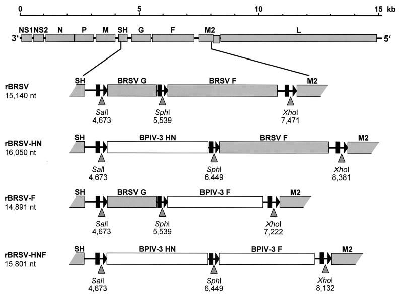
To direct transcription of the heterologous sequences by the BRSV polymerase it is necessary to add the BRSV gene start and gene end signals to the BPIV-3 ORFs. Therefore, PCR was done with primer pairs containing the conserved BRSV gene start and gene end signals. The transcription units were framed by SalI and SphI restriction sites (HN gene) and SphI and XhoI restriction sites (F gene). Primer sequences were (BPIV-3-specific sequences are in uppercase, nonspecific nucleotides are in lowercase, restriction sites are underlined, and BRSV transcription signals are in bold): BPIV-3 HN BRSV gene start 5′-ttctcgagtcgactggggcaaatacaagtATGGAATATTGGAAACACACAAAC-3′, BPIV-3 HN BRSV gene end 5′-aactcgagcatgcatatctttttaaataactatATGGGAGAGTCAACTTAACTAC-3′, BPIV-3 F BRSV gene start 5′-ttgaattcgcatgcaaaactggggcaaataagGATGATCACTATAGTTGCAACAAC-3′, and BPIV-3 F BRSV gene end 5′-aggaattctcgagaatatttttatataaCTTGATGTTTGGTGCTACATTTC-3′. PCR was carried out using the Expand high-fidelity PCR system (Roche Molecular Biochemicals). A 1,777-bp SalI-SphI fragment containing the HN ORF with BRSV transcription signals and a 1,684-bp SphI-XhoI fragment containing the BPIV-3 F ORF flanked by the BRSV transcription signals was cloned into full-length BRSV cDNA plasmid described previously (4), replacing the BRSV G gene or the BRSV F gene individually or both genes together, using the unique restriction sites SalI, SphI, and XhoI as depicted in Fig. 1.
FIG. 1.
Diagram of the rBRSV genome, illustrating the construction of chimeric rBRSV. The G gene alone was replaced with an artificial gene encoding the BPIV-3 HN to create rBRSV-HN, the F gene alone was replaced with an artificial gene encoding the BPIV-3 F protein to create rBRSV-F, or both BRSV G and F were replaced with BPIV-3 HN and F (rBRSV-HNF). The location of the ORFs is shown as shaded (BRSV) or open (BPIV-3) rectangles. The glycoprotein genes are shown as enlargements in which gene start signals are represented by triangles and gene end/polyadenylation signals are shown as bars. The synthetic restriction sites used for cloning are indicated as arrowheads, with the location in the respective genome noted underneath. The genome length of the recombinant viruses is noted on the left.
Transfection experiments.
Transfections were done as described before (1). Briefly, 32-mm-diameter dishes of subconfluent BHK T7/5 cells stably expressing T7 RNA polymerase were transfected with 5.5 μg of the respective full-length plasmids (pBRSV, pBRSV-HN, pBRSV-HNF, or pBRSV-F) and a set of four support plasmids (2 μg of pN, 2 μg of pP, 1 μg of pM2, and 1 μg of pL) from which the BRSV N, P, M2, and L proteins are expressed. All cDNA constructs were under control of a T7 promoter. Every 3 to 4 days, cells were split. The state of infection was verified by an indirect immunofluorescence assay. When cytopathic effect (CPE) was extensive, the medium was adjusted to 100 mM MgSO4 and 50 mM HEPES (pH 7.5) (14) and cells and medium were harvested and stored at −70°C.
RT-PCR.
To verify the identity of recombinant viruses, total RNA of infected MDBK cells (32-mm-diameter dishes) was prepared (RNeasy; Qiagen) and used for RT-PCR of regions containing synthetic restriction sites characteristic for the individual recombinant viruses.
Reverse transcription was done with a positive-sense primer which was complementary to the BRSV SH gene (strain ATue51908; GenBank Accession no. AF 092942; nucleotide [nt] 4,268 to 4,289), using SuperScript II reverse transcriptase (Life Technologies). PCR was carried out with the first-strand cDNA and the positive-sense primer described above and a negative-sense primer complementary to the BRSV M2 gene (ATue51908; nt 7,550 to 7,567), thus framing the heterologous sequences (Expand High Fidelity; Roche Molecular Biochemicals).
Northern hybridization of viral RNA.
Total RNA from MDBK cells was isolated 2 to 5 days postinfection (RNeasy; Qiagen). The RNA was analyzed by denaturing gel electrophoresis (32), blotted onto nitrocellulose, UV cross-linked, and hybridized with [α-32P]dCTP-labeled DNA fragments (nick translation kit, Amersham). Restriction fragments from BRSV cDNA plasmids were used to generate the BRSV G-specific probe (ATue51908; nt 4,673 to 5,539) and the BRSV F-specific probe (ATue51908; nt 5,539 to 7,471). Cloned and sequenced cDNA fragments comprising the complete ORFs of BPIV-3 HN and F were used to generate the BPIV-3-specific probes. Northern blots were exposed to Hyperfilm MP films (Amersham) at −70°C with an intensifying screen.
Virus purification.
rBRSV, the chimeras, and BPIV-3 were purified by sucrose gradient centrifugation (29). In short, MDBK cells grown to 90% confluency in 175-cm2 cell culture flasks were infected with the respective virus and split 36 and 72 h postinfection. Cell culture supernatants of the four resulting flasks were harvested 5 days postinfection, and cellular debris was removed by centrifugation for 15 min at 800 × g. Polyethylene glycol 6000 (Merck, Darmstadt, Germany) was added to a final concentration of 5%. After 90 min on ice, the precipitate was collected by centrifugation for 30 min at 3,200 × g. The pellet was resuspended in 250 μl of NTE buffer (10 mM Tris-HCl [pH 7.4], 150 mM NaCl, 1 mM EDTA), layered onto a preformed linear sucrose gradient (30 to 60% sucrose in NTE buffer), and centrifuged for 2 h at 35,000 rpm in an SW40 rotor (Beckman Instruments, Palo Alto, Calif.). The resulting virus band was drawn from the gradient after puncturing the centrifuge tube, diluted in NTE buffer, and sedimented in an SW40 rotor for 1 h at 30,000 rpm. The final pellet was resuspended in an adequate volume of NTE buffer (50 to 150 μl). The protein content of the purified virus preparations was determined by the bicinchoninic acid assay described by Stoschek (38).
Sodium dodecyl sulfate-polyacrylamide gel electrophoresis (SDS-PAGE) and immunoblotting.
Linear gradient Laemmli-type (25) gels (7.5 to 15% polyacrylamide) were run in Mini Protean II equipment (BioRad, Hercules, Calif.). Blotting to nitrocellulose membranes was performed as described by Towbin et al. (43). All molecular sizes were calculated from the relative mobilities of the respective protein bands using the 10-kDa protein ladder (Life Technologies, Karlsruhe, Germany) or SeeBlue (Invitrogen, Groningen, The Netherlands) marker proteins as references.
Indirect immunofluorescence assay and confocal laser scan analysis.
MDBK cells were infected with rBRSV or rBRSV/BPIV-3 chimeric viruses at an MOI of 0.1, incubated for 24 to 48 h, fixed with 80% acetone, and incubated with calf hyperimmune serum to BPIV-3 or a calf hyperimmune serum to BRSV, followed by staining with fluorescein-isothiocyanate (FITC)-conjugated secondary antibody (Dianova, Hamburg, Germany).
In order to show colocalization of BPIV-3 glycoproteins with the BRSV M protein, KNS-R cells (KNS-R CCLVRIE050, a permanent cell line generated from the nasal mucosa of a newborn calf, obtained from Roland Riebe, Insel Riems, Germany) were infected as described above, incubated for 3 days, fixed with 4% paraformaldehyde, and permeabilized with Triton X-100. Since monoclonal antibodies specific to the HN and F proteins of BPIV-3 strain INT2 were not available, cells were incubated with a hyperimmune serum against BPIV-3, which was specific to the INT2 glycoproteins, and in parallel with a rabbit hyperimmune serum directed against the BRSV M protein. Cells were stained with the FITC-conjugated goat anti-bovine antibody mentioned above and a Cy3-conjugated goat anti-rabbit antibody (Dianova, Hamburg, Germany). Monoclonal antibodies G66 and F9, directed to the BRSV G and F proteins, were used as control. Confocal laser scan analysis was performed using an LSM 510 (Zeiss). Each dye was excited and detected separately. The pinhole width was 100 μm for both channels.
Hemadsorption assay.
MDBK monolayers infected with rBRSV/BPIV-3 chimeric viruses or with rBRSV were incubated at 37°C for 3 days, washed with ice-cold phosphate-buffered saline (PBS), and incubated at 4°C with guinca pig erythrocytes (0.3% suspension in PBS) for 30 min. MDBK monolayers 16 h after infection with BPIV-3 strain INT2 were used as a control. The cells were washed four times with ice-cold PBS and then examined microscopically for adsorption of erythrocytes to the surface of infected cells.
Hemagglutination assay.
Purified virus suspensions were diluted in PBS to a protein concentration of 200 μg/ml. Twofold dilutions beginning with 100 μg of viral protein/ml were incubated in 96-well round bottom cell culture plates with a volume equal to that of a 1% suspension of human erythrocytes from a blood group O Rh− donor for 1 h and then checked for hemagglutination.
Serum neutralization assay.
A heat-inactivated calf hyperimmune serum against BPIV-3 was incubated in serial twofold dilutions with an equal volume of rBRSV/BPIV-3 chimeric viruses, BPIV-3 strain INT2, or rBRSV containing 100 infectious particles per 50 μl for 1 h at room temperature. A BRSV hyperimmune serum which was used as a control was prepared in gnotobiotic calves (a gift from Geraldine Taylor). Subsequently, 104 BSR T7/5 cells were added in a 0.1-ml volume. After 4 days, the 50% neutralization titer (ND50) was determined as the reciprocal serum dilution resulting in inhibition of CPE in 50% of parallel wells. Since BPIV-3 strain INT2 does not cause an obvious CPE in MDBK cells, the result was verified by indirect immunofluorescence.
Growth analysis.
Growth of rBRSV/BPIV-3 chimeric viruses in tissue culture was evaluated by infection of subconfluent MDBK monolayers at an MOI of 0.01 and incubation at 37°C. At various times after infection, 100 mM MgSO4–50 mM HEPES (pH 7.5) was added to the medium and samples were stored at −70°C. Titrations were done as described before (1).
RESULTS
Construction of cDNAs encoding BRSV/BPIV-3 chimeric antigenomes and sequence analysis of BPIV-3 strain INT2 HN and F ORFs.
A full-length BRSV cDNA (1) which was modified by insertion of five singular synthetic restriction sites was described previously (4). These singular restriction sites were used to replace the ORFs of the BRSV glycoproteins G and F with their BPIV-3 counterparts, HN and F, as depicted in Fig. 1, resulting in the substitution mutants rBRSV-HN, in which the BRSV G ORF is replaced by the BPIV-3 HN ORF, rBRSV-F, in which the BRSV F ORF is substituted by the BPIV-3 F ORF, and rBRSV-HNF, in which both BRSV glycoprotein ORFs are replaced by the two BPIV-3 glycoprotein ORFs. The genome lengths of the chimeras were 16,050 nt for rBRSV-HN, 14,891 nt for rBRSV-F, and 15,801 nt for rBRSV-HNF (Fig. 1).
The sequences of glycoprotein genes HN and F of BPIV-3 vaccine strain INT2 were determined from three cloned RT-PCR products, respectively. The amino acid sequences of the HN and F proteins were compared to the respective BPIV-3 sequences that were available from the GenBank, namely sequences of strains 910N, Ka, and SF. These three BPIV-3 strains are closely related with respect to the HN and F proteins, with amino acid identities of HN between 95 and 98% and amino acid identities of the F protein between 96 and 98%. Strain INT2 was more distantly related to these three strains, with an amino acid identity of the HN protein between 85 and 86% and of the F protein between 87 and 90%. The F2 (amino acid [aa] 19 to 109) and F1 (aa 110 to 493) subunits were highly conserved between strains 910N, Ka, and SF, with amino acid identities of at least 98%. The amino acid identities of the F1 and F2 subunits of these strains compared to those of strain INT2 ranged between 90 and 96%. Identities of the F proteins were lower in the putative N-terminal signal peptide (aa 1 to 18), the C-terminal membrane anchor (aa 494 to 516), and the cytoplasmic tail (aa 517 to 540), with identities of 33 to 44% for the signal peptide and 52 to 57% for membrane anchor and cytoplasmic tail.
Recovery and identification of substitution mutants.
Cotransfections of BSR T7/5 cells were done with a set of support plasmids and with pBRSV, pBRSV-HN, pBRSV-F, or pBRSV-HNF full-length cDNAs. The recovery rate of rBRSV, rBRSV-HN, and rBRSV-HNF was comparable, and the CPE was indistinguishable from that caused by BRSV, with enlarged and fused BSR T7/5 cells and foci of infected MDBK cells. However, no evidence of infectious virus could be found in rBRSV-F transfected cells in three independent transfection experiments. An indirect immunofluorescence assay was performed after each splitting of transfected cell cultures, with only a slight immunofluorescence signal of few single cells being detectable, with no visible spread to neighboring cells, and with no increase of infected cells during cell culture passages. The positive immunofluorescence signals were lost over time; 10 days after transfection, positive immunofluorescence signals could no longer be detected.
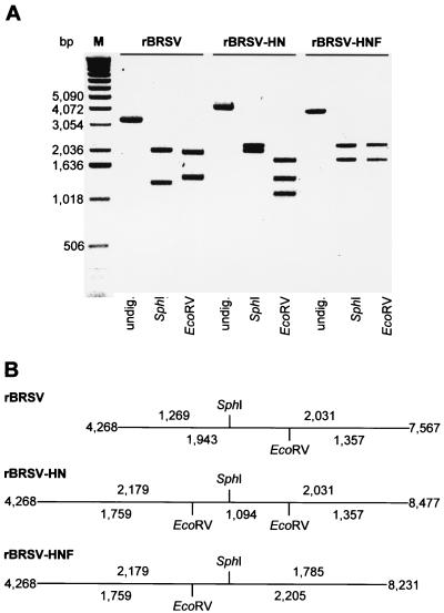
Stocks of rBRSV-HN and rBRSV-HNF were grown on MDBK cells, and total RNAs of rBRSV-HN- and rBRSV-HNF-infected MDBK cells were prepared for RT-PCR. The identity of the recombinant viruses was verified by restriction endonuclease digestion of the virus-specific RT-PCR fragments. All recombinant viruses contained a synthetic SphI restriction site at nt position 5,536 (rBRSV) or nt position 6,446 (rBRSV-HN and rBRSV-HNF), proving the recombinant origin of the viruses. The BPIV-3 HN ORF contained an EcoRV site at nt position 1,332, and the rBRSV F ORF contained an EcoRV site at nt position 641. Parallel digestion of the fragments yielded the expected restriction patterns (Fig. 2). No PCR products were generated in reactions from which the reverse transcriptase was omitted (not shown).
FIG. 2.
Demonstration of marker restriction sites in the genomes of rBRSV, rBRSV-HN, and rBRSV-HNF, confirming the identity of the recombinant viruses. (A) RT-PCR with primers encompassing the glycoprotein genes was performed on RNA of infected cells, submitted to restriction analysis, separated on a 0.8% agarose gel, and stained with ethidium bromide. M, 1-kb DNA ladder (Gibco BRL); fragment size is indicated in base pairs (bp). (B) Schematic diagram of amplified PCR products, with horizontal lines representing the amplified RT-PCR products and vertical bars representing the respective restriction sites. The sizes (in nucleotides) of the resulting DNA fragments are indicated above and below the lines. The positions of the fragments in the genomes of the parental or chimeric virus are indicated on the left and on the right.
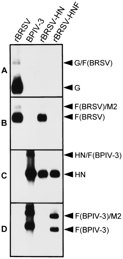
The BPIV-3 glycoprotein ORFs were flanked by BRSV transcription signals and thus are transcribed by the BRSV polymerase as monocistronic, capped, and polyadenylated mRNAs. To verify the correct expression of BPIV-3 transcription units in rBRSV, the presence of these mRNAs transcripts was examined by Northern blot hybridization of RNA isolated from infected MDBK cells. Hybridization with G (BRSV) (Fig. 3A)-, F (BRSV) (Fig. 3B)-, or HN (BPIV-3) (Fig. 3C)-specific probes revealed the presence of primarily monocistronic G (BRSV) mRNA in rBRSV-infected cells, monocistronic F (BRSV) mRNA in rBRSV or rBRSV-HN preparations, or HN (BPIV-3) mRNA in cells infected with BPIV-3, rBRSV-HN, and rBRSV-HNF. Using F (BPIV-3)-specific probes, monocistronic F (BPIV-3) mRNA was detected in BPIV-3- or rBRSV-HNF-infected cells (Fig. 3D). In rBRSV-HNF RNA preparations, a second mRNA corresponding to the size of bicistronic M2 (BRSV)/F (BPIV-3) readthrough products was present. The size differences of the mRNAs which contain the BPIV-3 F ORF are due to the large noncoding regions flanking the BPIV-3 ORF. These noncoding regions are present in the BPIV-3 genes but not in the synthetic BRSV/BPIV-3 genes.
FIG. 3.
Demonstration of viral transcripts by Northern hybridization. Total RNA of cells infected with the indicated viruses was isolated and separated by gel electrophoresis on a denaturing 1% agarose gel, blotted onto nitrocellulose, UV-cross-linked, and hybridized with [α-32P]dCTP-labeled specific probes. (A) BRSV G-specific probe; (B) BRSV F-specific probe; (C) BPIV-3 HN-specific probe; (D) BPIV-3 F-specific probe. mRNA transcripts are indicated by arrowheads on the right.
Characterization of recombinant virions.
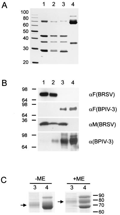
The recombinant virions and BPIV-3 were purified by sucrose gradient centrifugation. The virus preparations were checked for purity by negative-contrast electron microscopy. In preparations of rBRSV, rBRSV-HN, and rBRSV-HNF, pleomorphic virus particles were detected, with filaments of several micrometers in length being present in all preparations, whereas purified preparations of BPIV-3 exclusively contained spherical particles (not shown). The protein composition of purified rBRSV, rBRSV-HN, rBRSV-HNF, and BPIV-3 was analyzed by SDS-PAGE and immunoblotting. Coomassie staining (Fig. 4A and C) and reaction with specific antisera (Fig. 4B) confirmed that all virus preparations were of high purity and had the expected protein composition. BRSV F protein was present in rBRSV and rBRSV-HN, whereas BPIV-3 F protein was detected in rBRSV-HNF and BPIV-3. HN was detected in rBRSV-HN by an anti-BPIV-3 hyperimmune serum. Unfortunately, the sizes of HN and BPIV-3 F are very similar under nonreducing conditions and a reagent specific for HN under reducing conditions was not available, so the presence of HN in rBRSV-HNF could not be demonstrated by immunoblotting. However, after reduction with β-mercaptoethanol, the F protein disassembles into two fragments so that the HN is exposed, with a characteristic shift from 70 kDa to a slightly higher apparent molecular size (8) (Fig. 4C). Additional evidence for the presence of HN in the chimeras was obtained from a hemagglutination assay (HA) performed with dilutions of the purified virus preparations. As expected, no hemagglutination could be observed with rBRSV up to concentrations of 0.1 mg/ml. HA titers were high (1:4,096, corresponding to a virus protein concentration of 24 ng/ml) for BPIV-3. HA titers for rBRSV-HN were 1:32 (3.1 μg/ml) and 1:512 for rBRSV-HNF (0.19 μg/ml). Thus, BPIV-3 HN protein is not only expressed by cells infected with the chimeras but is also efficiently incorporated into the virion.
FIG. 4.
Protein composition of purified rBRSV, BPIV-3, and chimeras. Samples containing 10 μg of protein from purified rBRSV (lanes 1), rBRSV-HN (lanes 2), rBRSV-HNF (lanes 3), and BPIV-3 (lanes 4) were subjected to SDS-PAGE and stained with Coomassie brilliant blue R 250 (panels A and C) or processed for immunoblotting (panel B). Molecular sizes of marker proteins are indicated in kilodaltons. (A) Expected major structural proteins N (42 kDa), P (36 kDa), and M (28 kDa) of BRSV (lanes 1, 2, and 3). BRSV F migrates with an apparent molecular size of 71 kDa (lanes 1 and 2). HN, NP, and F protein of BPIV-3 migrate in the range of 65 to 75 kDa, and the M protein of BPIV-3 migrates at approximately 35 kDa. (B) Only rBRSV and rBRSV-HN react with a monoclonal antibody directed against BRSV F, whereas using the antiserum raised against BPIV-3 F, a specific band is detected in rBRSV-HNF and BPIV-3. Reaction with a BRSV M-specific antiserum yields a 28-kDa band exclusively in rBRSV and in the two chimeras. Among several bands that are stained by the calf hyperimmune serum directed against BPIV-3, one can be identified as the HN protein migrating at approximately 70 kDa in rBRSV-HN. Evidence for the presence of HN in rBRSV-HNF is gained from comparison of a Coomassie-stained gel (C) with samples of rBRSV-HNF and BPIV-3 run under nonreducing (−ME) and reducing (+ME) conditions. Reduction leads to disintegration of the F protein into the F1 and F2 subunits and exposes the diffuse HN band which shifts from approximately 70 kDa to a slightly higher apparent size (arrows). ME, mercaptoethanol.
Confocal laser scan analysis.
To demonstrate the localization of BPIV-3 glycoproteins HN and F which are expressed by the chimeric viruses, a bovine cell line originating from nasal mucosa, KNS-R, was used. For double-staining experiments, cells were fixed and permeabilized 72 h after infection with rBRSV, BPIV-3, rBRSV-HN, or rBRSV-HNF and incubated in parallel with a rabbit hyperimmune serum directed against the BRSV M protein and a calf hyperimmune serum specific for BPIV-3, followed by staining with Cy3-conjugated goat anti-rabbit antibody and FITC-conjugated goat anti-bovine antibody. Antibodies specific to BRSV G and F were used as controls.
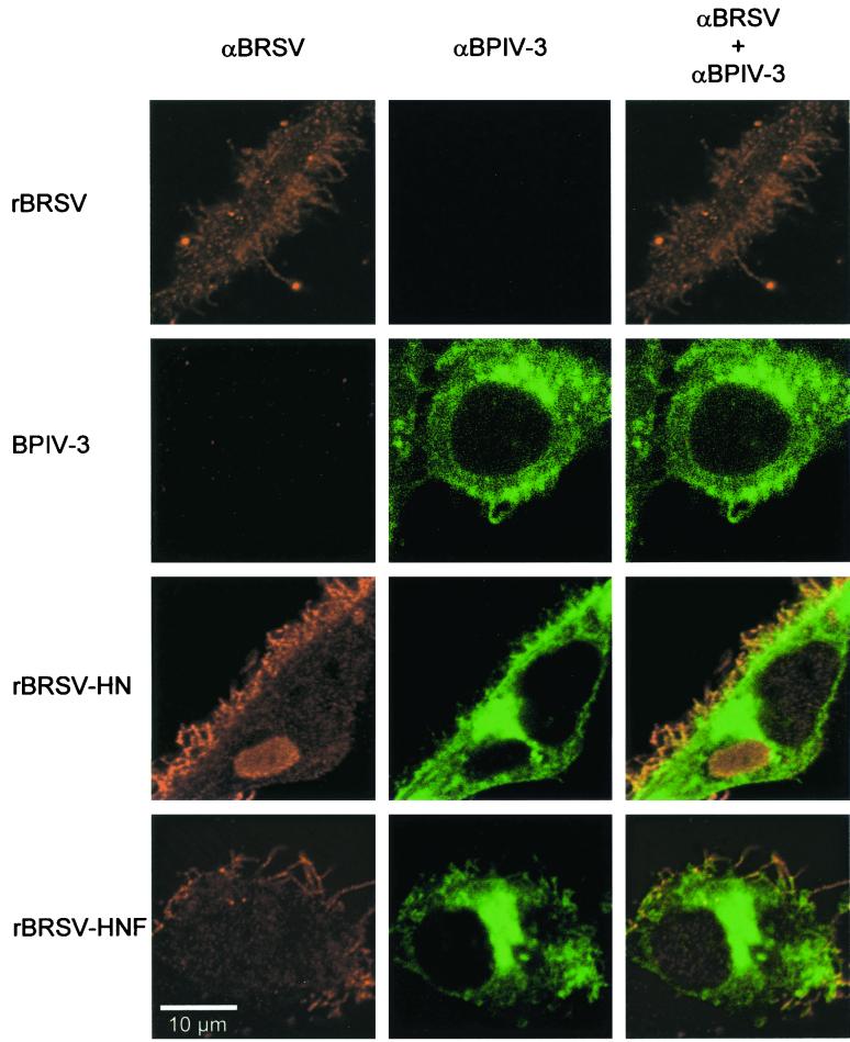
Confocal laser scan analysis showed that rBRSV, as well as rBRSV-HN and rBRSV-HNF, formed filamentous particles budding from the surface of infected cells. In the case of the chimeras, the filaments were double stained by antibodies specific to BRSV M, which colocalized with a fluorescence signal obtained from the BPIV-3-specific hyperimmune serum. The BPIV-3-specific hyperimmune serum (Fig. 5, middle panels) yielded an intracytoplasmic staining pattern in rBRSV-HN-infected cells, which might represent the location of BPIV-3 HN in the Golgi network, and a staining of filaments several micrometers in length. These filaments were also observed when a hyperimmune serum directed to BRSV M was used (Fig. 5, left panels). In addition, intracytoplasmic inclusion bodies typical for RSV were stained by the hyperimmune serum to M. A double staining of filaments was detected in the merged image (Fig. 5, right panels), indicating a colocalization of BPIV-3 proteins and BRSV M in the chimeric viruses, budding from infected cells. A similar colocalized staining of filaments was obtained from rBRSV-HN-infected controls which were double stained with a monoclonal antibody specific to BRSV F and M, but no signal was obtained from controls stained with a monoclonal antibody specific to BRSV G (not shown).
FIG. 5.
Colocalization of the BRSV M protein and of BPIV-3 glycoproteins in budding virus filaments. KNS-R cells were infected with the viruses indicated on the left, incubated for 3 days, fixed with 4% paraformaldehyde, and permeabilized with Triton X-100. Cells were incubated with a hyperimmune serum against BPIV-3 (αBPIV-3) and in parallel with a rabbit hyperimmune serum directed against the BRSV M protein (αBRSV) and stained with the FITC-conjugated goat anti-bovine antibody and a Cy3-conjugated goat anti-rabbit antibody. Cells were examined with a Zeiss LSM 510 confocal microscope. The merged image shows that BRSV M protein colocalizes with BPIV-3 proteins in filaments budding from the cell surface.
From cells 72 h after infection with rBRSV-HNF, no fluorescence signal was obtained using monoclonal antibodies to the BRSV G and F protein. No fluorescence staining was observed using the hyperimmune serum specific to BRSV M, the BPIV-3-specific hyperimmune serum, or using antibodies specific to BRSV F and G in cells infected with the heterologous parental virus (not shown).
Neutralization tests.
Neutralization assays were performed to characterize the role of the heterologous glycoproteins for infection (Table 1). Using a calf hyperimmune serum against BPIV-3, complement-independent neutralization of rBRSV-HNF and BPIV-3 strain INT2 was detected, with neutralizing titers against rBRSV-HNF (3,072) and BPIV-3 strain INT2 (6,144) being comparable. Using a calf hyperimmune serum specific to BRSV, both rBRSV and rBRSV-HN were neutralized with identical titers of 256, whereas no neutralizing activity for rBRSV-HNF and BPIV-3 strain INT2 was detected. This indicates that infection of rBRSV-HN, which is neutralized by a BRSV-specific hyperimmune serum but not by a hyperimmune serum specific to BPIV-3, is mediated by BRSV F. Moreover, when expressed in place of BRSV G, the BPIV-3 HN protein alone seems not to have a significant function in virus attachment and entry that is sensitive to neutralization. In contrast, in the case of rBRSV-HNF, infection is mediated by the BPIV-3 HN and F glycoproteins, giving evidence of correct incorporation in the BRSV envelope and of correct conformation and functionality of the heterologous glycoproteins. Taken together, these results indicate that BPIV-3 F and HN can cooperatively mediate infection of chimeric rBRSVs in vitro.
TABLE 1.
Neutralization titers (ND50)
| Virusa | Titer (ND50) using hyperimmune serum
|
|
|---|---|---|
| αBPIV-3b | αBRSVc | |
| rBRSV | 2 | 256 |
| BPIV-3 | 6,144 | 4 |
| rBRSV-HN | 2 | 256 |
| rBRSV-HNF | 3,072 | 4 |
A total of 100 tissue culture infectious doses was incubated with an equal volume of serum dilutions.
αBPIV-3, calf hyperimmune serum specific for BPIV-3.
αBRSV, hyperimmune serum specific to BRSV, prepared in a gnotobiotic calf.
BPIV-3 HN glycoprotein confers hemadsorption activity to BRSV.
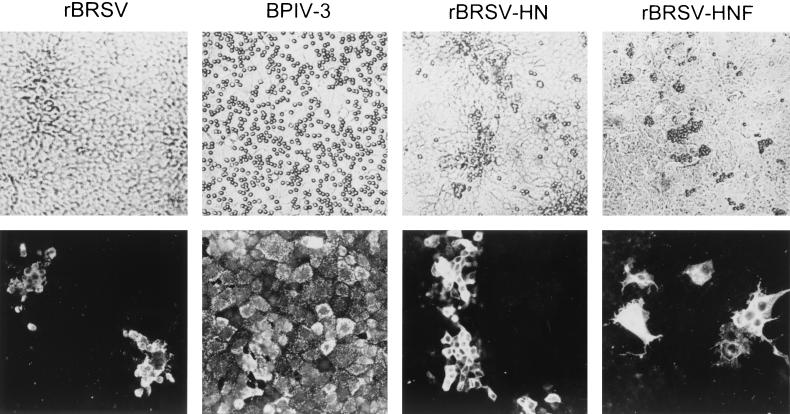
To demonstrate whether the BPIV-3 HN protein expressed by the substitution mutants was functionally active, hemadsorption of infected cells was examined. MDBK monolayers infected either with rBRSV-HN or rBRSV-HNF were incubated at 4°C with guinea pig erythrocytes. Cells infected with rBRSV-HN or rBRSV-HNF showed a similar hemadsorption pattern (Fig. 6, top), corresponding to the pattern of foci of infected cells as detected by indirect immunofluorescence using specific antisera (Fig. 6, bottom). BPIV-3 strain INT2, used as positive control, showed a more diffuse distribution of adsorbed erythrocytes, reflecting the distribution of infected cells in the monolayer. Bound erythrocytes were eluted from the monolayer infected with rBRSV-HN, rBRSV-HNF, or BPIV-3 strain INT2 when cells were incubated at 37°C for 1 additional h (data not shown), which can be taken as indirect evidence for neuraminidase activity of the HN protein, although a cellular neuraminidase activity cannot be excluded. Thus, the BPIV-3 HN protein is functional with respect to hemagglutination and, most likely, neuraminidase function in the background of chimeric BRSVs.
FIG. 6.
Hemadsorption of chimeric rBRSV expressing BPIV-3 HN protein. (Top) Three days after infection with rBRSV, BPIV-3, rBRSV-HN, or rBRSV-HNF, MDBK monolayers were incubated at 4°C with guinea pig erythrocytes. The cells were washed four times with ice-cold PBS. Cells were examined microscopically. Adsorption of bound erythrocytes was detected on the surface of cells infected with BPIV-3, rBRSV-HN, and rBRSV-HNF. (Bottom) Duplicates were fixed with 80% acetone and incubated with a calf hyperimmune serum to BRSV (rBRSV) or a calf hyperimmune serum to BPIV-3 (in the case of BPIV-3, rBRSV-HN, and rBRSV-HNF) and then stained with FITC-conjugated goat anti-bovine antibody. The distribution of bound erythrocytes corresponded to the distribution of infected cells in the monolayer.
In vitro growth of recombinant viruses.
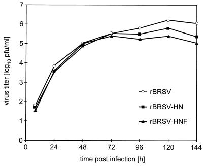
Virus replication characteristics were analyzed by multistep growth curves in MDBK cells. rBRSV-HN and rBRSV-HNF did not differ from rBRSV in their growth kinetics, with the exception of final infectious titers which were somewhat lower (Fig. 7). Thus, the chimeric viruses are competent for multicycle growth in cell culture, showing that BRSV glycoproteins G and F can be functionally replaced by their BPIV-3 counterparts, the BPIV-3 HN and F proteins.
FIG. 7.
Multicycle growth of chimeric BRSVs in MDBK cells. Duplicate MDBK monolayers in 24-well dishes were infected with rBRSV, rBRSV-HN, or rBRSV-HNF at an MOI of 0.01, harvested at the indicated times, and titrated in duplicate. Each value is the mean titer from two wells of infected cells.
DISCUSSION
Here, we described the generation of chimeric rBRSV in which the G gene alone, or the G and the F gene together, were replaced by their BPIV-3 counterparts, HN or both the HN and F genes. Surprisingly, when replaced together, the heterologous glycoproteins were not only expressed by BRSV, but were incorporated into the envelope of BRSV, and were functionally able to replace the BRSV glycoproteins, with the cell culture growth characteristics of the chimera being similar to the kinetics of parental BRSV. These results show that viral glycoproteins derived from a member of the Respirovirus genus can function in a Pneumovirus background. However, no viable virus could be obtained when the BRSV F gene alone was replaced by BPIV-3 F.
There are a number of reports of recombinant negative-strand RNA viruses which were designed to express heterologous viral glycoproteins in addition to or instead of the homologous envelope glycoprotein(s). These studies were done with two main intentions: first, to design novel vaccines, making use of members of the order Mononegavirales as vectors which express heterologous antigens (4, 12, 13, 35, 46). Second, the molecular mechanisms involved in particle formation, budding, and fusion of paramyxoviruses were characterized (19, 36). The glycoprotein substitution mutants presented in this work were used to characterize the interaction of BPIV-3 glycoproteins with BRSV as a first step on the way to develop an attenuated bivalent live vaccine against the two most important viral pathogens in the bovine respiratory tract, BRSV and BPIV-3.
We have shown that BRSV, like other members of the order Mononegavirales, is able to express heterologous sequences (4). Genome modifications are not dependent on the “rule of six,” which does not apply to members of the Pneumovirus genus (22). In the case of the chimeras presented here, the incorporation of artificial genes which comprise the conserved BRSV gene start signal, and a BPIV-3 ORF and a BRSV gene end signal led to viral mRNA transcription, as shown by Northern blotting, and protein expression, as demonstrated by indirect immunofluorescence assays with antibodies specific to the BPIV-3 proteins. RSV forms filamentous particles budding from infected cells (1), which can be visualized by confocal laser scan microscopy (20). In cell cultures infected with rBRSV or with the chimeras rBRSV-HN and rBRSV-HNF, similar filamentous cell protrusions were present. Moreover, colocalization of the BRSV M protein and of the respective heterologous glycoproteins was detected in the filamentous particles. When looking at the functions of the heterologous glycoproteins, we found that BPIV-3 HN which is expressed together with BRSV F is functional with respect to hemadsorption. From neutralization experiments using specific sera it could be concluded that in the case of rBRSV-HN, infection is mediated primarily by BRSV F, with no detectable additional role for BPIV-3 HN. In contrast, neutralization of rBRSV-HNF was only achieved by hyperimmune sera specific to BPIV-3, indicating that BPIV-3 HN and F are functional and mediate infection of BRSV when expressed together. However, BPIV-3 F seems not to be functional if expressed alone instead of BRSV F, since the mutant rBRSV-F could not be recovered from cDNA.
The current model of negative-strand RNA virus envelope protein interactions is that the matrix proteins specifically interact with cytoplasmic tails of the envelope glycoproteins (31) which are incorporated into the host cell-derived viral envelope. Between the matrix proteins of BRSV and BPIV-3, regions of sequence homology are absent, something that is also the case in glycoprotein cytoplasmic tails. The RSV G protein does not share structural or functional homologies with attachment glycoproteins of other members of the order Mononegavirales (33, 47). Therefore, it is surprising that Respirovirus glycoproteins HN and F are not only incorporated into the BRSV envelope in the absence of BRSV G and F proteins but that they are fully functional in a BRSV background. The BRSV F protein is essential for BRSV, whereas the BRSV G gene can be deleted with only little influence on cell culture growth of BRSV (20). Thus, in the absence of the BRSV F protein, functions necessary for adsorption and entry have to be provided by the BPIV-3 glycoproteins. The underlying mechanism of interaction seems to give rise to a relatively stable viral envelope. It is conceivable that there is an interaction between the BRSV M protein and the BPIV-3 glycoprotein cytoplasmic tails which is purely conformational, representing a conserved structure rather than a conserved sequence, allowing the functional incorporation of glycoproteins of a distantly related virus. The interactions of BPIV-3 F and HN proteins with BRSV M protein are currently under investigation. Also, a possible additional role of actin with respect to interaction with M protein and glycoprotein cytoplasmic tails has to be considered, as actin filaments are known to be involved in budding of RSV (5) and paramyxoviruses (11), and actin is found to be present in purified RSV (5) as well as in paramyxoviruses (11).
There are reports of chimeric paramyxoviruses successfully recovered from cDNA that bear double glycoprotein replacements from members of the Paramyxovirinae subfamily. For example, BPIV-3 with glycoproteins replaced by glycoproteins of HPIV-3 and HPIV-3 glycoproteins replaced by glycoproteins of HPIV-1 gave rise to replication-competent viruses with replication kinetics similar to those of the parental virus (35, 40). When both surface glycoproteins of peste des petits ruminants virus were expressed in rinderpest virus, the resulting chimeric virus was heavily attenuated, even though the two species are closely related and even though there are amino acid homologies between the matrix proteins and the cytoplasmic tails of HN and F (12). When rinderpest virus was designed to express heterologous membrane-anchored marker proteins, it was found that these markers were excluded from the viral envelope (46). Also, in another report it was shown that when expressed by HPIV-3, measles virus HA was not incorporated into virions (13). Taken together, these data suggest that there is a rather specific mechanism of interaction between the Paramyxovirus envelope and glycoproteins. However, our work shows that a set of Paramyxovirus envelope glycoproteins, lacking any amino acid similarities to RSV glycoproteins and derived from a different subfamily of the order Mononegavirales, can functionally completely replace the Pneumovirus glycoproteins.
One characteristic feature of paramyxoviruses is that a highly type-specific interaction between F and HN is crucial for fusion activity (39, 42, 48). So far, the only exceptions are that the Morbillivirus glycoproteins of canine distemper virus and measles virus are functional for fusion when transiently expressed in heterotypic combinations (37); fusion was also observed after coexpression of the heterologous Respirovirus glycoprotein pair of HPIV-1 HN and Sendai virus F (2, 48). Due to the type-specific interaction of parainfluenza virus F and HN, it was not possible to generate viable recombinant virus from cDNA with HN and F derived from heterotypic parainfluenza virus serotypes (12). We show that expression of BPIV-3 HN in combination with BRSV F leads to viable virus, with infection mediated by BRSV F and hemadsorption conferred by expression of BPIV-3 HN. The confocal laser scan analysis and analysis of the protein composition of the virus particles suggest that the HN protein is incorporated into the chimeric envelope but from neutralization experiments a possible role of HN in attachment seems rather unlikely. Whether the BPIV-3 HN protein in combination with BRSV F is of any function for BRSV virions remains unclear, since it was shown that the RSV F alone is necessary and sufficient to yield infection-competent virus (20, 21).
This work represents the starting point toward the development of a bivalent live vaccine against the two most important viral causes of respiratory disease in cattle. In a first step, we were able to show that the BPIV-3 glycoproteins are expressed by BRSV and that they can, due to a functional incorporation into the viral envelope, replace the homologous BRSV glycoproteins. In a next step, we aim to combine both sets of glycoproteins to address the question whether homologous glycoproteins will be preferentially incorporated into recombinant virions. The in vitro characteristics of the chimeras will be studied, and the immune response that is produced in cattle will be evaluated.
ACKNOWLEDGMENTS
We thank Geraldine Taylor, Compton, United Kingdom, José Antonio Melero, Madrid, Spain, and Horst Schirrmeier, Insel Riems, Germany, for gifts of monoclonal antibodies and antisera. We thank Matthias Lenk, Insel Riems, for assistance with confocal microscopy and Thomas C. Mettenleiter, Insel Riems, for helpful comments on the manuscript. We thank Harald Granzow, Insel Riems, for the electron microscopy.
This work was supported by the Intervet International B.V., The Netherlands.
REFERENCES
- 1.Bächi T. Direct observation of the budding and fusion of an enveloped virus by video microscopy of viable cells. J Cell Biol. 1988;107:1689–1695. doi: 10.1083/jcb.107.5.1689. [DOI] [PMC free article] [PubMed] [Google Scholar]
- 2.Bousse T, Takimoto T, Gorman W L, Takahashi T, Portner A. Regions on the hemagglutinin-neuraminidase proteins of human parainfluenza virus type-1 and Sendai virus important for membrane fusion. Virology. 1994;204:506–514. doi: 10.1006/viro.1994.1564. [DOI] [PubMed] [Google Scholar]
- 3.Buchholz U J, Finke S, Conzelmann K-K. Generation of bovine respiratory syncytial virus (BRSV) from cDNA: BRSV NS2 is not essential for virus replication in tissue culture, and the human RSV leader region acts as a functional BRSV genome promoter. J Virol. 1999;73:251–259. doi: 10.1128/jvi.73.1.251-259.1999. [DOI] [PMC free article] [PubMed] [Google Scholar]
- 4.Buchholz U J, Granzow H, Schuldt K, Whitehead S S, Murphy B R, Collins P L. Chimeric bovine respiratory syncytial virus with glycoprotein gene substitution from human respiratory syncytial virus (HRSV): effects on host range and evaluation as a live-attenuated HRSV Vaccine. J Virol. 2000;74:1187–1199. doi: 10.1128/jvi.74.3.1187-1199.2000. [DOI] [PMC free article] [PubMed] [Google Scholar]
- 5.Burke E, Dupuy L, Wall C, Barik S. Role of cellular actin in the gene expression and morphogenesis of human respiratory syncytial virus. Virology. 1998;252:137–148. doi: 10.1006/viro.1998.9471. [DOI] [PubMed] [Google Scholar]
- 6.Collins P L, McIntosh K, Chanock R M. Respiratory syncytial virus. In: Fields B N, Knipe D M, Howley P M, Chanock R M, Melnick J L, Monath T P, Roizman B, Straus S E, editors. Fields virology. 3rd ed. Vol. 2. Philadelphia, Pa: Lippincott-Raven; 1996. pp. 1313–1352. [Google Scholar]
- 7.Collins P L, Hill M G, Camargo E, Grosfeld H, Chanock R M, Murphy B R. Production of infectious human respiratory syncytial virus from cloned cDNA confirms an essential role for the transcription elongation factor from the 5′ proximal open reading frame of the M2 mRNA in gene expression and provides a capability for vaccine development. Proc Natl Acad Sci USA. 1995;92:11663–11667. doi: 10.1073/pnas.92.25.11563. [DOI] [PMC free article] [PubMed] [Google Scholar]
- 8.Collins P L, Mottet G. Homo-oligomerization of the hemagglutinin-neuraminidase glycoprotein of human parainfluenza virus type 3 occurs before the acquisition of correct intramolecular disulfide bonds and mature immunoreactivity. J Virol. 1991;65:2362–2371. doi: 10.1128/jvi.65.5.2362-2371.1991. [DOI] [PMC free article] [PubMed] [Google Scholar]
- 9.Collins P L, Hill M G, Cristina J, Grosfeld H. Transcription elongation factor of respiratory syncytial virus, a nonsegmented negative-strand RNA virus. Proc Natl Acad Sci USA. 1996;93:81–85. doi: 10.1073/pnas.93.1.81. [DOI] [PMC free article] [PubMed] [Google Scholar]
- 10.Conzelmann K-K. Nonsegmented negative-strand RNA viruses: genetics and manipulation of viral genomes. Annu Rev Genet. 1998;32:123–162. doi: 10.1146/annurev.genet.32.1.123. [DOI] [PubMed] [Google Scholar]
- 11.Cudmore S, Reckmann I, Way M. Viral manipulations of the actin cytoskeleton. Trends Microbiol. 1997;5:142–148. doi: 10.1016/S0966-842X(97)01011-1. [DOI] [PubMed] [Google Scholar]
- 12.Das S C, Baron M D, Barrett T. Recovery and characterization of a chimeric rinderpest virus with the glycoproteins of peste-des-petits-ruminants virus: homologous F and H proteins are required for virus viability. J Virol. 2000;74:9039–9047. doi: 10.1128/jvi.74.19.9039-9047.2000. [DOI] [PMC free article] [PubMed] [Google Scholar]
- 13.Durbin A, Skiadopulos M H, McAuliffe J M, Riggs J M, Surman S R, Collins P L, Murphy B R. Human parainfluenza virus type 3 (PIV3) expressing the hemagglutinin protein of measles virus provides a potential method for immunization against measles virus and PIV3 in early infancy. J Virol. 2000;74:6821–6831. doi: 10.1128/jvi.74.15.6821-6831.2000. [DOI] [PMC free article] [PubMed] [Google Scholar]
- 14.Fernie B F, Gerin J L. The stabilization and purification of respiratory syncytial virus using MgSO4. Virology. 1980;106:141–144. doi: 10.1016/0042-6822(80)90229-9. [DOI] [PubMed] [Google Scholar]
- 15.Furze J, Wertz G, Lerch R, Taylor G. Antigenic heterogeneity of the attachment protein of bovine respiratory syncytial virus. J Gen Virol. 1994;75:363–370. doi: 10.1099/0022-1317-75-2-363. [DOI] [PubMed] [Google Scholar]
- 16.García-Barreno B, Palomo C, Penas C, Delgado T, Perez-Brena P, Melero J A. Marked differences in the antigenic structure of human respiratory syncytial virus F and G glycoproteins. J Virol. 1989;63:925–932. doi: 10.1128/jvi.63.2.925-932.1989. [DOI] [PMC free article] [PubMed] [Google Scholar]
- 17.Grosfeld H, Hill M G, Collins P L. RNA replication by respiratory syncytial virus (RSV) is directed by the N, P, and L proteins; transcription also occurs under these conditions but requires RSV superinfection for efficient synthesis of full-length mRNA. J Virol. 1995;69:5677–5686. doi: 10.1128/jvi.69.9.5677-5686.1995. [DOI] [PMC free article] [PubMed] [Google Scholar]
- 18.Hardy R W, Wertz G W. The product of the respiratory syncytial virus M2 gene ORF1 enhances readthrough of intergenic junctions during viral transcription. J Virol. 1998;72:520–526. doi: 10.1128/jvi.72.1.520-526.1998. [DOI] [PMC free article] [PubMed] [Google Scholar]
- 19.Kahn J S, Schnell M J, Buonocore L, Rose J K. Recombinant vesicular stomatitis virus expressing respiratory syncytial virus (RSV) glycoproteins: RSV fusion protein can mediate infection and cell fusion. Virology. 1999;254:81–91. doi: 10.1006/viro.1998.9535. [DOI] [PubMed] [Google Scholar]
- 20.Karger A, Schmidt U, Buchholz U J. Recombinant bovine respiratory syncytial virus with deletions of the G or SH genes: G and F proteins bind heparin. J Gen Virol. 2001;82:631–640. doi: 10.1099/0022-1317-82-3-631. [DOI] [PubMed] [Google Scholar]
- 21.Karron R A, Buonagurio D A, Georgiu A G, Whitehead S S, Adamus J E, Clements-Mann M L, Harris D O, Randolph V B, Udem S A, Murphy B R, Sidhu M S. Respiratory syncytial virus (RSV) SH and G proteins are not essential for viral replication in vitro: clinical evaluation and molecular characterization of a cold-passaged, attenuated RSV subgroup B mutant. Proc Natl Acad Sci USA. 1997;94:13961–13966. doi: 10.1073/pnas.94.25.13961. [DOI] [PMC free article] [PubMed] [Google Scholar]
- 22.Kolakofsky D, Pelet T, Garcin D, Hausmann S, Curran J, Roux L. Paramyxovirus RNA synthesis and the requirement for hexamer genome length: the rule of six revisited. J Virol. 1998;72:891–899. doi: 10.1128/jvi.72.2.891-899.1998. [DOI] [PMC free article] [PubMed] [Google Scholar]
- 23.Kuo L, Grosfeld H, Cristina J, Hill M G, Collins P L. Effect of mutations in the gene-start and gene-end sequence motifs on transcription of monocistronic and dicistronic minigenomes of respiratory syncytial virus. J Virol. 1996;70:6892–6901. doi: 10.1128/jvi.70.10.6892-6901.1996. [DOI] [PMC free article] [PubMed] [Google Scholar]
- 24.Kuo L, Fearns R, Collins P L. Analysis of the gene start and gene end signals of human respiratory syncytial virus: quasi-templated initiation at position 1 of the encoded mRNA. J Virol. 1997;71:4944–4953. doi: 10.1128/jvi.71.7.4944-4953.1997. [DOI] [PMC free article] [PubMed] [Google Scholar]
- 25.Laemmli U K. Cleavage of structural proteins during the assembly of the head of bacteriophage T4. Nature. 1970;227:680–685. doi: 10.1038/227680a0. [DOI] [PubMed] [Google Scholar]
- 26.Langedijk J P M, Meloen R H, Taylor G, Furze J M, van Oirschot J T. Antigenic structure of the central conserved region of protein G of bovine respiratory syncytial virus. J Virol. 1997;71:4055–4061. doi: 10.1128/jvi.71.5.4055-4061.1997. [DOI] [PMC free article] [PubMed] [Google Scholar]
- 27.Lerch R A, Anderson K, Wertz G W. Nucleotide sequence analysis and expression from recombinant vectors demonstrate that the attachment protein G of bovine respiratory syncytial virus is distinct from that of human respiratory syncytial virus. J Virol. 1990;64:5559–5569. doi: 10.1128/jvi.64.11.5559-5569.1990. [DOI] [PMC free article] [PubMed] [Google Scholar]
- 28.Levine S, Klaiber-Franco R, Paradiso P R. Demonstration that glycoprotein G is the attachment protein of respiratory syncytial virus. J Gen Virol. 1987;68:2521–2524. doi: 10.1099/0022-1317-68-9-2521. [DOI] [PubMed] [Google Scholar]
- 29.Mbiguino A, Menezes J. Purification of human respiratory syncytial virus: superiority of sucrose gradient over percoll, renografin, and metrizamide gradients. J Virol Methods. 1991;31:161–170. doi: 10.1016/0166-0934(91)90154-r. [DOI] [PubMed] [Google Scholar]
- 30.Palese P, Zheng H, Engelhardt O G, Pleschak S, Garcia-Sastre A. Negative-strand RNA viruses: genetic engineering and applications. Proc Natl Acad Sci USA. 1996;93:11354–11358. doi: 10.1073/pnas.93.21.11354. [DOI] [PMC free article] [PubMed] [Google Scholar]
- 31.Peeples M E. Paramyxovirus M proteins: pulling it all together and taking it on the road. In: Kingsbury D W, editor. The paramyxoviruses. New York, N.Y: Plenum Press; 1991. [Google Scholar]
- 32.Sambrook J, Fritsch E F, Maniatis T. Molecular cloning: a laboratory manual. Cold Spring Harbor, N.Y: Cold Spring Harbor Laboratory Press; 1989. [Google Scholar]
- 33.Satake M, Coligan J E, Elango N, Norrby E, Venkatesan S. Respiratory syncytial virus envelope glycoprotein (G) has a novel structure. Nucleic Acids Res. 1985;13:7795–7812. doi: 10.1093/nar/13.21.7795. [DOI] [PMC free article] [PubMed] [Google Scholar]
- 34.Schlender J, Bossert B, Buchholz U, Conzelmann K-K. Bovine respiratory syncytial virus nonstructural proteins NS1 and NS2 cooperatively antagonize alpha/beta interferon-induced antiviral response. J Virol. 2000;74:8234–8242. doi: 10.1128/jvi.74.18.8234-8242.2000. [DOI] [PMC free article] [PubMed] [Google Scholar]
- 35.Schmidt A C, McAuliffe J M, Huang A, Surman S R, Bailly J E, Elkins W R, Collins P L, Murphy B R, Skiadopoulos M H. Bovine parainfluenza virus type 3 (BPIV3) fusion and hemagglutinin-neuraminidase glycoproteins make an important contribution to the restricted replication of BPIV3 in primates. J Virol. 2000;74:8922–8929. doi: 10.1128/jvi.74.19.8922-8929.2000. [DOI] [PMC free article] [PubMed] [Google Scholar]
- 36.Spielhofer P, Bächi T, Fehr T, Christiansen G, Cattaneo R, Kaelin K, Billeter M A, Naim H Y. Chimeric measles viruses with a foreign envelope. J Virol. 1998;72:2160–2169. doi: 10.1128/jvi.72.3.2150-2159.1998. [DOI] [PMC free article] [PubMed] [Google Scholar]
- 37.Stern L B, Greenberg M, Gershoni J M, Rozenblatt S. The hemagglutinin envelope protein of canine distemper virus (CDV) confers cell tropism as illustrated by CDV and measles virus complementation analysis. J Virol. 1995;69:1661–1668. doi: 10.1128/jvi.69.3.1661-1668.1995. [DOI] [PMC free article] [PubMed] [Google Scholar]
- 38.Stoschek C M. Quantitation of protein. Methods Enzymol. 1990;182:50–68. doi: 10.1016/0076-6879(90)82008-p. [DOI] [PubMed] [Google Scholar]
- 39.Tanabayashi K, Compans R W. Functional interaction of paramyxovirus glycoproteins: identification of a domain in Sendai virus HN which promotes cell fusion. J Virol. 1996;70:6112–6118. doi: 10.1128/jvi.70.9.6112-6118.1996. [DOI] [PMC free article] [PubMed] [Google Scholar]
- 40.Tao T, Durbin A P, Whitehead S S, Davoodi F, Collins P L, Murphy B R. Recovery of a fully viable chimeric human parainfluenza virus (PIV) type 3 in which the hemagglutinin-neuraminidase and fusion glycoproteins have been replaced by those of PIV type 1. J Virol. 1998;72:2955–2961. doi: 10.1128/jvi.72.4.2955-2961.1998. [DOI] [PMC free article] [PubMed] [Google Scholar]
- 41.Teng M N, Collins P L. Identification of the respiratory syncytial virus proteins required for formation and passage of helper-dependent infectious particles. J Virol. 1998;72:5707–5716. doi: 10.1128/jvi.72.7.5707-5716.1998. [DOI] [PMC free article] [PubMed] [Google Scholar]
- 42.Tong S, Compans R W. Alternative mechanisms of interaction between homotypic and heterotypic parainfluenza virus HN and F proteins. J Gen Virol. 1999;80:107–115. doi: 10.1099/0022-1317-80-1-107. [DOI] [PubMed] [Google Scholar]
- 43.Towbin H, Staehelin T, Gordon J. Electrophoretic transfer of proteins from polyacrylamide gels to nitrocellulose sheets: procedure and some applications. Proc Natl Acad Sci USA. 1979;76:4350–4354. doi: 10.1073/pnas.76.9.4350. [DOI] [PMC free article] [PubMed] [Google Scholar]
- 44.Van der Poel W H, Brand A, Kramps J A, van Oirschot J T. Respiratory syncytial virus infections in human beings and in cattle. An epidemiological review. J Infect. 1994;29:216–228. doi: 10.1016/s0163-4453(94)90866-4. [DOI] [PubMed] [Google Scholar]
- 45.Walsh E E, Hruska J. Monoclonal antibodies to respiratory syncytial virus proteins: identification of the fusion protein. J Virol. 1983;47:171–177. doi: 10.1128/jvi.47.1.171-177.1983. [DOI] [PMC free article] [PubMed] [Google Scholar]
- 46.Walsh E P, Baron M D, Rennie L F, Monaghan P, Anderson J, Barrett T. Recombinant rinderpest vaccines expressing membrane-anchored proteins as genetic markers: evidence of exclusion of marker protein from the virus envelope. J Virol. 2000;74:10165–10175. doi: 10.1128/jvi.74.21.10165-10175.2000. [DOI] [PMC free article] [PubMed] [Google Scholar]
- 47.Wertz G W, Collins P L, Huang Y, Gruber C, Levine S, Ball L A. Nucleotide sequence of the G protein gene of human respiratory syncytial virus reveals an unusual type of viral membrane protein. Proc Natl Acad Sci USA. 1985;82:4075–4079. doi: 10.1073/pnas.82.12.4075. [DOI] [PMC free article] [PubMed] [Google Scholar]
- 48.Yao Q, Hu X, Compans R W. Association of the parainfluenza virus fusion and hemagglutinin-neuraminidase glycoproteins on cell surfaces. J Virol. 1997;71:650–656. doi: 10.1128/jvi.71.1.650-656.1997. [DOI] [PMC free article] [PubMed] [Google Scholar]
- 49.Yu Q, Hardy R W, Wertz G W. Functional cDNA clones of the human respiratory syncytial (RS) virus N, P, and L proteins support replication of RS virus genomic RNA analogs and define minimal trans-acting requirements for RNA replication. J Virol. 1995;69:2412–2419. doi: 10.1128/jvi.69.4.2412-2419.1995. [DOI] [PMC free article] [PubMed] [Google Scholar]
- 50.Zamora M, Samal S K. Gene junction sequences of bovine respiratory syncytial virus. Virus Res. 1992;24:116–121. doi: 10.1016/0168-1702(92)90035-8. [DOI] [PubMed] [Google Scholar]


