



Abstract
Background
Organophosphate-Induced Delayed Neuropathy (OPIDN) is a rare neurological disorder triggered by exposure to organophosphorus compounds. These compounds exert their neurotoxic effects by impacting the nervous system, leading to systemic manifestations. Urinary system symptoms are infrequently observed in clinical settings. Currently, effective therapeutic interventions for OPIDN-related urinary symptoms are lacking. Sacral nerve modulation therapy, an FDA-approved approach for managing lower urinary tract symptoms, presents as a promising option. Herein, we present a case of OPIDN-induced lower urinary tract obstruction successfully treated with sacral nerve modulation therapy, resulting in substantial symptom relief.
Case report
A 27-year-old male patient presented with severe bilateral hydronephrosis, attributed to low bladder compliance and accompanied by a fever persisting for 6 days. The patient’s medical history revealed accidental ingestion of organophosphate pesticide (Dimethoate) with no concomitant underlying diseases. In consideration of the potential for OPIDN, surgical intervention in the form of sacral neuromodulation (phase I) was undertaken. Subsequent evaluation one month post-surgery revealed notable improvements in both bladder compliance and bilateral hydronephrosis, necessitating sacral neuromodulation (phase II). Presently, following a 5-month follow-up period, the patient remains asymptomatic and in favorable health.
Conclusion
This patient achieved long-term relief using sacral neuromodulation.
Keywords: Organophosphate-induced delayed neuropathy (OPIDN), Sacral neuromodulation, Neurogenic bladder, Organophosphorus compounds, Peripheral nerve axons
Background
Organophosphorus compounds are a category of chemical substances widely utilized in agricultural and industrial sectors, garnering significant attention due to their inherent toxicity. Organophosphate poisoning manifests both as an acute cholinergic crisis and in an intermediate phase, with certain individuals being particularly vulnerable to organophosphate-induced delayed neuropathy (OPIDN). This neuropathy, triggered by organophosphorus compounds, entails neural damage extending beyond the confines of the central nervous system, implicating the peripheral nervous system as well. The regulation of lower urinary tract function primarily hinges on three sets of peripheral nerves, and any impairment or affliction of the nervous system can lead to lower urinary tract dysfunction.
In the presented case, sacral nerve modulation effectively transitioned the patient’s small-capacity, high-pressure bladder into a large-capacity, low-pressure bladder, thereby alleviating the patient’s symptoms. Currently, research on organophosphate-induced delayed neuropathy concerning the urinary system remains relatively limited. This paper aims to provide additional insights into understanding and addressing this clinical challenge, while also fostering advancements in related research endeavors geared towards enhancing the quality of life for affected individuals.
Case report
A 27-year-old male, presented with intermittent low back pain and fever persisting for six days. Six days earlier, the patient had undergone a CT scan of the upper abdomen, along with routine blood and urine tests at a local hospital, due to back pain and fever. The results indicated severe hydronephrosis with ureteral dilatation in both kidneys, alongside elevated leukocyte counts. The diagnosis was bilateral hydronephrosis accompanied by a urinary tract infection. Despite receiving anti-infection treatment, the patient’s symptoms persisted, necessitating admission to our hospital. Upon admission, the patient was administered anti-inflammatory treatment and fitted with an indwelling catheter for one week. One week later, a follow-up urologic CT examination revealed persistent hydronephrosis in both kidneys and the bilateral ureters, along with bladder wall thickening (Fig. 1). Further urodynamic evaluation revealed several issues: increased bladder sensitivity, decreased compliance, and reduced detrusor muscle contraction, which required abdominal pressure to aid urination. During the storage phase, the bladder’s initial sensation was noted at 36 ml, with a strong urge at 54 ml and a maximum infusion volume of 140 ml. The patient was instructed to void, but no urine was produced. After the removal of the pressure catheter, the maximum urine flow rate was 8 ml/s, and the voided volume was 50 ml. Following catheter insertion, 300 ml of urine was drained, likely due to ureteral expansion (Fig. 2A). Urethrocystography revealed a characteristic “pear-shaped” bladder pattern (Fig. 3). The physical examination did not reveal any specific signs. The patient’s medical history was not reported. At age 14, the patient accidentally ingested a poison known as Dimethoate and underwent dialysis treatment at a local hospital. Currently, the patient’s renal function is normal.
Fig. 1.

Pretreatment CT-Abdomen demonstrating hydronephrosis in both kidneys and the bilateral ureters (A) and bladder wall thickening and bladder capacity reduction(B)
Fig. 2.

Pre-test urodynamics showed the following: During the storage phase, the bladder’s initial sensation occurred at 36 ml, with a strong urge at 54 ml and a maximum infusion volume of 140 ml. The patient was instructed to urinate, but no urine was produced. After removing the pressure catheter, the maximum urine flow rate was 8 ml/s, with a urine volume of 50 ml. Following catheter insertion, 300 ml of urine was drained, likely due to the expansion of the ureter (A).Post-second-stage implantation, urodynamic review showed the following: During the storage phase, the bladder’s initial sensation occurred at 134 ml, with a strong urge at 269 ml and a maximum infusion volume of 316 ml. The maximum urine flow rate was 16 ml/s, with a total urine volume of 310 ml and a residual urine volume of 10 ml (B)
Fig. 3.
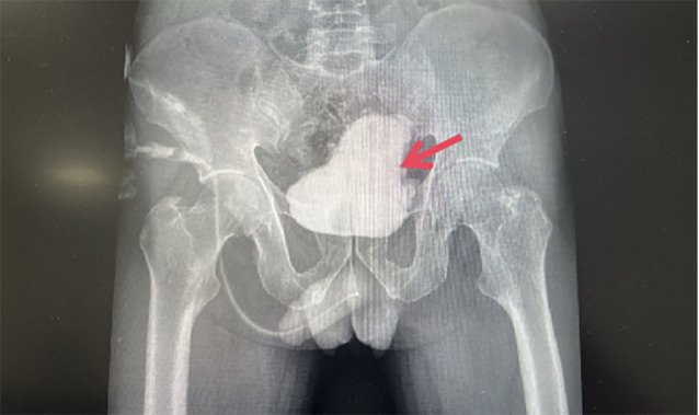
Cystography demonstrating pear bladder
Based on medical history and auxiliary examinations, the patient was diagnosed with a neurogenic bladder, low-compliance bladder, and vesicoureteral reflux. To resolve the patient’s bilateral hydronephrosis, it was necessary to improve bladder compliance. In July 2023, our institution performed a phase I (test phase) sacral nerve electrical stimulation implantation on the patient (Fig. 4). Postoperative parameters recorded included a pulse width of 210 µs, a voltage of 0.5 V, and a frequency of 14 Hz. One month post-surgery, follow-up imaging and urodynamic studies revealed the following results: During the storage phase, the bladder’s initial sensation was at 134 ml, with a strong urge occurring at 269 ml and a maximum infusion volume of 316 ml. The voided urine volume was 310 ml, and the residual bladder volume was 10 ml. Bladder capacity increased from 150 ml before the procedure to 320 ml afterward. There was no evidence of ureteral reflux, and the maximum urine flow rate improved from 8 ml/s prior to the procedure to 16 ml/s postoperatively (Fig. 2B). A comparison of cystography and urological CT findings indicated a reduction in bladder wall thickness and significant increases in bladder capacity and compliance post-test (Fig. 5). Additionally, the characteristic ‘pear-shaped’ bladder morphology was no longer present. The patient experienced significant symptomatic relief following the evaluation and was referred for the second stage of permanent stimulator implantation. As of now, after five months of follow-up, the patient has shown symptom improvement and positive outcomes.
Fig. 4.
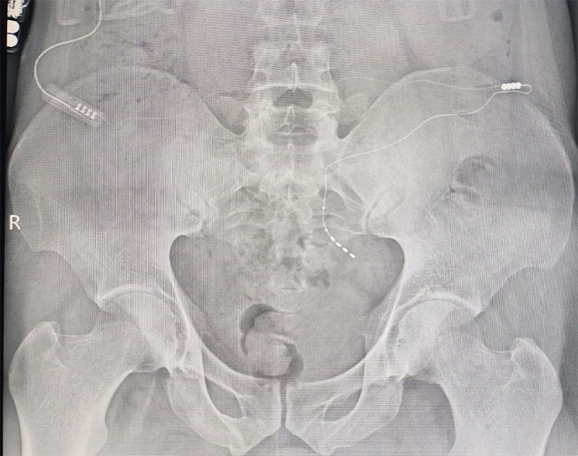
After electrical stimulation of sacral nerve implantation
Fig. 5.

Posttreatment CT-Abdomen demonstrating hydronephrosis relief in both. kidneys and the bilateral ureters (A) and bladder wall thinning and bladder capacity. Expanding (B). Cystography demonstrating pear bladder disappeared (C)
Discussion
Organophosphate-induced delayed neuropathy (OPIDN) is a neurological disorder induced by exposure to organophosphorus compounds. The pathogenesis of delayed-onset peripheral neuropathy remains incompletely understood. Some researchers propose that exposure to organophosphorus compounds inhibits neurotoxic esterase (NTE) activity in axons, leading to their aging and disrupted energy metabolism in axonal transport. This disruption can impair transport, induce degenerative changes in axons, and potentially cause demyelinating disease [1]. Additionally, organophosphorus compounds are thought to disrupt calcium ion/calmodulin kinase II activity, resulting in disturbed calcium homeostasis and the degeneration of peripheral nerve axons [2].
Research indicates that exposure to organophosphorus compounds may result in peripheral nerve damage, manifesting after a latency period of 1–8 weeks as chronic progressive lesions [3]. A cohort epidemiological study using vibration sensitivity testing found that motor nerve conduction velocity (NCV) decreases following exposure to organophosphorus compounds [4], before the appearance of clinical signs of peripheral neuropathy or muscle weakness. The literature suggests that OPIDN begins with a latency period of 1 to 3 weeks, leading to a progressive phase characterized by motor-sensory neuropathy. This phase involves motor or sensory-motor weakness in the lower extremities. Subsequently, a resting phase occurs, allowing for partial recovery of motor functions. Ultimately, symptoms indicating the involvement of cones and the central nervous system intensify. Although less frequently discussed [5], OPIDN can also impact the urinary system.
Urine storage and release are dependent on the activity of smooth and striated muscles in the bladder, urethra, and external urethral sphincter [6]. However, the coordination of these functions is regulated by a complex neural control system in the brain, spinal cord, and peripheral ganglia [7]. Regulation of these muscles is primarily managed by three groups of peripheral nerves: sacral parasympathetic, thoracolumbar sympathetic, and somatic nerves distributed bilaterally. Lower urinary tract dysfunction can result from nervous system injuries or diseases [8]. Urinary function is susceptible to a range of injuries, diseases, and chemicals impacting the nervous system [9]. Bladder dysfunction is often attributed to the degeneration of axons in peripheral and central nerves [10]. Thus, neurological mechanisms must be considered in the diagnosis and treatment of voiding disorders.
The patient had a history of Dimethoate usage. Cystoscopy revealed no bladder outlet obstruction or other underlying conditions. Despite the absence of overt clinical symptoms, imaging findings indicated a chronic progression. Consequently, the patient’s condition was deemed closely associated with Dimethoate use. The patient displayed high bladder pressure during storage and low urine flow rate during voiding, typical of a neurogenic bladder.
Sacral neuromodulation, involving electrode placement in the S3 or S4 sacral foramen and continuous low-frequency electrical stimulation of the sacral nerves, is a therapeutic technique leading to the denervation of the bladder, urethral sphincter, and pelvic floor muscles [11]. It is currently FDA-approved for treating refractory overactive bladder (OAB), non-obstructive urinary retention (NOR), and fecal incontinence [12], and has shown effectiveness in treating neurogenic lower urinary tract dysfunction (NLUTD), interstitial cystitis, bladder pain syndrome (IC/BPS), and other bowel dysfunctions [13, 14]. Neurogenic bladder, however, is a complex syndrome with various etiologies and factors, leading to diverse urodynamic and clinical manifestations. Thus, a thorough preoperative evaluation of neurogenic bladder across different clinical manifestations is crucial for positive postoperative outcomes. The primary goal in treating neurogenic bladder is to preserve upper urinary tract function [15]. Consequently, the main treatment objective was to enhance bladder compliance. In this instance, severe bladder hypo-compliance resulted in reduced bladder capacity and the development of bilateral hydronephrosis. The study noted an increase in maximum bladder capacity from 150 ml to 320 ml pre- and post-test period. Additionally, there was an 8 ml/s increase in the maximum urinary flow rate pre- and post-test period. CT scans demonstrated a significant reduction in bilateral hydronephrosis pre- and post-test periods. Bladder elasticity significantly improved, with no ureteral reflux observed. Consequently, progression to the second stage of permanent stimulator implantation was warranted.
Ensuring treatment longevity necessitates strict management of the post-sacral neurotomy regimen. While there was an increase in bladder capacity and a decrease in residual urine volume post-test, studies indicate that patients previously experiencing chronic urinary retention, despite spontaneous urination post-implantation, continue to depend on abdominal pressure to void and remain at risk for hydronephrosis and vesicoureteral reflux [16]. Post-operatively, patients were advised to consume 1500 ml of water daily, maintain a urinary voiding diary, and undergo regular follow-ups and urodynamic evaluations. To prevent transient ureteric reflux, intermittent catheterization was recommended. Follow-up results were positively noted.
Conclusion
In this instance of OPIDN presenting as a neurogenic lower urinary tract disorder, sacral neuromodulation (SNM) effectively transformed the patient’s small, high-pressure bladder into one with a larger capacity and lower pressure. This outcome, in conjunction with postoperative management, mitigated the patient’s bilateral renal and ureteral hydronephrosis caused by reflux, thereby safeguarding the upper urinary tract. Consequently, SNM proves to be a safe and efficacious therapy for neurogenic bladder in these circumstances.
Acknowledgements
Thank for the patients in this research, thank for all the scholars in this article. Thank for all the teammates for supporting this research. We are also particularly grateful to our colleagues in The Second Affiliated Hospital of Jilin University for their contributions.
Abbreviations
- OPIDN Organophosphate
Induced Delayed Neuropathy
- FDA
Food and Drug Administration
- CT
Computed tomography
- ML
Milliliter
- US
Microsecond
- V
Volt
- HZ
Hertz
- NTE
Neurotoxic esterase
- NCV
Nerve conduction velocity
- NOR
Non-obstructive urinary retention
- NLUTD
Neurogenic lower urinary tract dysfunction
- IC/BPS
Interstitial cystitis/Bladder pain syndrome
- SNM
Sacral neuromodulation
Author contributions
JJH, DLZ, ZHD: preparing manuscript. JJH, XSY, YCW, ZS, ZYD: patient management, providing diagnostic and treatment results. SQF, YZ: providing diagnostic and treatment results. RWL: Article Guidance. All authors contributed to the article and approved the submitted version.
Funding
Not applicable.
Data availability
All data generated or analyzed during this study are included in this published article.
Declarations
Ethics approval and consent to participate
The study was conducted according to the guidelines of the Declaration of Helsinki and approved by the Ethics Committee of The Second Hospital of Jilin University (Changchun, China). Written informed consent was obtained from the patient.
Consent for publication
Written informed consent was obtained from the patient for publication of the data and images in this case report.
Competing interests
The authors declare no competing interests.
Footnotes
Publisher’s note
Springer Nature remains neutral with regard to jurisdictional claims in published maps and institutional affiliations.
Contributor Information
Yi Zhang, Email: yizhang0502@jlu.edu.cn.
Ranwei Li, Email: ranwei1968@sina.com.
References
- 1.Richardson RJ, Fink JK, Glynn P, Hufnagel RB, Makhaeva GF, Wijeyesakere SJ. Neuropathy target esterase (NTE/PNPLA6) and organophosphorus compound-induced delayed neurotoxicity (OPIDN). Adv Neurotoxicol. 2020;4:1–78. [DOI] [PMC free article] [PubMed] [Google Scholar]
- 2.Ding Q, Fang S, Chen X, Wang Y, Li J, Tian F, et al. TRPA1 channel mediates organophosphate-induced delayed neuropathy. Cell Discovery. 2017;3:17024. [DOI] [PMC free article] [PubMed] [Google Scholar]
- 3.Mendes PA, Pereira TC, Pina R, Santos R. Chlorpyrifos-induced delayed neurotoxicity with a rare presentation of flaccid quadriplegia: a diagnostic challenge. Eur J Case Rep Intern Med. 2018;5:751. [DOI] [PMC free article] [PubMed] [Google Scholar]
- 4.Pannu AK, Bhalla A, Vishnu RI, Dhibar DP, Sharma N, Vijayvergiya R. Organophosphate induced delayed neuropathy after an acute cholinergic crisis in self-poisoning. Clin Toxicol (Phila Pa). 2021;59:488–92. [DOI] [PubMed] [Google Scholar]
- 5.Nayak P, Mallick AK, Mishra S, Panigrahy D. Organophosphorus-induced toxic myeloneuropathy: series of three adolescent patients with short review. J Pediatr Neurosci. 2019;14:42–5. [DOI] [PMC free article] [PubMed] [Google Scholar]
- 6.Panicker JN, Fowler CJ, Kessler TM. Lower urinary tract dysfunction in the neurological patient: clinical assessment and management. Lancet Neurol. 2015;14:720–32. [DOI] [PubMed] [Google Scholar]
- 7.Tudor KI, Sakakibara R, Panicker JN. Neurogenic lower urinary tract dysfunction: evaluation and management. J Neurol. 2016;263:2555–64. [DOI] [PubMed] [Google Scholar]
- 8.Podnar S, Vodušek DB. Lower urinary tract dysfunction in patients with peripheral nervous system lesions. Handb Clin Neurol. 2015;130:203–24. [DOI] [PubMed] [Google Scholar]
- 9.New frontiers of basic science research in. neurogenic lower urinary tract dysfunction - PubMed. https://pubmed.ncbi.nlm.nih.gov/28716328/. Accessed 13 Apr 2024.
- 10.Smith PP, DeAngelis A, Kuchel GA. Detrusor expulsive strength is preserved, but responsiveness to bladder filling and urinary sensitivity is diminished in the aging mouse. Am J Physiol Regul Integr Comp Physiol. 2012;302:R577–586. [DOI] [PMC free article] [PubMed] [Google Scholar]
- 11.Barboglio Romo PG, Gupta P. Peripheral and sacral neuromodulation in the treatment of neurogenic lower urinary tract dysfunction. Urol Clin N Am. 2017;44:453–61. [DOI] [PubMed] [Google Scholar]
- 12.Tanagho EA, Schmidt RA. Bladder pacemaker: scientific basis and clinical future. Urology. 1982;20:614–9. [DOI] [PubMed] [Google Scholar]
- 13.Hanno PM, Erickson D, Moldwin R, Faraday MM, American Urological Association. Diagnosis and treatment of interstitial cystitis/bladder pain syndrome: AUA guideline amendment. J Urol. 2015;193:1545–53. [DOI] [PubMed] [Google Scholar]
- 14.Moore CK, Rueb JJ, Derisavifard S. What is new in neuromodulation? Curr Urol Rep. 2019;20:55. [DOI] [PubMed] [Google Scholar]
- 15.Wiener JS, Chaudhry R. Neurogenic lower urinary tract dysfunction. Urol Clin N Am. 2023;50:415–32. [DOI] [PubMed] [Google Scholar]
- 16.Chen G, Liao L, Deng H. The effect of sacral neuromodulation in ambulatory spina bifida patients with neurogenic bladder and bowel dysfunction. Urology. 2021;153:345–50. [DOI] [PubMed] [Google Scholar]
Associated Data
This section collects any data citations, data availability statements, or supplementary materials included in this article.
Data Availability Statement
All data generated or analyzed during this study are included in this published article.