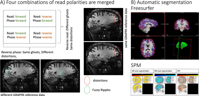
Fig. 2: Merging four combinations of read and phase polarity imaging for tissue-type segmentation.
A) Example image showing artifacts that are mitigated by combining read and phase polarities. The ellipses highlight artifact levels that are benignly inverted by reversing read and phase polarity. Green ellipses indicate “fuzzy ripples”, a low spatial frequency artifact caused by gradient trajectory imperfections at EPI ramp-sampling corners. For an animated version of this figure that more clearly depicts the fuzzy ripples, see here: https://github.com/layerfMRI/repository/blob/master/T1234/animation_2.gif
B-C) After combining the four images, the data can be processed through mainstream automatic segmentation pipelines. Representative results from FreeSurfer and SPM are shown, respectively.

